Introduction
Immunoglobulin G4 related disease is a recently described clinicopathological entity characterised by extensive tissue infiltration by immunoglobulin G4 (IgG4) positive plasma cells and T lymphocytes.Reference Kamisawa and Okamoto1 It is known to affect various organs including the pancreas, liver and lungs, as well as the salivary and lacrimal glands and pituitary gland in the head and neck region.Reference Kamisawa and Okamoto1, Reference Cheuk and Chan2 The sinonasal cavity is an extremely rare site for this disease and has previously only been reported to be involved in association with other sites.Reference Cheuk and Chan2
Case report
This report was approved by the institutional review boards of the Western Sydney Local Health District Human Research Ethics Committee and the Westmead Scientific Advisory Committee. Informed consent was obtained from the patient, prior to chart review.
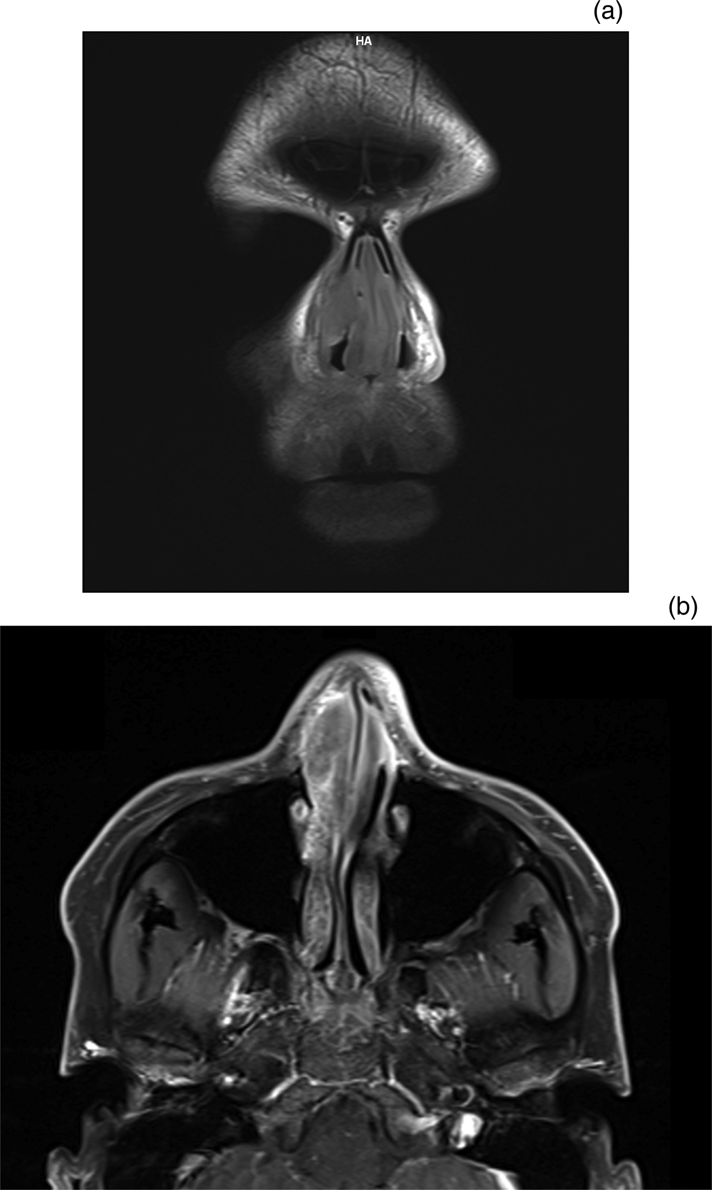
A 34-year-old man presented with a 2-year history of progressively increasing, painless, bilateral nasal obstruction and right external nasal swelling, with no other sinonasal symptoms. Clinical examination, computed tomography and magnetic resonance imaging findings (Figure 1) demonstrated a firm submucosal mass, present bilaterally over the anterior septum, with intact underlying cartilage. A synchronous, firm, subcutaneous mass was identified over the right lateral nasal wall. Initial conservative biopsy results were inconclusive. Extensive submucosal and mucosal tissue, and septal cartilage, were dissected under general anaesthesia and sent for histology.
Coronal, T1-weighted (a) and axial, T1-weighted, fluid-attenuated inversion recovery (b) magnetic resonance imaging scans, demonstrating a contrast-enhanced submucosal mass over the anterior septum bilaterally. H = head; A = anterior

Histopathology
Microscopic examination revealed fibrosclerosing tissue containing dense aggregates of mixed inflammatory cells, particularly lymphocytes and plasma cells, along with histiocytes and scattered eosinophils, often centred around prominent endothelial cells. These findings indicated obliterative phlebitis (Figure 2). Based on these characteristic histological findings, immunostaining for IgG4 and immunoglobulin G (IgG) was undertaken. Results revealed numerous plasma cells positive for IgG4 (more than 50 cells per high power field) (Figure 3). The ratio of IgG4-positive to IgG-positive cells was over 40 per cent, confirming the diagnosis of IgG4-related disease.Reference Cheuk and Chan2, Reference Deshpande, Zen, Chan, Yi, Sato and Yoshino3
(a) Low power photomicrograph demonstrating fibrosclerosis (black arrow) with mixed inflammatory cells (white arrow) (H&E; ×100), and (b) high power photomicrograph demonstrating inflammatory cells (black arrow indicates a typical plasma cell within a cluster of plasma cells) (H&E; ×400).
Immunostaining of plasma cells for immunoglobulin G4 (IgG4) (a) was compared with immunostaining for total immunoglobulin G (IgG) (b); the ratio of IgG4-positive to IgG-positive cells was over 40 per cent. (×400)
Management
Clinical examination and imaging did not reveal any further lesions within the abdominal cavity or elsewhere. Serum IgG4 was within the normal range at 2.30 g/l (normal range, 0.06–2.56 g/l). The patient was referred for immunology review. The patient initially responded to high-dose oral steroids (40 mg prednisone daily), but relapsed when the dose was weaned. Long-term treatment with methotrexate (10 mg per week increasing to 20 mg per week), with folic acid (0.5 mg twice daily), resulted in good symptomatic relief. Repeat imaging at 18 months demonstrated a reduction in the volume of the involved tissue.
Discussion
This report demonstrates a case of IgG4-related disease isolated solely to the nasal cavity. Immunoglobulin G4 is the least common of the four subclasses of IgG, typically representing 3–6 per cent of the entire IgG fraction.Reference Hamano, Kawa, Horiuchi, Unno, Furuya and Akamastsu4 It is thought to play a significant role in allergic reactions and parasitic infestations.Reference Cheuk and Chan2 Immunoglobulin G4 related disease was first proposed as a new clinicopathological entity in 2001, following the identification of high serum IgG4 concentrations in patients with autoimmune pancreatitis.Reference Kamisawa and Okamoto1 This led to the identification of pancreatic and extrapancreatic fibro-inflammatory lesions rich in IgG4-bearing cells. This rare condition has since been characterised by extensive tissue infiltration of various organs by IgG4-positive plasma cells and T lymphocytes. The aetiology remains unknown.Reference Cheuk and Chan2
The main pathological findings in IgG4-related disease include a dense lymphoplasmacytic infiltrate, a storiform pattern of fibrosis and obliterative phlebitis.Reference Cheuk and Chan2, Reference Deshpande, Zen, Chan, Yi, Sato and Yoshino3 Diagnosis relies on identification of these characteristic changes, along with high tissue IgG4 levels and/or an elevated IgG4 to total IgG ratio. Immunostaining for IgG4 is essential for diagnosis; however, the cut-off for the required number of IgG4 cells per high power field varies from 10 to 200, according to the site of the lesion and the degree of fibrosis.Reference Deshpande, Zen, Chan, Yi, Sato and Yoshino3 The IgG4-positive to IgG-positive ratio is a more specific measure, with a consensus figure of more than 40 per cent considered diagnostic.Reference Deshpande, Zen, Chan, Yi, Sato and Yoshino3 However, a raised IgG4-positive to IgG-positive ratio alone, without the associated characteristic histological findings, is insufficient for diagnosis. Our case meets all the established criteria for diagnosis.
Whilst it has been noted that increased serum IgG4 may assist in identification of the disease, it is not essential for or specific to this condition, with up to 40 per cent of patients demonstrating normal serum IgG4 levels.Reference Cheuk and Chan2, Reference Deshpande, Zen, Chan, Yi, Sato and Yoshino3, Reference Moteki, Yasuo, Hamano, Uehara and Usami5 Serum IgG4 has been correlated with disease activity and the number of involved organs.Reference Cheuk and Chan2 It is therefore unsurprising that our case, with disease isolated to the nasal cavity, did not demonstrate raised serum IgG4.
Immunoglobulin G4 related disease is most commonly found to affect the pancreas, gallbladder and bile duct. As regards the head and neck region, IgG4-related disease has been described in the submandibular gland (Kuttner's tumour), lacrimal and parotid glands (Mikulicz's disease), and pituitary gland (IgG4-related infundibulo-hypophysitis).Reference Cheuk and Chan2 With regard to the sinonasal region, two recent papers have reported cases of IgG4-related rhinosinusitis.Reference Moteki, Yasuo, Hamano, Uehara and Usami5, Reference Ikeda, Awataguchi, Shoji and Oshima6 To date, these cases of sinonasal involvement have been described in association with disease in other sites and all with raised serum IgG4 levels.Reference Moteki, Yasuo, Hamano, Uehara and Usami5, Reference Ikeda, Awataguchi, Shoji and Oshima6 The predominant symptoms were those of chronic rhinosinusitis, including rhinorrhoea (70 per cent) and postnasal drip (80 per cent), with only 30 per cent complaining of nasal obstruction. To our knowledge, there have been no previous reports of IgG4-related disease isolated solely to the nasal cavity.
The natural history of the condition typically, but not exclusively, results in multiple sites of involvement over time.Reference Cheuk and Chan2 The function of the involved tissues may be affected as a result of the inflammatory and infiltrative components of the condition. Immunoglobulin G4 related disease has been found to respond well to immunosuppressive medical management, namely steroid therapy.Reference Kamisawa and Okamoto1, Reference Cheuk and Chan2 Surgical intervention is thus largely limited to diagnostic evaluation via tissue biopsy.Reference Kamisawa and Okamoto1 Loss of function has been reported to have a component of reversibility with therapy; however, relapse is typical with steroid weaning or cessation.Reference Cheuk and Chan2 Accordingly, long-term therapy with steroid-sparing anti-metabolites, such as methotrexate, is often required.
• Immunoglobulin G4 (IgG4) related disease is characterised by extensive tissue infiltration by IgG4-positive plasma cells and T lymphocytes
• Presenting signs and symptoms vary according to the infiltration site
• In the sinonasal region, isolated deposits may result in nasal obstruction, whereas generalised infiltration may result in chronic rhinosinusitis symptoms
• Diagnosis is based on strict histological criteria and IgG4 immunostaining findings
• Immunosuppressive treatments, including oral steroids (short-term) and anti-metabolites (long-term, steroid-sparing), are effective in controlling this condition
Conclusion
To the best of our knowledge, this case represents the first report in the literature of IgG4-related disease isolated to the nasal cavity.
Acknowledgements
The authors would like to thank Professor Nicholas Manolios, from the Department of Rheumatology, Westmead Hospital, for his assistance and continued care of this patient.