Bovine mastitis is the most important disease in dairy cattle worldwide because of its economic impact. Mastitis causes inflammation of the mammary gland and usually originates with bacterial infection, commonly by Staphylococcus aureus, Streptococcus agalactiae, Escherichia coli, Streptococcus uberis, and coagulase-negative staphylococci. Mastitis-associated pathogens possess multiple virulence factors, among these, adherence to host epithelial cells is particularly important for the establishment of infection (Raspanti et al., Reference Raspanti, Bonetto, Vissio, Pellegrino, Reinoso, Dieser, Bogni, Larriestra and Odierno2016).
Dairy farms rely heavily on antibiotics to control and prevent bovine mastitis. However, their indiscriminate use poses substantial risks like residues in milk, and can lead to the selection and possible transmission of resistant bacterial strains. This has encouraged research into new antimicrobial compounds that may improve udder health, where metallic nanoparticles have recently become more relevant thanks to their unique properties (Nagarajan et al., Reference Nagarajan, Thamarai, Kamaraj, Al-Ghanim, Subramaniam and Malafaia2024). In particular, silver nanoparticles (AgNPs) are characterized by their potent activity against both gram-positive and gram-negative microorganisms and their safety to eukaryotic cells (Hussain et al., Reference Hussain, Singh, Singh, Singh and Singh2016). AgNPs can be obtained by physical and chemical methods but are expensive and require compounds that can be toxic to the environment. For this reason, biological or green synthesis of nanoparticles from plant extracts is a simple and eco-friendly method, since such extracts are non-toxic and contain reducing and stabilizing compounds (Velgosova et al., Reference Velgosova, Dolinská, Podolská, Mačák and Čižmárová2024).
In this work, green silver nanoparticles (G-AgNPs) were produced with an aqueous extract of chicory, and studied in terms of their activity against bovine mastitis-associated strains and their ability to inhibit the adherence capacity of these pathogens.
Materials and methods
Species identification of staphylococcus isolates by groEL PCR-RFLP
Nine pathogens corresponding to the genus Staphylococcus, which had been previously isolated from bovine milk samples (Dieser et al., Reference Dieser, Vissio, Lasagno, Bogni, Larriestra and Odierno2014), were selected and identified by PCR-RFLP analysis of a partial groEL gene sequence, as described by (Raspanti et al., Reference Raspanti, Bonetto, Vissio, Pellegrino, Reinoso, Dieser, Bogni, Larriestra and Odierno2016) and the online Supplementary Material.
Green synthesis of AgNPs
Green silver nanoparticles (G-AgNPs) were synthesized from AgNO3 (Cicarelli, Argentina) using an aqueous extract of Cichorium intybus (chicory) as a reducing and stabilizing agent, as described by (Ferreyra Maillard et al., Reference Ferreyra Maillard, Dalmasso, de Mishima Ba and Hollmann2018) and the online Supplementary Material.
Assessment of antibacterial activity by G-AgNPs
The antimicrobial activity of the G-AgNPs was evaluated by the disc diffusion method and the determination of the minimum inhibitory concentration. The antimicrobial activity was evaluated against the Staphylococcus species identified in the previous step and other previously isolated (Supplementary Material).
Bacterial adherence and inhibition of adherence assays
To assess the adherence ability of the bovine mastitis pathogens, assays were performed following (Almeida et al., Reference Almeida, Luther, Kumar, Calvinho, Bronze and Oliver1996) with modifications. The ability of G-AgNPs to inhibit pathogen adherence was evaluated on MAC-T cell monolayers preincubated with a G-AgNP concentration below the MIC (see online Supplementary Material).
Statistical analysis
The data from three independent adherence assays were expressed as the mean ± standard error. Comparisons between treatments were evaluated via ANOVA with a post-hoc Fisher's LSD test on the software InfoStat. A p-value < 0.05 was indicative of a statistically significant difference.
Results
Species identification of staphylococcus isolates by groEL PCR-RFLP analysis
An expected 550 bp fragment was obtained by amplifying genomic DNA from Staphylococcus isolates (Figure S1A). After digestion of the PCR products with AluI, clear species-specific RFLP patterns came up for nine isolates (Figure S1B,). Five strains were thus identified as S. aureus (size fragments: 14/78/204/258), two as S. simulans (size fragments: 46/114/147/246), and one as S. haemolyticus (size fragments: 9/107/438). Since the pattern for another isolate was compatible with S. capitis, S. hyicus, and S. chromogenes, a double enzymatic digestion was performed with HindIII/PvuII. In this manner, the isolate was finally identified as S. hyicus (size fragments: 14/539) (Figure S1C).
Green synthesis of AgNPs
The phytosynthesis of G-AgNPs was first confirmed visually by the colour change of colorless AgNO3 solution to yellowish brown (G-AgNPs) after the addition of the chicory extract (Fig. 1A). Further corroboration was provided by UV-vis extinction spectra: a single extinction band between 350-450 nm appeared, which corresponds to the characteristic surface plasmon resonance profile of spherical nanosilver (Fig. 1B). Through transmission electron microscropy (TEM), spherical G-AgNPs averaging 50 nm in size were observed (Fig. 1C). These results demonstrate that the aqueous extract of chicory leaves reduced the silver ions into uniformly sized nanoparticles and successfully stabilized silver colloidal dispersion.
(A) G-AgNPs synthesized with chicory extract. (B) uv-vis spectrum of G-AgNPs. (C) representative TEM micrograph of G-AgNPs.

Antimicrobial activity
The G-AgNPs had an inhibitory effect on all the strains evaluated: 65% of the strains were highly inhibited (++) and 35% were moderately inhibited (+). Among the Staphylococcus species, 82% were highly inhibited, whereas only 50% of the Streptococcus spp. were inhibited to this degree (Figure S2). For all the strains assessed, the MIC values ranged between 21 and 30 pM (Table S1). The MIC for S. aureus was higher than for the other Staphylococcus strains. In contrast, all the streptococcocci were inhibited by a similar concentration of G-AgNPs. As in the case of S. aureus, the MIC values for the E. coli strains were at the higher end of the range (28-30 pM).
Bacterial adherence
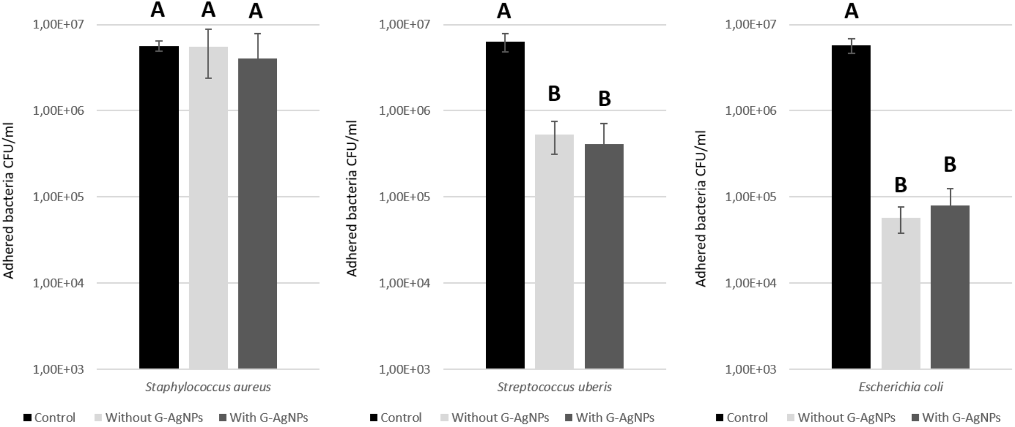
The highest level of adherence, 5.56 × 10⁶ (± 3.2) CFU/mL (98%), corresponded to S. aureus RC108. Lower values were recorded for the other pathogens: 5.31 × 10⁵ (± 2.2) CFU/mL (8.0%) for S. uberis and only 5.71 × 10⁴ (± 1.9) CFU/mL (1.0%) for E. coli (Fig. 2).
Mean values for bacterial strain adherence to MAC-T epithelial cells in the presence or absence of g-agnps. Each bar represents the arithmetic mean ± standard error (SEM) of the mean of three independent experiments performed in triplicate, expressed as cfu/ml.

Adherence was then tested in MAC-T cells that had been preincubated with the G-AgNPs at a sub-MIC concentration. Under these conditions, the value for S. aureus RC108 was 4.06 × 10⁶ (± 3.8) CFU/mL (73%), which is 25% lower than the adhesion observed for the S. aureus RC108 without G-AgNPs. For S. uberis, adherence in the presence of the nanoparticles was 4.13 × 10⁵ (± 3.0) CFU/mL (6.2%), which is a 1.8% reduction with respect to S. uberis without G-AgNPs. In contrast, the adhesion of E. coli was actually higher in the presence of the G-AgNPs than in their absence. Nevertheless, none of these observations constituted a statistically significant change in comparison with pathogens without G-AgNPs (p > 0.05).
Discussion
As a preliminary step, staphylococcal strains that had been previously isolated from milk samples of cows with mastitis were identified genotypically at species level, through a PCR-RFLP analysis of the groEL gene The PCR-RFLP analysis of the groEL gene, in particular, is a useful tool to correctly identify coagulase-negative staphylococci (Onni et al., Reference Onni, Vidili, Bandino, Marogna, Schianchi and Tola2012). In this study, the number and size of the fragments resulting from enzymatic digestion enabled the identification of the isolates as S. aureus, S. hyicus, S. haemolyticus and S. simulans, in agreement with other researchers (Barros et al., Reference Barros, Iório, Bastos, Dos Santos and Giambiagi-demarval2007; Raspanti et al., Reference Raspanti, Bonetto, Vissio, Pellegrino, Reinoso, Dieser, Bogni, Larriestra and Odierno2016).
Next, the bottom-up phytosynthesis of silver nanoparticles was carried out using an aqueous extract of chicory leaves as a reducing/stabilizing agent in a silver solution. The biomolecules in this extract, mainly polyphenols and flavonoids (Innocenti et al., Reference Innocenti, Gallori, Giaccherini, Ieri, Vincieri and Mulinacci2005), are responsible for the bioreduction of metal ions (Ag+ to Ag0), their growth, and their stabilization into a nanoform (Mittal et al., Reference Mittal, Chisti and Banerjee2013). The nanoparticles thus produced (G-AgNPs) had a strong inhibitory effect against the evaluated strains, at significantly low concentrations (of the order of picomolars, pM). Considering there was no appreciable antimicrobial activity in the control assays with synthesis precursors (aqueous leaf extract and 1 µM AgNO3 solution, data not shown), the inhibition measured when the nanoparticles were used is clearly attributable to them. Other researchers have similarly reported that biosynthesized AgNPs have broad-spectrum activity against gram-negative and gram-positive bacteria (Ferreyra Maillard et al., Reference Ferreyra Maillard, Dalmasso, de Mishima Ba and Hollmann2018; Salayová et al., Reference Salayová, Bedlovičová, Daneu, Baláž, Lukáčová Bujňáková, Balážová and Tkáčiková2021).
Various mechanisms have been proposed to explain the antibacterial activity of biosynthesized AgNPs, e.g. their interfacial affinity and interaction with bacterial lipid membranes (Ferreyra Maillard et al., Reference Ferreyra Maillard, Dalmasso, de Mishima Ba and Hollmann2018) andwere able to attach to and disrupt the bacterial envelope of S. aureus and E. coli strains, with the effect being more noticeable in E. coli (Ferreyra Maillard et al., Reference Ferreyra Maillard, Gonçalves, Santos, de Mishima Ba, Dalmasso and Hollmann2019). This indicates that bacterial lipid membranes could be one of the targets of nanosilver and may explain the inhibition observed in the present work. Moreover, (Quinteros et al., Reference Quinteros, Cano Aristizábal, Dalmasso, Paraje and Páez2016) demonstrated that exposure to biosynthesized AgNPs was linked to the generation of ROS in different bacteria. This means that the nanoparticles may produce the oxidation of macromolecules such as lipids, proteins and DNA, and thus lead to bacterial death.
An important factor in the development of bovine mastitis is the ability of pathogens to adhere to host cells, a virulence factor that has been reported for S. aureus (Al-Mebairik et al., Reference Al-Mebairik, El-Kersh, Al-Sheikh and Marie2016) and S. uberis (Fessia and Odierno, Reference Fessia and Odierno2021). However, the G-AgNPs did not have a significant effect on adherence to MAC-T cells in any the pathogens evaluated here. This could be due to the fact that the nanoparticles have a high affinity with bacterial membranes but not with eukaryotic cell membranes, as demonstrated in model membranes (Ferreyra Maillard et al., Reference Ferreyra Maillard, Dalmasso, de Mishima Ba and Hollmann2018).
In conclusion, the present study identified bovine mastitis-associated bacteria at the species level using a molecular technique. It also successfully synthesized AgNPs from a chicory leaf extract. Picomolar concentrations of these nanoparticles showed broad-spectrum action against all the pathogens evaluated. Although they could not significantly reduce bacterial adherence to MAC-T bovine mammary epithelial cells under the experimental conditions, their potent antibacterial effect at very low concentrations makes them a promising alternative for the control of bovine mastitis.
Supplementary material
The supplementary material for this article can be found at https://doi.org/10.1017/S0022029925101209.
Acknowledgements
This work was supported by grants from the Agencia Nacional de Promoción Científica y Tecnológica (ANPCyT-FonCyT). M. Pellegrino and P. Dalmasso are Career Researchers of the Consejo Nacional de Investigaciones Científicas y Técnicas (CONICET). A. P.V. Ferreyra Maillard and A. Fili are recipients of a fellowship from CONICET.
Competing interests
The authors have no conflicts of interest to declare.
Author contributions
APVFM and MSP conceived the study and participated in its design. APVFM carried out the experimental work and wrote the manuscript. PRD and MSP revised the manuscript. CR contributed specialist knowledge to the design of the molecular identification assay, and AF provided expertise in cell culturing. All the authors read and approved the final manuscript.