INTRODUCTION
Microsporidia are obligate intracellular fungal parasites, approximately 14 species of which infect humans (Didier and Weiss, Reference Didier and Weiss2006). The 1200 species that make up the phylum, Microsporidia, share a common stage, a spore that contains a unique coiled tube used to impale target cells and inject infectious sporoplasm (Keohane and Weiss, Reference Keohane, Weiss, Whittner and Weiss1999). Deployment of the tube is known as germination. Spores may also be phagocytosed, with germination taking place within the cell (Couzinet et al. Reference Couzinet, Cejas, Schittny, Deplazes, Weber and Zimmerli2000; Franzen, Reference Franzen2004). In the gastrointestinal tract, where the majority of human microsporidia infections occur (Kotler and Orenstein, Reference Kotler and Orenstein1998; Didier and Weiss, Reference Didier and Weiss2006), infection of differentiated mucosal epithelial cells most likely results from impalement via spores that germinate in the lumen in close proximity to the target cells (Leitch et al. Reference Leitch, Ward, Shaw and Newman2005), and inhibition of this germination process is an attractive target for therapy (Leitch et al. Reference Leitch, Visvesvara and He1993; Keohane and Weiss, Reference Keohane, Weiss, Whittner and Weiss1999).
In humans, environmental spores infect the gastrointestinal tract, nasopharynx and lungs, eyes and skin (Mathis et al. Reference Mathis, Weber and Deplazes2005; Didier and Weiss, Reference Didier and Weiss2006), all of which are protected by an arsenal of antimicrobial peptides (Hancock and Scott, Reference Hancock and Scott2000). While these peptides have been extensively studied for their antimicrobial effects against bacteria, some protozoa, viruses and other fungi, they have not been studied in the microsporidia. The present study employed spores from clinical isolates of 3 microsporidia species, Encephalitozoon intestinalis, the second most common cause of intestinal microsporidiosis (Kotler and Orenstein, Reference Kotler and Orenstein1998), a second Encephalitozoon species, E. hellem, known to cause ocular and disseminated infections (Didier and Weiss, Reference Didier and Weiss2006), and Anncaliia (Brachiola, Nosema) algerae, an insect parasite that has caused several human infections (Coyle et al. Reference Coyle, Weiss, Rhodes, Cali, Takvorian, Brown, Visvesvara, Xiao, Naktin, Young, Gareca, Colasante and Wittner2004; Visvesvara et al. Reference Visvesvara, Moura, Leitch, Schwartz and Xiao2005).
A germination assay and a cultured intestinal epithelial cell infection assay were used to determine whether antimicrobial peptides that are typically found in the gastrointestinal tract inhibited infection, whether any inhibition could be attributed to an inhibition of spore germination, and whether antimicrobial peptides account for the fact that only certain microsporidia species cause intestinal microsporidiosis.
MATERIALS AND METHODS
Microsporidia culture and spore isolation
Clinical isolates of Encephalitozoon intestinalis, Encephalitozoon hellem and Anncaliia algerae were used in this study. The E. intestinalis and E. hellem isolates were obtained from American Type Culture Collection (ATCC 50651 and 50451 respectively), while the A. algerae isolate (CDC: V422) was obtained from the Centers for Disease Control and Prevention, Atlanta, GA. The microsporidia cultures were maintained as described elsewhere using green monkey kidney, Vero cells, cultured at 37°C (Visvesvara et al. Reference Visvesvara, Moura, Leitch, Schwartz, Whittner and Weiss1999; Leitch and Ceballos, Reference Leitch and Ceballos2008), except that the A. algerae-infected cultures were maintained at 33°C to improve spore yield and because we found that human isolates of this species cultured at 30°C readily infected mammalian cells maintained at 37°C (Kucerova et al. Reference Kucerova, Moura, Visvesvara and Leitch2004). Spores were purified as described previously (Leitch et al. Reference Leitch, Ward, Shaw and Newman2005) and maintained in water at 4°C until used within 1 week of purification.
Germination assay
Purified spores were pre-treated with test solutions for 30 min at 37°C. Test solutions contained Lf (human milk), lactoferricin B or Lz (human milk) (Sigma Chemical Co., St Louis, MO) or recombinant human defensins (Peptides International, Louisville, KY). Peptide concentrations are given in μg/ml, except Lz which is given in thousand units/ml (concentration used, ⩾30 μg protein/ml). This pre-treatment was followed by exposure to a germination solution for an additional 30 min at 37°C and fixation with 5% neutral formalin. Fixed spore samples were then placed on chambered cover-slips, the spores allowed to settle overnight, and the percentage of germinated spores determined by phase-contrast microscopy as described previously (Leitch and Ceballos, Reference Leitch and Ceballos2008). Two germination solutions were used, most commonly a 140 mm NaCl solution buffered to pH 7·5 with 20 mm Hepes to which was added H2O2 to a final concentration of 5%. In some A. algerae experiments parallel assays were performed in which a 140 mm NaCl, pH 9·5, glycine-buffered solution was used. The high pH germination solution was previously used to germinate Brachiola (Anncaliia) algerae spores (Frixione et al. Reference Frixione, Ruiz, Cerbon and Undeen1997). However, we found no qualitative difference between the two germination solutions and have used the 5% H2O2 solution whenever we compared germination in A. algerae and the two Encephalitozoon species.
Infection assay
The Caco2 clone, C2Bbel (ATCC CRL-2102), was used in all infection assays as described elsewhere (Leitch and Ceballos, Reference Leitch and Ceballos2008). Briefly, cells were cultured on collagen-coated chamber slides and at 7 days post-confluence monolayers were infected with 4×10⁵ spores per well of the 8 well-chamber slides. After 24 h post-infection (p.i.) excess spores were removed by 2 washes with Opti-MEM containing 1 mg/ml chondroitin sulfate, and the wells refilled with medium. At 3 days p.i., cells were fixed with paraformaldehyde and stained with calcofluor to visualize parasite sporogonial stages (Didier et al. Reference Didier, Orenstein, Aldras, Bertucci, Rogers and Janney1995) and propidium iodide to detect host cell and parasite nuclei. Fluorescent microscopy was used to visualize 2300 μm2 fields and images captured and analysed as described previously (Leitch and Ceballos, Reference Leitch and Ceballos2008).
Statistical evaluation
In all germination assays differences between control and experimental samples from the same stock of purified spores were evaluated using paired t-tests of a minimum of 4 replicate experiments for each treatment. Results are given as percentage change in germination, negative values indicating inhibition. In infection assays an average of 15 fields were evaluated for the number of infected cells and the significance of differences between means determined by one-way ANOVA of repeated measures using aggregate data sets of a minimum of 4 replicate experiments. Post-hoc analyses were performed using Tukey's tests, and P values of <0·05 were considered significant.
RESULTS
Effects of antimicrobial peptides on microsporidia spore germination
Preliminary spore germination experiments were performed using A. algerae spores stimulated to germinate with the pH 9·5 germination buffer (Frixione et al. Reference Frixione, Ruiz, Cerbon and Undeen1997). No qualitative differences were found between A. algerae spore germination assays when the high pH germination buffer was used or when 5% H2O2 was added to the pH 7·5 germination buffer. In all experiments where germination was compared between the 3 microsporidia species the H2O2-containing germination buffer was used.
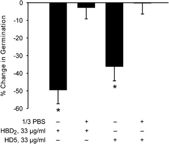
The antimicrobial effects of many peptides are notoriously salt sensitive (e.g. Vylkova et al. Reference Vylkova, Nayyar, Li and Edgerton2007). To test whether this was the case with inhibition of microsporidia spore germination a preliminary experiment was undertaken in which A. algerae germination assays were performed with the defensin pre-treatment taking place in either water or in 1/3 PBS. Exposure to 33 μg/ml HBD2 or HD5 in water significantly inhibited germination, but even 1/3 PBS completely eliminated this inhibition (Fig. 1). Because the electrolyte composition of the intestinal lumen is sodium-rich all subsequent germination experiments were performed with the spore defensin exposure occurring in PBS.
Inhibition of Anncaliia algerae spore germination in response to pH 9·5 germination solution. Spores were pre-treated with HBD2 or HD5 in water or in 1/3PBS. Mean and s.e.m. values. *Different from control spore germination, P<0·05.

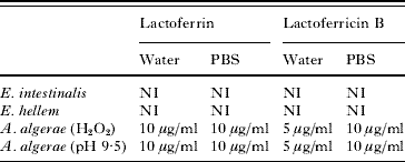
Preliminary experiments showed that A. algerae spore germination was significantly inhibited by exposure to Lf. Table 1 summarizes minimum inhibitory concentration (MIC) data obtained when spores of the 3 microsporidia species under study were exposed to Lf and its pepsin-digestion product, lactoferricin B. This exposure took place in water or in PBS. The A. algerae germination was stimulated by the high pH germination solution or by 5% H2O2 in pH 7·5 germination solution, while the Encephalitozoon species spore germination assays only employed the H2O2 stimulation. The peptide concentration used in these experiments was in the range 2 μg/ml to 2 mg/ml, the higher concentration of Lf being in the range of that reported in tears (Levay and Viljoen, Reference Levay and Viljoen1995). The A. algerae MIC for Lf was 10 μg/ml using peptide exposure in either water or PBS, while the Encephalitozoon species were unaffected by Lf up to a concentration of 2 mg/ml. With the smaller lactoferricin B the MIC for A. algerae germination was in the range of 5–10 μg/ml.
Microsporidia spore germination lactoferrin and lactoferricin B minimum inhibitory concentrations (MIC)
(Spores were exposed to agents in water or PBS at concentrations ranging from 2 μg/ml to 2 mg/ml prior to exposure to germination solution. NI, no inhibition at the concentration range tested.)
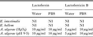
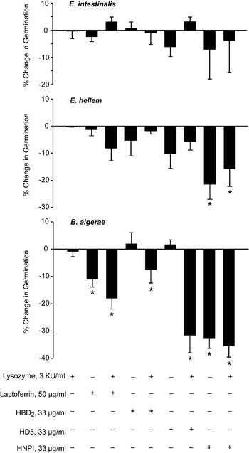
Figure 2 summarizes data obtained when assessing the effects of Lz, Lf and the 3 defensins on spore germination. Lysozyme alone had no effect on germination in the 3 microsporidia species. Because there are reports that Lz has synergistic antimicrobial effects with other peptides (Hancock and Scott, Reference Hancock and Scott2000; Chen et al. Reference Chen, Niyonsaba, Ushio, Okuda, Nagaoka, Ikeda, Okumura and Ogawa2005) experiments were conducted in which the Lf and defensin exposure occurred in the presence and absence of Lz. All peptide exposure took place in PBS and the germination solution used contained H2O2. At the concentrations used, Lf, HBD2, HD5 and HNP1, and these peptides in combination with Lz were without effect on E. intestinalis spore germination, while E. hellem spore germination was significantly inhibited by HNP1, with or without Lz. In the case of A. algerae, HBD2 and HD5 significantly inhibited germination, but only in the presence of Lz, while both Lf and HNP1 significantly inhibited germination in both the presence and absence of Lz.
Effect of prior exposure of Encephalitozoon intestinalis, E. hellem and Anncaliia algerae spores to a panel of antimicrobial peptides in the presence and absence of lysozyme on 5% H2O2-stimulated spore germination. Mean and s.e.m. values. *Different from control spore germination, P<0·05.

Effect of antimicrobial peptides on microsporidia infection of enterocytes
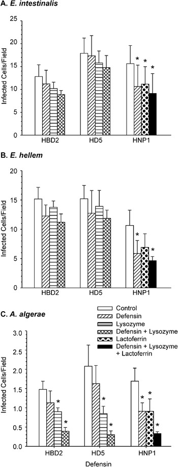
Figure 3 illustrates the effects of Lz and Lf alone, and HBD2 and HD5 in the presence or absence of Lz, and HNP1 alone and in the presence of both Lz and Lf on the infection of differentiated enterocytes by the 3 microsporidia species studied. The results of these infection assay experiments are similar but not identical to the germination assay results illustrated in Fig. 2. In these infection assays the variance was high because infection tended to be greater in those randomly chosen fields with more differentiated enterocytes that formed villus-like crests. Trends were observed with all 3 parasite species that suggested that all the antimicrobial peptides used reduced infection. However, as with the germination assay, there were no statistically significant effects of Lz alone or when combined with HBD2 or HD5 on E. intestinalis or E. hellem enterocyte infection.
Effect of antimicrobial peptides on the infection of differentiated C2Bbe1 cells 3 days after exposure to Encephalitozoon intestinalis, E. hellem or Anncaliia algerae spores in the presence or absence of antimicrobial peptides, alone or in combination. The peptide concentrations were the same as those in the germination experiment (Fig. 2). Mean and s.e.m. values. *Different from control, P<0·05.

In the case of A. algerae infections HBD2 and HD5 also had no statistically significant effect on infection while, unlike spore germination, Lz alone inhibited infection. Combining either of these 2 defensins with Lz further reduced infection, but this effect was only statistically significant in the case of HD5. With all 3 microsporidia species HNP1 alone inhibited enterocyte infection, as did Lf alone. However, the Lf inhibition was not statistically significant in the case of E. hellem. Combining HNP1 with both Lf and Lz resulted in a greater decrease in infection than with HNP1 alone. This additional effect was only statistically significant with A. algerae.
DISCUSSION
Multiple microsporidia species can be cultured in mammalian cells (Visvesvara et al. Reference Visvesvara, Moura, Leitch, Schwartz, Whittner and Weiss1999), yet only 2 species, Enterocytozoon bieneusi and Encepahalitozoon intestinalis, generally infect the human gastrointestinal tract (Didier and Weiss, Reference Didier and Weiss2006). Although relatively rare, there have been reports of other microsporidia species being recovered from patient stool samples (e.g. del Aguila et al. Reference del Aguila, Moura, Fenoy, Navajas, Lopez-Velez, Li, Xiao, Leitch, da Silva, Pieniazek, Lal and Visvesvara2001; Muller et al. Reference Muller, Bialek, Kamper, Fatkenheuer, Salzberger and Franzen2001). Franzen et al. (Reference Franzen, Hosl, Salzberger and Hartmann2005) concluded that the route of infection was a key factor in determining the target organ(s) in clinical microsporidiosis.
Of the 3 parasite species used in the present study only E. intestinalis commonly infects the human gastrointestinal tract of immunodeficient individuals. In a previous study we used the same parasite species and found that modelled gastric and duodenal chemical environments did not inhibit either spore germination or infection of C2Bbe1 cells (Leitch and Ceballos, Reference Leitch and Ceballos2008). The turnover time for human intestinal epithelial cells averages about 3 days (Jones and Gore, Reference Jones and Gore1997). The 2 Encephalitozoon species completed their life cycles well within this time-frame. However, the insect microsporidia, A. algerae, required 3–5 days to complete its life cycle in C2Bbe1 cells, suggesting that the mammalian body temperature is a limiting factor in the production of a sustained intestinal infection by this parasite species (Leitch and Ceballos, Reference Leitch and Ceballos2008).
Antimicrobial peptides constitute a major part of the innate immune system (Dann and Eckman, Reference Dann and Eckmann2007) and could potentially explain why only Enterocytozoon bieneusi and E. intestinalis infect the human gastrointestinal tract. Two peptides, Lf and Lz, were chosen that are ubiquitous in the GI tract, and 3 defensins were chosen to represent those found on the intestinal villus surface (HBD2), in the lumen of the crypt of Lieberkuhn (HD5), and in lamina propria neutrophils (HNP1) (Hancock and Scott, Reference Hancock and Scott2000; Cunliffe, Reference Cunliffe2003; Ouellette, Reference Ouellette2006).
A total of 6 antimicrobial peptides were tested for their effect on spore germination and enterocyte infection using concentrations that were chosen based on antimicrobial data in the literature (Ganz et al. Reference Ganz, Selsted, Szklarek, Harwig, Daher, Bainton and Lehrer1985; Nagoak et al. Reference Nagaoka, Hirota, Yamogida, Ohwada and Hirata2000; Newman et al. Reference Newman, Gootee, Gabay and Selsted2000; Samaranayke et al. Reference Samaranayke, Samaranayake, Pow, Beena and Yeung2001; Joly et al. Reference Joly, Maze, McCray and Guthmiller2004; Chen et al. Reference Chen, Niyonsaba, Ushio, Okuda, Nagaoka, Ikeda, Okumura and Ogawa2005; Vylkova et al. Reference Vylkova, Nayyar, Li and Edgerton2007). The peptides were the multifunctional Lf, lactoferricin B, HBD2, HD5, HNP1 and Lz. Lactoferrin was employed at a concentration above that generally reported in saliva and duodenum (Abrink et al. Reference Abrink, Larsson, Gobl and Hellman2000; Jentsch et al. Reference Jentsch, Sievert and Gocke2004), but less than that reported in human or bovine milk, or in tears (Levay and Viljoen, Reference Levay and Viljoen1995; Bard et al. Reference Bard, Laibe, Bettinger, Riethmuller, Buchle, Seilles and Meillet2003). The Lz concentration used was also in excess of that of saliva (Rudney and Smith, Reference Rudney and Smith1985), but less than that found in human milk (Montagne et al. Reference Montagne, Cuilliere, Mole, Rene and Faure1998). It is difficult to know the concentrations of defensins in the mucus unstirred layer of the intestine (Lievin-Le Moal and Servin, Reference Lievin-Le Moal and Servin2006), but in the confined volume of the mouse crypt lumen the alpha defensin concentration has been estimated to be as high as 10 mg/ml following Paneth cell stimulation (Ayabe et al. Reference Ayabe, Satchell, Wilson, Parks, Selsted and Ouellette2000).
The germination assay pointed to significant differences between the 3 parasite species in the response of spores to the antimicrobial peptides tested. E. intestinalis spore germination was not affected by any of the peptides and E. hellem spore germination was only inhibited by HNP1. Anncaliia algerae spore germination was inhibited by Lf and lactoferricin B at concentrations above 10 μg/ml and by HNP1 when peptide exposure occurred in PBS. The pepsin digest product of Lf, lactoferricin B, lacks the iron-binding domain of the parent peptide, suggesting that the N-terminal cationic antimicrobial domains were responsible for the observed inhibition (Gifford et al. Reference Gifford, Hunter and Vogel2005). Human β defensin 2 and HD5 only inhibited A. algerae spore germination when exposure occurred in water, but could occur in PBS if Lz was present. Lysozyme has been shown to have additive or synergistic antimicrobial effects with other antimicrobial peptides (Chen et al. Reference Chen, Niyonsaba, Ushio, Okuda, Nagaoka, Ikeda, Okumura and Ogawa2005; Nagoaka et al. Reference Nagaoka, Hirota, Yamogida, Ohwada and Hirata2000), and salt sensitivity is a common feature of the effect of many antimicrobial peptides (Vylkova et al. Reference Vylkova, Nayyar, Li and Edgerton2007).
Infection assays were conducted using differentiated C2Bbe1 cells in an assay we believe favoured infection via spore germination and target cell impalement (Leitch et al. Reference Leitch, Ward, Shaw and Newman2005). All 3 microsporidia species infected differentiated enterocytes in culture, although the A. algerae infection was only about 10% that of the 2 Encephalitozoon species (Leitch and Ceballos, Reference Leitch and Ceballos2008).
In both the germination and infection assays A. algerae was clearly more sensitive to the actions of antimicrobial peptides than were either of the Encephalitozoon species. However, there were some differences between the effects of these peptides on germination and on infection. Lactoferrin did not inhibit E. intestinalis spore germination but it significantly inhibited enterocyte infection. The large variance seen with the infection assay may have obscured any Lf effect on E. hellem infection. Given the multifunctional nature of this and other antimicrobial peptides (Levay and Viljoen, Reference Levay and Viljoen1995; Gifford et al. Reference Gifford, Hunter and Vogel2005) it is perhaps not surprising that there might be some limited uncoupling of the antimicrobial effects when comparing a short-term germination assay and a cell infection assay assessed 72 h after an initial 24 h spore exposure to peptide. Lactoferrin increases Caco2 cell proliferation (Buccigrossi et al. Reference Buccigrossi, de Marco, Bruzzese, Ombrato, Bracale, Polito and Guarino2007), which might have contributed to a reduction in the count of infected cells, and the binding of Lf to enterocyte glycosaminoglycans (El Yazidi-Belkoura et al. Reference El Yazidi-Belkoura, Legrand, Nuijens, Slomiany, van Berkel and Spik2001) may have interfered with spore adherence to the target cells (Hayman et al. Reference Hayman, Southern and Nash2005). Additionally, Lz had no effect on A. algerae germination in our standard germination assay, yet it unmasked the salt-sensitive inhibition by HBD2 and HD5, and inhibited infection by this species. It is not uncommon for Lz to lack antimicrobial effects on it own, yet increase the effect of other antimicrobial peptides (e.g. Singh et al. Reference Singh, Tack, McCray and Welsh2000).
The neutrophil defensin, HNP1, alone or in combination with Ly and Lz, two peptides also produced by neutrophils (Peeters and Vantrappen, Reference Peeters and Vantrappen1975; Pryzwansky et al. Reference Pryzwansky, Rausch, Spitznagel and Herion1979), inhibited enterocyte infection by the 3 microsporidia species tested. Thus, neutrophil peptides could constitute an important line of defence against microsporidia infection in the intestinal lamina propria.
Antimicrobial peptides have multiple ways of killing or disabling microorganisms, in addition to a porin action (Brogden, Reference Brogden2005; Vylkova et al. Reference Vylkova, Nayyar, Li and Edgerton2007). We have shown that the inhibition of spore germination is one way in which some antimicrobial peptides can protect the intestine against microsporidia infection by some parasite species. Differences in spore, germination apparatus, and sporoplasm ultrastructure (Vavra and Larsson, Reference Vavra, Larsson, Whittner and Weiss1999; Cali et al. Reference Cali, Weiss and Takvorian2002) may underlie some of the species-specific effects we observed in peptide inhibition of germination and infection.
This study suggests that intestinal antimicrobial peptides play a role in determining if an ingested microsporidia spore will infect the intestinal epithelium, with a principal effect of the peptides being on the spore germination process. The greater susceptibility of A. algerae spores to the antimicrobial peptides tested may help account for the fact that this species has not been implicated in intestinal microsporidiosis. However, E. intestinalis and E. hellem were similarly affected by the peptides used in this study, suggesting that other peptides, peptide concentrations, or other factors are responsible for the fact that only the former Encephalitozoon species is important in infecting the gastrointestinal tract, particularly in immunocompromised individuals.
This work was supported by U.S. PHS grant R21 DK64573-A1.