Implications
Today chicken management is highly specialized and directed toward meat and egg production. Chicks of both sexes (meat breeds) are used for meat production, whereas egg production is based upon pullet chicks. The elimination of unwanted day-old cockerel-chicks poses a problem for industrial hatcheries. The meat of capons (surgically castrated male chickens) is considered a delicacy of high nutritional value. Increased capon production could contribute to rational management of male chicks of dual-purpose breeds and layer-type chicks which are considered ‘waste products’ in the egg industry.
Introduction
In many countries, the production of capons (surgically castrated male chickens) is deeply rooted in local tradition (Hsu and Lin, Reference Hsu and Lin2003; Miguel et al., Reference Miguel, Ciria, Asenjo and Calvo2008; Díaz et al., Reference Díaz, Rodríguez, Torres and Cobos2010). Chicken capons are most popular, but males of other poultry species, such as pheasants, are also caponized (Mašek et al., Reference Mašek, Severin, Kos, Janicki, Filipović, Kozacinski, Cvrtila and Dzaka2010). Castration is a hormonal intervention which permanently influences metabolic processes in birds (Rikimaru et al., Reference Rikimaru, Takahashi and Nicholas2011; Symeon et al., Reference Symeon, Charismiadou, Mantis, Bizelis, Kominakis and Rogdakis2013; Chen et al., Reference Chen, Li, Tsai, Lo and Chen2014). Due to reduced synthesis of sex steroids, caponization increases fat deposition in the carcass, which was confirmed by postmortem analyses and chemical analyses of muscles in castrated birds (Díaz et al., Reference Díaz, Rodríguez, Torres and Cobos2010; Symeon et al., Reference Symeon, Mantis, Bizelis, Kominakis and Rogdakis2012).
Caponization changes the blood lipoprotein profile and stimulates fat accumulation in adipose, subcutaneous and muscle tissues. Chen et al. (Reference Chen, Chen, Lin and Chiou2007a) reported changes in liver lipid composition after caponization. However, there is a scarcity of published data on histological changes, in particular fatty degeneration, in the liver of capons.
A few studies only have investigated changes in blood testosterone levels in growing cockerels of various breeds, and their effects on the growth rate of birds (Rikimaru et al., Reference Rikimaru, Shiji, Komastu and Ishizuka2009; Symeon et al., Reference Symeon, Mantis, Bizelis, Kominakis and Rogdakis2010). This is an important consideration as the above changes may have potential consequences for bird behavior. In some chicken breeds, roosters become aggressive as they reach sexual maturity, which poses a problem for breeders and producers (Essien et al., Reference Essien, Akpet, Ukorebi, Orok, Akinola, Ayuk and Adejumo2012). Capons are more docile and gentle than roosters, but castration influences metabolism, thus affecting the health status of birds and feed conversion. Feed consumption per unit of gain is the most important determinant of animal production profitability, but this aspect of capon production has been scarcely researched. Very few studies conducted in recent years have analyzed feed efficiency in caponized birds (Lin and Hsu, Reference Lin and Hsu2003; Mašek et al., Reference Mašek, Severin, Gottstein, Filipović, Stojević and Mikulec2013).
The objective of this study was to determine the effect of castration and age on the growth rates of tissue components and selected organs, carcass quality parameters, feed conversion and histopathological changes in the liver of Green-legged Partridge roosters and capons.
Material and methods
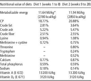
The experimental material comprised 200 Green-legged Partridge (old traditional Polish chicken breed) cockerels. One-day-old birds were weighed, marked with wing tags and randomly distributed to pens in the experimental center of the Department of Commodity Science and Animal Improvement of the University of Warmia and Mazury in Olsztyn, Poland. The birds were raised to 28 weeks of age, and were fed commercial diets ad libitum (Table 1). At 8 weeks of age, 100 birds were surgically castrated by a qualified veterinarian in accordance with Commission Regulation (EC) No. 543/2008. All birds were weighed at 4-week intervals throughout the experiment. Total feed consumption was recorded. Bird behavior was monitored daily. The data, acquired during feeding in the morning (0700 to 0730 h) and in the afternoon (0500 to 0530 h), were not analyzed statistically.
Composition of experimental diets composition of diets 1 and 2: ground cereal grains (cornmeal, ground wheat), soybean meal (line 40-3-2), calcium carbonate, monocalcium phosphate, sodium chloride

Additives: Microelements: Cu, 8 mg/kg; Fe, 116 mg/kg; Mn, 80 mg/kg; Zn, 100 mg/kg; J, 0.80 mg/kg; Se, 0.20 mg/kg. Vitamins: vitamin E, 68 mg/kg; vitamin K3, 4.80 mg/kg; vitamin B1, 2.2 mg/kg; vitamin B2, 7.2 mg/kg; vitamin B6, 5.0 mg/kg; vitamin B12, 44 mcg/kg; vitamin H (biotin), 136 mcg/kg; niacin, 44.0 mg/kg; Ca-d-pantothenate, 18.0 mg/kg.
* Metabolizable energy content estimated based on the percentages of selected ingredients determined analytically.
At 4 and 8 weeks of age, blood samples were collected from 10 randomly selected birds to determine testosterone levels. Between weeks 12 and 28, at 4-week intervals, blood samples were collected from 10 intact cockerels and 10 capons, and the birds were slaughtered (electrical stunning followed by cutting the jugular vein). Carcasses were chilled for ~18 h at a temperature of 4°C, and carcass dissection was performed.
The birds were fasted for 10 h before blood sampling and slaughter. Blood was collected into test tubes containing heparin. Freshly collected blood was centrifuged twice (MPW-350R centrifuge; MPW Med. Instruments, Warsaw, Poland; 5 min, 10 000 r.p.m.), and each time the supernatant was transferred to 1.5 ml Eppendorf Safe-Lock micro test tubes with an Eppendorf automatic electronic pipette (Eppendorf, Polfa Warsaw Ltd, Poland). Plasma samples were frozen at −72°C in 1.5 ml Eppendorf Safe-Lock micro test tubes in the Kaltis 390 ultra low temperature laboratory freezer. The concentrations of testosterone, triacylglycerols (TG), total cholesterol (TC) and high-density lipoprotein (HDL) cholesterol were determined. Testosterone levels were analyzed by radioimmunoassay (RIA) with the use of commercial kits supplied by DIAsource – Testo, RIA CT (DIAsource ImmunoAssays S.A., Louvain-la-Neuve, Belgium). The plasma concentrations of TG, TC and HDL cholesterol were determined with the use of commercial kits supplied by Pointe Scientific (Poland). Low-density lipoprotein (LDL) cholesterol levels were determined based on the formula developed by Friedewald et al. (Reference Friedewald, Levy and Fredrickson1972).
Histological analysis of the liver. During post-slaughter processing, liver samples collected from six intact cockerels and six capons selected randomly from age groups were fixed in 10% neutralized formalin and embedded in paraffin blocks. Paraffin sections of liver (5 µm) were stained with hematoxylin–eosin (HE). Liver samples were immersed in 30% saccharose solution with the addition of sodium azide, frozen samples were cut into 8 µm sections, and were stained with Oil Red O (Bio-Optica, Milano, Italy) to detect lipids. Histological analyses were performed by two pathologists. Each section was imaged using the MIDI 3DHISTECH panoramic scanner (Hungary). Lipid concentrations in hepatocytes were determined on a three-point scale: (+) low lipid content; (++) moderate lipid content; (+++) high lipid content.
The statistical analysis involved the determination of arithmetic means (
 $$\bar{x}$$
) and standard deviations. Data were subjected to two-way or one-way ANOVA. The significance of differences in mean values between age groups was determined by Duncan’s D-test. The results were processed using Statistica 2010 software. Significance was set at P<0.05. The experiment was approved by the Local Ethics Committee.
Results
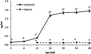
Average plasma testosterone levels in 4-week-old birds reached 0.15 ng/ml, and increased to 1.94 ng/ml in control group (uncastrated) cockerels at 28 weeks of age. The highest increase in testosterone concentrations (by 1.28 ng/ml) was noted between weeks 12 and 16 (Figure 1; P<0.05). At 8 weeks of age, when selected birds were caponized, average plasma testosterone levels reached 0.20 ng/ml, whereas testosterone concentration of 0.13 ng/ml was noted in 12-week-old castrated males, i.e. 4 weeks after caponization. This value was significantly lower than that determined in control group birds (Figure 1; P<0.05). In caponized birds, plasma testosterone levels remained stable until the end of the experiment.
Blood testosterone levels in intact cockerels and capons (ng/ml; mean±SD). a,b,cValues followed by different letters (age) or * (sex category) differ significantly; α=0.05.

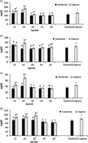
Surgical castration had a significant effect on the concentrations of TC, HDL cholesterol, LDL cholesterol and TG (Figure 2a to d; P<0.05). In intact cockerels, TC levels increased from 12 to 16 weeks of age, and decreased from 16 to 20 weeks of age (P<0.05). A similar trend was noted in caponized birds, but their TC concentrations were higher than those of uncastrated males by 28.4, 45.9 and 14.0 mg/dl in weeks 12, 16 and 20, respectively (Figure 2a; P<0.05). At 12 weeks of age, HDL cholesterol levels were comparable in birds of both groups. Between weeks 12 and 16, they decreased in intact cockerels (by 15.6 mg/dl) and increased in capons (by 9.30 mg/dl, Figure 2b; P<0.05). At 12 and 16 weeks of age, LDL cholesterol concentrations were by ~12.5 and 27.3 mg/dl higher, respectively, in castrated males than in uncastrated birds (Figure 2c; P<0.05). Between 16 and 20 weeks of age, LDL cholesterol concentrations decreased in both cockerels (by 4.2 mg/dl) and capons (by 33.2 mg/dl). In weeks 12, 16 and 20, plasma TG levels were higher in capons than in uncastrated males, by 13.8, 9.40 and 21.1 mg/dl, respectively (Figure 2d; P<0.05).
The serum concentrations of (a) total cholesterol, (b) high-density lipoprotein-cholesterol, (c) low-density lipoprotein-cholesterol and (d) triacylglycerols of intact cockerels and capons (mg/dl; mean±SD). a,b,c,dValues followed by different letters (age) or * (sex category) differ significantly; α=0.05.

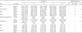
At 8 weeks of age, before caponization, the average BW of birds was 579.7 g. From 12 weeks of age, capons were characterized by greater BW than intact cockerels, but statistically significant differences were found only in week 28 (Table 2; P<0.05). A similar trend was observed in carcass weight, but caponization had no significant effect on carcass weight, which was affected only by age (Table 2; P<0.05).
BW, carcass weight, weights of tissue components and organs in Green-legged Partridge cockerels and capons (g; mean±SD)

ns=non-significant differences.
ANOVA (significance), *significant differences, α=0.05.
a,b,c,dMean values in a row (age) followed by different letters are significantly different at α=0.05.
† Mean values in the age groups of intact cockerels and capons are significantly different at α=0.05.
Average bone weight in the carcasses of 12-week-old cockerels and capons was 155.9 and 155.4 g, respectively, and it increased to 239.0g and 240.6 g, respectively, at 28 weeks of age (Table 2; P<0.05). Caponization had no significant influence on bone weight, except in week 16 (Table 2; P<0.05).
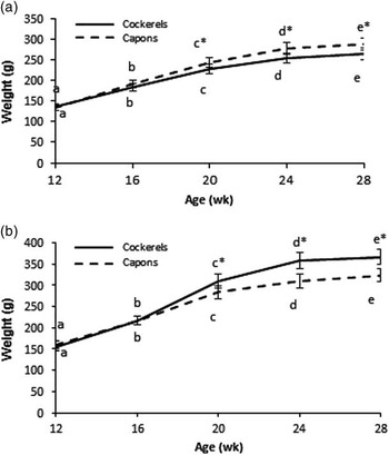
An increase in meat weight (muscles and intermuscular fat) was observed in intact cockerels until week 24, and in capons until week 28 (Table 2; P<0.05). At 12 weeks of age, average meat weight in cockerels and capons was 432.9 and 412.6 g, respectively, and it increased to 954.8 and 931.1 g, respectively, at 28 weeks of age. Surgical castration had no effect on total meat weight in the carcasses of intact cockerels and capons (Table 2). However, significant differences were noted in the weights of breast muscles and leg muscles. From 20 weeks of age, capons were characterized by higher breast muscle weight than uncastrated males (by 17.1, 24.2 and 23.9 g in weeks 20, 24 and 28, respectively; Figure 3a; P<0.05). Leg muscle weight was lower in capons than in intact cockerels, by 26.6, 52.1 and 42.8 g in weeks 20, 24 and 28, respectively (Figure 3b; P<0.05).
Weight of (a) breast muscles and (b) leg muscles in intact cockerels and capons (g; mean±SD). a,b,c,dValues followed by different letters (age) or * (sex category) differ significantly; α=0.05.

Capon carcasses were characterized by increased fat deposition (Table 2; P<0.05). At 12 and 28 weeks of age, the weight of skin with subcutaneous fat was higher in capons than in uncastrated birds, by 24.2 and 30.8 g, respectively (Table 2; P<0.05), and the weight of abdominal fat was higher by 10.7 and 37.4 g, respectively (Table 2; P<0.05).
Heart weight increased significantly to 16 weeks of age in capons and to 24 weeks of age in intact cockerels. From 20 weeks of age, heart weight was higher in cockerels than in capons (Table 2; P<0.05). Gizzard weight was higher in capons than in intact cockerels, except in week 12 (Table 2; P<0.05). An increase in gizzard weight was observed to 20 weeks of age in uncastrated cockerels, and to 16 weeks of age in capons. At 12 weeks of age, the liver weight of capons was 22.51 g, and it increased to 27.64 g in week 28 (Table 2; P<0.05). In intact cockerels, liver weight remained at a similar level throughout the experiment (21.26 g in week 12, 22.12 g in week 28, Table 2). From 20 weeks of age, liver weight was significantly higher in capons than in cockerels (Table 2; P<0.05).
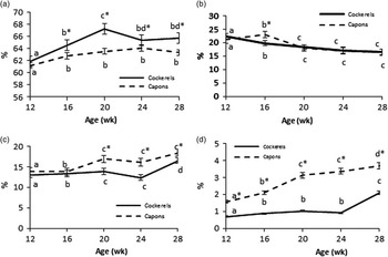
To better illustrate age-related changes in Green-legged Partridge capons and cockerels, and to compare them with those reported for other breeds, the weights of tissue components were expressed as a percentage of total carcass weight (Figure 4a to c) and total BW (Figure 4c). The carcasses of caponized birds contained less meat (by 2.3% in week 28, Figure 4a; P<0.05) and more skin with subcutaneous fat (by 1.9% in week 28, Figure 4c; P<0.05) than the carcasses of uncastrated males. Abdominal fat accounted for 1.6% and 0.7% of total BW in 12-week-old capons and cockerels, respectively (P<0.05), and it increased to 3.7% and 2.1%, respectively, at 28 weeks of age (Figure 4d; P<0.05).
Percentage of (a) meat (muscles with intermuscular fat), (b) bones, (c) skin with subcutaneous fat in total carcass weight and (d) abdominal fat in the total BW of intact cockerels and capons (%; mean±SD). a,b,c,dValues followed by different letters (age) or * (sex category) differ significantly; α=0.05.

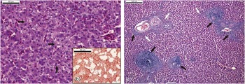
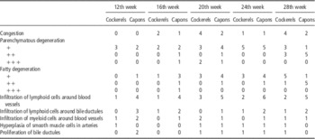
Table 3 shows histopathological changes in the livers of intact cockerels and capons at different stages of fattening. At 28 weeks of age, moderate fatty degeneration was noted in the hepatocytes of five capons and mild fatty degeneration was observed in one capon (Figure 5a), whereas in intact cockerels, one case of moderate fatty degeneration and five cases of mild fatty degeneration were noted. Red Oil O staining revealed small and medium-sized cytoplasmic lipid vacuoles within hepatocytes (Figure 5b), which were encountered more frequently in capons than in intact cockerels from 16 weeks of age. The analysis also revealed infiltration of lymphoid cells around blood vessels, which was observed more frequently in capons than in uncastrated males (Figure 5c) and differences were noted from 12 weeks of age.
Capon, liver: (a) fatty degeneration of hepatocytes, small and medium droplets of fat (arrows). hematoxylin–eosin (HE) staining (16th week); (b) small and medium vacuoles of lipids in the cytoplasm of hepatocytes. Red Oil O staining (16th week); (c) infiltration of lymphoid cells around blood vessels (black arrows), infiltration of myeloid cells around blood vessels (white arrows) HE staining (28th week).

Histopathological changes in the liver in intact cockerels and capons at different stages of fattening (n=6)

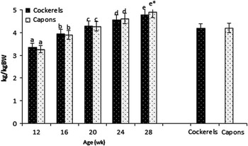
Average feed intake was similar in both uncastrated and castrated birds (4.19 and 4.20 kg/kg BW from day 1 to week 28). However, from 24 weeks of age, capons tended to consume more feed per kg BW than control group birds (Figure 6).
Feed conversion of cockerels and capons (kg/kg BW; mean±SD) a,b,c,d,eValues followed by different letters (age) or * (sex category) differ significantly: α=0.05.

Capons did not display antagonistic behaviors, whereas uncastrated cockerels were characterized by increased motor activity from 12 weeks of age, accompanied by ruffling feathers, chasing and pushing other birds around, or pecking one another.
Discussion
Some research findings have pointed to a beneficial influence of castration on the BW of birds (Tor et al., Reference Tor, Estany, Villalba, Molina and Cubilo2002; Chen et al., Reference Chen, Hsieh and Chiou2006; Kwiecień et al., Reference Kwiecień, Kasperek and Grela2015), whereas other studies (Miguel et al., Reference Miguel, Ciria, Asenjo and Calvo2008; Symeon et al., Reference Symeon, Mantis, Bizelis, Kominakis and Rogdakis2012; Adamski et al., Reference Adamski, Kuźnicka, Banaszak and Wegner2016) have not confirmed such a correlation. In the present study, a tendency toward a higher growth rate was observed in capons already 4 weeks after castration, but a statistically significant difference relative to the control group was noted only in week 28, i.e. 20 weeks after castration (Table 2). In experiments performed by Calik et al. (Reference Calik, Połtowicz, Świątkiewicz, Krawczyk and Nowak2015) and Kwiecień et al. (Reference Kwiecień, Kasperek and Grela2015), Green-legged Partridge capons were characterized by higher BW than roosters. Cockerels of various breeds are caponized, and technologies and systems of capon production differ in terms of age considered optimal for castration and slaughter, housing conditions and feeding regime (Hsu and Lin, Reference Hsu and Lin2003; Duran, Reference Duran2004; Chen et al., Reference Chen, Chen, Lin and Chiou2007a; Miguel et al., Reference Miguel, Ciria, Asenjo and Calvo2008; Shao et al., Reference Shao, Wu, Li and Zhao2009; Díaz et al., Reference Díaz, Rodríguez, Torres and Cobos2010; Rikimaru et al., Reference Rikimaru, Takahashi and Nicholas2011). Interactions are also observed between the above factors.
In our study, carcass weight was lower in capons than in intact cockerels in all age groups (Table 2). Similar results were reported by Tor et al. (Reference Tor, Estany, Villalba, Molina and Cubilo2002) who found no differences in carcass weight between roosters and capons despite higher BW of the latter. Increased deposition of fat, in particular mesenteric fat, in the body cavity is observed in caponized birds. During carcass evisceration, fat is removed together with the intestines. Therefore, total offal weight may be higher in capons than in uncastrated males, which affects carcass dressing percentage (Zawacka et al., Reference Zawacka, Murawska and Michalik2014). According to Shao et al. (Reference Shao, Wu, Li and Zhao2009), caponized birds are characterized by a faster rate of adipose tissue deposition in the abdominal cavity, which was also observed in our study. Abdominal fat weight was 2.5-fold higher in capons than in control group birds already 4 weeks after castration (Table 2), and abdominal fat accounted for nearly 1.6% of total BW (Figure 4d).
Studies conducted by other authors (Hsu and Lin, Reference Hsu and Lin2003; Chen et al., Reference Chen, Hsieh and Chiou2006) revealed that capon carcasses were characterized by increased deposition of subcutaneous and intermuscular fat. In our study, the carcasses of caponized birds contained more skin with subcutaneous fat already in week 12, and the rate of adipose tissue deposition increased from 20 weeks of age (Table 2, Figure 4c). A preliminary morphological evaluation of skeletal muscles in intact cockerels and capons, conducted in our previous study (Gesek et al., Reference Gesek, Zawacka, Murawska, Charuta, Mika and Otrocka-Domagała2015), revealed the accumulation of lipids in tissue structures: in the perimysium between fascicles, in the endomysium between fibers, around blood vessels and in the sarcoplasm of capons. Its higher fat content is considered an advantage because it enhances palatability (Calik et al., Reference Calik, Połtowicz, Świątkiewicz, Krawczyk and Nowak2015; Amorim et al., Reference Amorim, Rodrigues, Pereira, Valentim and Teixeira2016).
According to Chen et al. (Reference Chen, Chen, Lin and Chiou2007a), testosterone – which stimulates protein synthesis and contributes to increasing muscle weight in mammals – may exert inhibitory effects in prepubertal birds. The cited authors demonstrated that birds caponized at an early age (3 weeks) were characterized by higher BW and a higher percentage of breast muscles in the carcass than uncastrated males, but such relationships were not noted when birds were castrated at a later age (12 weeks; raising to 26 weeks). Other authors (Tor et al., Reference Tor, Estany, Villalba, Molina and Cubilo2002; Mahmud et al., Reference Mahmud, Shaba, Gana, Yisa and Ndagimba2013; Calik et al., Reference Calik, Połtowicz, Świątkiewicz, Krawczyk and Nowak2015) also observed higher breast muscle weight in capons castrated at 4 or 8 weeks of age. Tor et al. (Reference Tor, Estany, Villalba, Molina and Cubilo2002) and Shao et al. (Reference Shao, Wu, Li and Zhao2009) noted lower leg muscle weight in caponized birds, compared with roosters, which resulted from varied effects exerted by androgens on the growth rates of different muscle groups. Our findings regarding the growth of muscle and leg muscles in intact cockerels and capons throughout the experimental period, and the analysis of changes in testosterone concentrations indicate that this hormone affects the development of those muscle groups (Table 2; Figure 1; Figure 3a and b). Between 12 and 16 weeks of age, a rapid increase in blood testosterone levels was noted in control group birds. As a result, from 16 weeks of age, uncastrated cockerels were characterized by an increased growth rate of leg muscles and a decreased growth rate of breast muscles. It appears that the ‘investment’ in leg muscles, at the expense of breast muscles, observed in roosters, is biologically justified as it prepares the birds for the mating season.
Research shows that caponization affects the weight, length and histology of bones (Lin and Hsu, Reference Lin and Hsu2003; Chen et al., Reference Chen, Li, Tsai, Lo and Chen2014). In the present study, caponization had no significant effect on total bone weight or bone proportion in the carcass, but an age×sex interaction was observed between 12 and 16 weeks of age, when capons were characterized by rapid bone growth.
Shao et al. (Reference Shao, Wu, Li and Zhao2009) noted higher TG and TC concentrations in the blood of caponized birds. Guo et al. (Reference Guo, Nan, Shi, Zhou, Wan, Zhou, Geng, Chen and Jiang2015) observed higher levels of TC, HDL and LDL in castrated birds. Similar trends were found in our study (Figure 2d). Lipid metabolism in the liver involves the synthesis, elongation and degradation of fatty acids, and synthesis of TG. Cholesterol and lipoproteins are produced by hepatocytes (Lieberman and Marks Reference Lieberman and Marks2009). The male sex and obesity are regarded as risk factors that contribute to many diseases, including steatosis. An imbalance between the inflow of fatty acids, lipid synthesis in the liver and β-oxidation can intensify fat deposition in hepatocytes (Kargulewicz et al., Reference Kargulewicz, Stankowiak-Kulpa and Grzymisławski2010). In our study, an increase in liver weight was observed in capons from 20 weeks of age (Table 2), which could result from increased lipid deposition confirmed by histological analysis (Table 3, Figure 5a to c). Fatty degeneration in hepatocytes was more advanced in capons from 16 weeks of age. Lipid droplets observed in hepatocytes confirm other authors’ suggestions concerning changes in lipid metabolism. Chen et al. (Reference Chen, Chi, Chu, Chen and Chiou2007b) noted increased hepatic lipid composition and higher total lipid level in capons. The cited authors also observed higher levels of cholesterol, TG and phospholipids, and lower concentrations of non-esterified fatty acids. Thus, increased hepatic lipid production leads to fatty degeneration in hepatocytes, which was demonstrated in our study. Low lipid concentrations in hepatocytes are commonly encountered in cockerels fed a well-balanced diet, whereas inadequate dietary protein and energy levels lead to fatty degeneration in hepatocytes (Gesek et al., Reference Gesek, Szarek, Szweda and Babińska2010 and Reference Gesek, Szarek, Otrocka-Domagała, Babińska, Paździor, Szweda, Andrzejewska and Szynaka2013). Interestingly, castration caused changes in lymphoid cell infiltration. From 12 weeks of age, infiltration was more frequently observed in capons, which suggests that high testosterone levels contribute to a decrease in the counts of inflammatory cells as a component of lymphoid tissue in internal organs. Similar changes were noted in lymphoid organs such as the spleen, bursa of Fabricius and thymus (Gesek et al., unpublished data).
Higher liver weight in capons was also reported by Rahman et al. (Reference Rahman, Islam, Ali, Khondaker and Hossain2004) and Symeon et al. (Reference Symeon, Mantis, Bizelis, Kominakis and Rogdakis2010). In a study by Chen et al. (Reference Chen, Hsieh and Chiou2006) capons had higher weights of the liver and gizzard. Mašek et al. (Reference Mašek, Severin, Gottstein, Filipović, Stojević and Mikulec2013) and Kwiecień et al. (Reference Kwiecień, Kasperek and Grela2015) studies revealed that castration had no influence on hearth weight in capons. In our study, however, intact cockerels were characterized by higher heart weight than capons from 20 weeks of age. Our results could suggest that the growth of cardiac muscle tissue is affected by androgens (Table 2, Figure 1), and this effect can be further enhanced by increased activity of cockerels, observed from 12 weeks of age. Other authors (Miguel et al., Reference Miguel, Ciria, Asenjo and Calvo2008; Rikimaru et al., Reference Rikimaru, Shiji, Komastu and Ishizuka2009; Symeon et al., Reference Symeon, Mantis, Bizelis, Kominakis and Rogdakis2012) also noted higher heart weight in roosters than in caponized birds.
As already mentioned in the Introduction section, capon production systems differ widely, making comparison of feed efficiency in capons and roosters across studies very difficult. Mašek et al. (Reference Mašek, Severin, Gottstein, Filipović, Stojević and Mikulec2013) noted a beneficial influence of castration, carried out at 18 days of age, on the feed conversion ratio in Ross 308 chickens raised to 55 days of age. Mas et al. (Reference Mas, Šerman, Horvat, Strakova, Valpotić, Mikulec, Mašek, Suchy and Vince2013) who analyzed Ross 308 chickens caponized at 28 days of age, reported similar total feed efficiency (from day 1 to week 22) in roosters and capons, but in week 22 the feed conversion ratio was higher in capons than in control group birds. In a study by Lin and Hsu (Reference Lin and Hsu2003), Taiwan country chicken cockerels (Taiwan Livestock Research Institute (TLRI) native chicken Taishi meat No. 13) caponized at 10 weeks of age and raised to 28 weeks, were characterized by lower feed consumption per unit of gain until 24 weeks of age; however, total feed efficiency between 10 and 28 weeks was comparable in caponized and intact cockerels. A similar trend was noted in our study. The feed conversion ratio (kg/kg BW) calculated for the entire experimental period (from day 1 to week 28) was comparable in both groups, but from 24 weeks of age, it increased to a higher extent in capons than in uncastrated birds (Figure 6). A decrease in testosterone levels, observed in caponized birds, leads to changes in their behavior, including reduced motor activity (Zuk and Johnsen, Reference Zuk and Johnsen2000; Essien et al., Reference Essien, Akpet, Ukorebi, Orok, Akinola, Ayuk and Adejumo2012; Mahmud et al., Reference Mahmud, Shaba, Gana, Yisa and Ndagimba2013), which was noted also in our study. Caponized birds became more docile and less aggressive, and their maintenance energy requirements decreased considerably.
The majority of day-old male layer-type chicks and dual-purpose breeds are euthanized (Bruijnis et al., Reference Bruijnis, Blok, Stassen and Gremmen2015). Increased capon production could contribute to rational management of male chicks which are considered ‘waste products’ in the egg industry. In Europe, capon meat is appreciated by consumers for its tenderness and flavor, and it is more expensive than meat from broiler chickens and organic chickens (Duran, Reference Duran2004).
Conclusions
Caponization had a significant effect on carcass tissue composition and bird behavior, but it did not lead to significant differences in feed conversion. Total meat weight in the carcasses of intact cockerels and capons was similar, but from 20 weeks of age, the weight of breast muscles was higher, and the weight of leg muscles was lower in capons than in cockerels. The optimal slaughter age of capons should be determined based on carcass quality and economically important factors. The values of carcass quality parameters and feed conversion ratio as well as histopathological changes in the liver indicate that Green-legged Partridge capons should be slaughtered at 20 to 24 weeks of age.
Acknowledgement
The authors thank Dr M. Mika with the Department of Animal Physiology at University of Agriculture in Krakow, for help in research and comments that strengthened the manuscript.