Introduction
The optic nerve has an intimate relationship to the posterior ethmoid cells and sphenoid sinus. Protrusion of the optic canal into the sphenoid and/or ethmoid sinus (e.g. in an Onodi cell, or due to an atypical extracranial course of the optic nerve) has considerable clinical significance. Damage of the optic nerve is one of the most serious complications of endoscopic sinus surgery.
It is essential that clinicians performing sinus surgery have a detailed knowledge of the paranasal sinuses and also of the adjacent anatomical structures and their known variations. It is important to review such knowledge pre-operatively, so that unintentional damage to these structures can be avoided.
Case report
A 39-year-old woman with Turner syndrome presented with a recent episode of severe headache associated with visual disturbances.
Ophthalmological examination revealed papilloedema in both eyes, with no other pathology. Other than Turner syndrome, the patient's medical history was unremarkable.
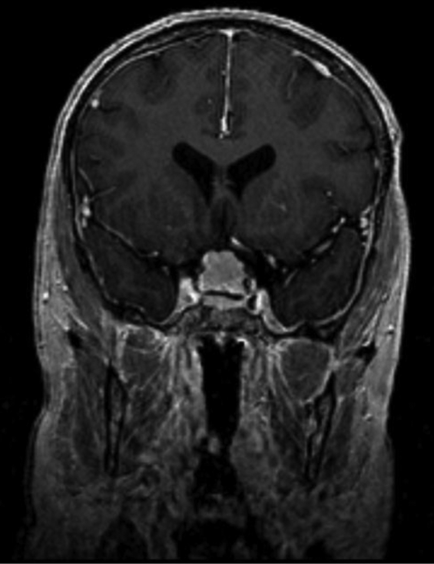
Magnetic resonance imaging of the brain revealed a large (19 × 6 mm), well circumscribed mass in the region of the optic chiasm on the diaphragma sellae. The lesion was hyperintense on T1-weighted images with gadolinium contrast (Figure 1) and isointense on T2-weighted images. The mass was presumed to be a meningioma.
Coronal, T1-weighted magnetic resonance imaging head scan with gadolinium contrast, showing the hyperintense lesion.

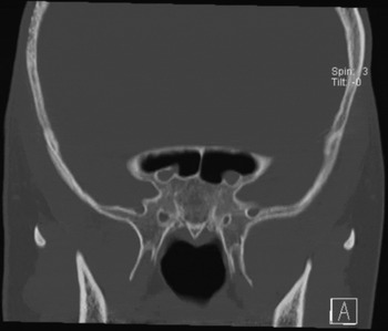
A computed tomography (CT) scan of the paranasal sinuses was undertaken to help plan surgical removal via an endoscopic trans-sphenoidal approach (Figure 2). Surprisingly, it revealed a previously unreported anatomical variation of the extracranial course of the optic nerve, riding on the floor of the sphenoid sinus.
Coronal computed tomography scan of the paranasal sinuses, showing a previously unreported anatomical variation of the extracranial course of the optic nerve, riding on the floor of the sphenoid sinus.

Surgery was uneventful.
Histopathological examination confirmed that the lesion was a sella meningioma.
Discussion
Turner syndrome is a common chromosomal abnormality characterised by complete or partial X chromosome monosomy. Schoemaker et al. Reference Schoemaker, Swerdlow, Higgins, Wright and Jacobs1 performed a cohort study of Turner syndrome patients, and observed increased prevalence of various neoplastic disorders including meningioma, childhood brain tumour and gonadoblastoma. Interestingly, these patients showed a decreased risk of breast cancer. Such risks are postulated to relate either to direct genetic factors or to hormonal therapies used to treat the condition.
• Optic nerve damage is one of the worst complications of endoscopic sinus surgery
• In the presented case, the optic nerve coursed along the floor of the sphenoid sinus
• This anatomical variation has not been previously reported
• Pre-operative computed tomography is mandatory prior to endoscopic sinus surgery, to detect anatomical variation
Patients undergoing endoscopic sinus surgery must receive pre-operative CT scanning, particularly when a trans-sphenoidal approach is anticipated. It is usually advised that these pre-operative CT scans be evaluated using a checklist.Reference Simmen and Schuknecht2 Such CT scans provide a wealth of information on the patient's bony anatomy, giving surgeons a detailed map with which to plan their approach. Knowledge of anatomical variations may be crucial. In particular, knowledge of the anatomical course of the optic nerve is of great importance in order to avoid injury while performing an endoscopic trans-sphenoidal approach. The usual anatomical course of the optic nerve is within the supero-lateral aspect of the wall of the sphenoid sinus.
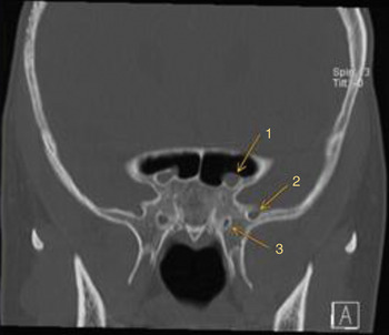
The presented patient's variant anatomy prompted us to search the literature, using the PubMed database, to identify previous reports of anatomical variation of the extracranial course of the optic nerve. Variant courses have been previously reported, including cases in which the optic nerve bulged into the sinus or even traversed it.Reference Heskova, Mellova, Holomanova, Vybohova, Kunertova and Marcekova3–Reference Dessi, Moulin, Chagnaud and Cannoni5 However, we could find no previous report of the optic nerve occupying such a low position within the floor of the sinus. Figures 3 and 4 show further detail of this previously unreported anatomical variation.
Coronal computed tomography scan showing anatomical variation of the optic nerve course. 1 = optic nerve; 2 = maxillary nerve; 3 = vidian nerve

Composite presentation of axial, sagittal and coronal computed tomography scans centred on the optic nerve, showing its variant course in three dimensions.