INTRODUCTION
Leishmaniasis is a vector-borne disease caused by protozoan haemoflagellate parasites of the genus Leishmania. Over 20 species of Leishmania both from the New and Old World are able to cause one or more types of leishmaniasis in humans. An estimated 350 million people in over 88 countries are at risk of infection, with an annual incidence of 500 000 cases for the lethal visceral form and 1·5 million for the cutaneous forms (WHO, 2010). No effective vaccines are available to date and current chemotherapy for leishmaniasis is reliant predominantly on pentavalent antimonials and miltefosine as first-line drugs, with amphotericin B and pentamidine following as second-line alternatives (Zucca and Savoia, Reference Zucca and Savoia2011; Ejazi and Ali, Reference Ejazi and Ali2013).
Although Leishmania species are morphologically indistinguishable and show little genetic and expression profile variability, species-specific parasite factors are known to cause differences in both virulence and pathogenicity in the host (Rogers et al. Reference Rogers, Hilley, Dickens, Wilkes, Bates, Depledge, Harris, Her, Herzyk, Imamura, Otto, Sanders, Seeger, Dujardin, Berriman, Smith, Hertz-Fowler and Mottram2011). Differences associated with each species and strains are also a recognized cause of variable susceptibility to drugs and vaccines (Croft et al. Reference Croft, Yardley and Kendrick2002, Reference Croft, Seifert and Yardley2006; Melby, Reference Melby2002). This implies that investigations of the processes behind Leishmania infection as well as identification of novel treatments and/or vaccines must be based on models that include a variety of species. Such studies are slowed down by the necessity of constantly monitoring infection mostly via time-consuming and laborious procedures based on Giemsa staining and microscopic counting (Nwaka and Hudson, Reference Nwaka and Hudson2006).
In the present study, we established a fast and sensitive method to quantify infection of four Leishmania species in terminally differentiated THP-1 cells. This method is based on the development and validation of stable expression of GFP in Old and New World Leishmania species (Leishmania mexicana, Leishmania aethiopica, Leishmania tropica and Leishmania major) through homologous recombination in the 18 s rRNA locus. All four transgenic species were able to express stable level of GFP without any antibiotic pressure. Expression of GFP did not significantly impair viability or infection ratio in vitro. They were successfully used to set up a flow cytometry-based semi-automated in vitro assay for quantification of infected cells. Transgenic GFP parasites were generated for the first time in L. tropica and L. aethiopica and proved to be excellent models for further studies. Both species are able to cause a range of diseases (LCL, DCL and MCL by L. aethiopica and VL and CL by L. tropica) and the generation of such infection models will greatly contribute to further understanding of disease development as well as to the drug discovery process.
MATERIALS AND METHODS
Culturing of Leishmania species and mammalian THP-1 cells
Four Leishmania species, L. mexicana (MNYC/BZ/62/M379), L. aethiopica (MHOM/ET/72/L100), L. tropica (MHOM/SU/58/OD) and L. major (MHOM/SU/73/5ASKH) were maintained as promastigotes at 24 °C in Schneider (Gibco-Invitrogen) medium supplemented with 23% (v/v) heat-inactivated bovine serum and 1× penicillin-streptomycin-glutamine (Gibco-Invitrogen). For selection and transfection procedures, parasite species were maintained in the same medium above but supplemented with 700 μg mL‒1 G418 (Sigma).
Human acute monocytic leukaemia (THP-1) cell line (ECACC, catalogue code: 88081201) was routinely cultured at a density of 2·5×105 cells mL‒1 in complete RPMI medium supplemented with 10% (v/v) heat-inactivated bovine serum and 1× penicillin-streptomycin-glutamine at 37 °C in a humidified CO2 (5%) incubator.
GFP construct
The reporter gene, GFP, obtained as a vector construct, pRib1.2αNeoαGFP (a generous gift from Dr Papadopoulou, Infectious Diseases Research Center, CHUL Research Center, Laval University, Quebec, Canada), for genomic integration of GFP downstream of promoter of the 18S rRNA in all four species described above was used.
To facilitate genomic integration, the construct was linearized with restriction enzyme BstXI (Fermentas), phenol-chloroform isoamyl alcohol treated, precipitated in ethanol with 0·3 m sodium acetate and pellet resuspended in nuclease-free deionized distilled water.
Transfection of Leishmania species with pRib1.2αNeoαGFP vector construct
Transfections of L. mexicana, L. aethiopica, L. tropica and L. major were carried out as previously described (Robinson and Beverley, Reference Robinson and Beverley2003). Clonal selection was carried out by limiting dilution procedures in 96-well plates isolating high GFP-expressing parasites, followed by expansion of desired clones in 24-well plates and subsequently in T-25 flasks.
PCR analysis of genomic integration of pRib1.2αNeoαGFP construct in 18S ribosomal RNA locus
Genomic DNA was isolated from promastigotes of all four species expressing GFP using the DNAeasy® Blood and Tissue kit (Qiagen), according to manufacturer's protocol. Primers were designed for full length GFP amplification (5′-GFP 829 – GTGAGCAAGGGC GAGGAGCT and 3′-GFP 1543 – ACTTGTACAGCTCGTCCATGC) and to assess site-specific homologous recombination of the reporter gene (5′−18SRibo. – GCATTGCTTCGCGTGTGAGT and 3′-sequence primer, as above) at the 18S ribosomal locus of all four species. Amplification was achieved using the REDtaq® ReadyMix™ PCR reaction mix (Sigma) under the following conditions: 94 °C 2 min, 30 cycles of 94 °C 30 s, 64 °C 30 s and 72 °C 2 min followed by an extended 72 °C 10 min elongation cycle. Amplification conditions to assess site-specific genomic integration of GFP at 18S ribosomal locus were 94 °C 2 min, 30 cycles of 94 °C 30 s, 50 °C 30 s and 72 °C 2 min followed by an extended 72 °C 10 min elongation cycle.
Western blot analysis of GFP expression
Approximately 4×10⁶ parasites mL‒1 were harvested from log phase promastigotes at 1000 g for 10 min. Parasite pellets for each of the four species were washed twice in ice-cold PBS and resuspended in cells lysis buffer (930 mg EDTA, 4350 mg NaCl, 3028·5 mg Tris, 2·5 mL IGEPAL® CA-630 and 497·5 mL H2O). Parasites were snap-frozen in liquid nitrogen and immediately thawed at 80 °C for 3 cycles to lyse the cells. Lysates were centrifuged at a maximum speed (12 500 g ) for 10 min to remove cell debris. Protein concentration was determined using the Bradford reagent (Sigma) according to manufacturer's instructions.
Approximately 40 μg of total proteins were separated on precast SDS-Tris-Glycine (Pierce) gels and transferred to HYBOND (Amersham) membranes by electroblotting. Membranes were blocked overnight at 4 °C in blocking solution (PBS, 0·01% (w/v) Tween and 5% (v/v) milk). Subsequently, membranes were incubated with rabbit anti-GFP immunoglobulin (1 : 1000 dilution) at 37 °C for 1 h followed by three 5-min washes each in PBS with 0·01% (w/v) Tween. A secondary ECL™ peroxidase labelled anti-rabbit immunoglobulin was applied to the membranes incubated under the same conditions as the primary antibody and washed as above to remove unbound excess antibody. Detection was performed using The ECL™ Plus Western Blotting Detection Reagents (GE Healthcare).
Fluorescent microscopy of transgenic Leishmania species
A total of 5 μL of log phase transgenic promastigotes or infected THP-1 cells were independently spotted onto glass microscopy slides. Log phase promastigotes were previously fixed with a 0·02% (v/v) formaldehyde solution. Slides were allowed to stand still for 2 min before microscopy analysis. Images were acquired using the Nikon ECLIPSE 90i overhead epifluorescent microscope attached to a Nikon digital camera (DS-Qi1Nc) and a computer running Nikon NIS-Elements Advanced Research software. The principal objective used for fluorescent imaging was an oil immersion CFI Plan Apochromat VC 60X N2 (NA1·4, WD 0·13 mm).
Infection of THP-1 cells with transgenic GFP-expressing Leishmania parasites
Stationary phase metacyclic promastigotes were isolated via peanut agglutination from transgenic GFP-expressing parasites of all four species as previously reported (Ready and Smith, Reference Ready and Smith1988) and used to infect retinoic acid-treated THP-1 cells at a ratio of 10 : 1.
Percentage of infection was monitored over a period of 96 h by microscopic counting of infected THP-1 cells through visualization of GFP fluorescence combined with flow cytometry analysis.
Antileishmanial activity assay
Promastigote stages: Stationary phase promastigotes of wild-type and GFP-expressing species were seeded in 96-well plates at a final concentration of 1×10⁶ parasites mL‒1 in triplicate, and treated with appropriate concentrations of amphotericin B (0·0169–1·08 μ m) and miltefosine (6·25–400 μ m). Plates were incubated for 24 h at 24 °C and inhibition of promastigote growth was determined by MTS assay as described previously (Getti et al. Reference Getti, Durgadoss, Domínguez-Carmona, Martín-Quintal, Peraza-Sánchez, Peña-Rodrguez and Humber2009).
Intracellular amastigote stages: Infection of retinoic acid-treated THP-1 cells was prepared as described above. Following 24 h infections with all four transgenic GFP-expressing Leishmania species, THP-1 cultures of infected and uninfected controls were centrifuged at 100 g for 5 min and washed twice in pre warmed complete RPMI medium to remove extracellular promastigotes. The remaining cells were then transferred to a 96-well plate at a final concentration of 2·5×105 cells mL‒1 and treated with amphotericin B (0·039–5 μ m) and miltefosine (0·0312–80 μ m). Plates were incubated at 37 °C in a CO2, humidified incubator until analysis. Drug activity was detected at 24 and 72 h after treatment by assessing the decrease in the percentage of THP-1 cells infected with fluorescent parasites via flow cytometry (C6 Accuri, BD Bioscience).
Flow cytometry analysis of GFP-expressing promastigotes and intracellular amastigotes
GFP-expressing promastigotes from all four transgenic Leishmania species were analysed on the Accuri C6 flow cytometer for GFP expression using a solid state blue laser with a 488 nm excitation spectrum and a detector of FL1 path with filter 530/30 nm. A minimum of 10 000 gated events was acquired from the promastigote population and data analysed using the Accuri C6 software.
The same parameters as above were used to analyse THP-1 cells infected with GFP-expressing Leishmania species, independently over a time course of 96 h (at 4, 24, 48, 72 and 96 h time points). Gates were set based on size and granularity of infected THP-1 cells. A minimum of 10 000 gated events was acquired for the total population of viable THP-1 cells.
To determine inhibition of antileishmanial drugs on the amastigote stage of GFP-expressing Leishmania species in retinoic acid-treated THP-1 cells as a measure of decrease in fluorescent intensity, the same parameters as above were used. A minimum of 10 000 gated events was acquired for the total population of viable THP-1 cells with a cell density of 1×10⁶ cells mL‒1.
Data and statistical analyses
All numerical data was analysed using GraphPad Prism 4. IC50 values were extrapolated from dose–response curves using the sigmoidal–dose response (variable) model.
To establish statistical significance, the unpaired student t-test validated by the Mann–Whitney U test was applied using GraphPad Prism 4.
RESULTS
GFP expression in Leishmania species
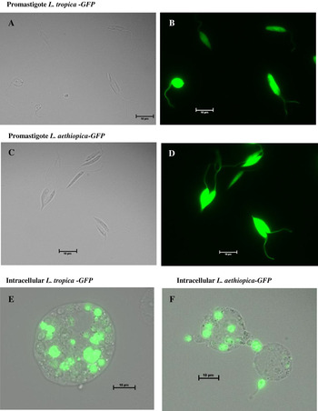
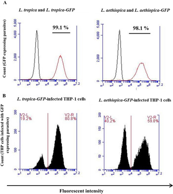
Successful transfection and expression of GFP in L. mexicana and L. major has been demonstrated previously (Bolhassani et al. Reference Bolhassani, Taheri, Taslimi, Zamanilui, Zahedifard, Seyed, Torkashvand, Vaziri and Rafati2011) using various expression cassettes and has been reproduced in this study as controls for subsequent experiments. Fluorescence of GFP was observed throughout the cytoplasm and flagella in viable extracellular promastigotes (Fig. 1B and C) and throughout the cytosol in intracellular amastigotes (Fig. 1E and F). Clonal selection procedure enabled isolation of a homogeneous population of several clones expressing GFP for each of the species. In this study, four represented clones are shown for each species except L. major for which a single clone was isolated. Flow cytometry and fluorescent microscopy analyses demonstrated a distinct and high level of GFP fluorescence both for the promastigotes and the intracellular amastigotes (L. aethiopica and L. tropica shown in Fig. 2).
Detection of GFP expression in transgenic parasites by epi-fluorescence microscopy; fluorescent microscopic images show expression of GFP in transfected L. tropica (before (A) and after (B) glinting of fluorescence) and in L. aethiopica (before (C) and after (D) glinting of fluorescence) during logarithmic phase of growth. Bright field images (A and C) for each species are shown to represent same field view. Bright field images of infected THP-1 cells were overlapped with corresponding FITC images; intracellular parasites were clearly visible 48 h following infection with (E) GFP-expressing L. tropica-GFP and (F) L. aethiopica-GFP. All images were captured under a 60× oil immersion lens.

Flow cytometry analysis of GFP expressing parasites. (A) Extracellular promastigotes of L. tropica-GFP and L. aethiopica -GFP at logarithmic phase of growth. The figure shows two samples overlapping. Black histogram represents non-transfected parasites and red histogram show homogeneous population of transfected parasites. Over 98% of transfected parasites show high expression of GFP; (B) THP-1 cells following 24 h infection with L. tropica-GFP and L. aethiopica -GFP. The first peak represents THP-1 cells not affected by presence of GFP expressing parasites, therefore considered uninfected; the second peak represents THP-1 cells showing high degree of fluorescence therefore considered infected with GFP-expressing parasites, the two populations are clearly distinguishable.

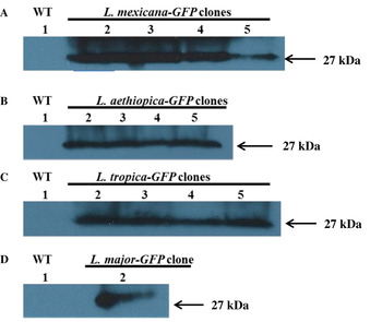
The presence of GFP in all four species (L. mexicana, L. major, L. aethiopica and L. tropica) was further validated by Western blot analysis. Green fluorescent protein was detectable in whole cell lysates of transgenic parasites using polyclonal rabbit antiserum. An expected dominant band of 27 kDa protein was visible in all transgenic clones while no protein of this molecular weight was detected in the negative controls, i.e. whole cell lysates prepared from wild-type promastigotes of all four species (Fig. 3).
Western blot analysis of GFP expression in represented clones of (A) L. mexicana-GFP (clones 2, 3, 4 and 5); (B)L. aethiopica-GFP (clones 2, 3, 4 and 5); (C) L. tropica-GFP (clones 2, 3, 4 and 5); and (D) L. major-GFP (clone 2). Arrows on the right-hand side indicates the molecular weight of GFP (27 kDa).

Chromosomal integration of GFP in the 18S rRNA locus
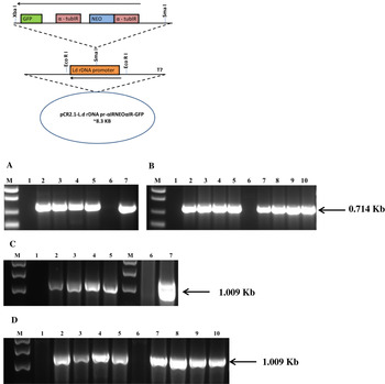
Integration of the complete GFP gene was confirmed by PCR amplification in the genome of each of the transfected species mediated via site-specific integration (Fig. 4). Transfection of L. mexicana, L. major, L. aethiopica and L. tropica with linearized pRib1.2αNEOαGFP enabled chromosomal integration of the complete GFP gene at the 18S rRNA locus downstream of the DNA Pol I-driven promoter.
Top: A schematic representation of GFP integration mediated by the pRib1.2αNeoαGFP vector construct at the 18S rRNA locus downstream of the DNA Pol I-driven promoter in the parasite genome. Bottom: PCR analysis to confirm integration of the full length GFP gene (panel A and B) present at the 18 s ribosomal locus (panel C and D) of Leishmania species. In panel A, lane M: 1 kB plus marker; lane 1: L. mexicana; lanes 2–5: L. mexicana-GFP clones; lane 6: L. major and lane 7: L. major-GFP clone. In panel B, lane M: 1 kB plus marker; lane 1: L. aethiopica; lanes 2–5: L. aethiopica-GFP clones; lane 6: L. tropica; and lanes 7–10: L. tropica-GFP clones. In panel C, lane M: 1 kB marker; lane 1: L. mexicana; lanes 2–5: L. mexicana-GFP clones; second lane M: 1 kB marker, lane 6: L. major and lane 7: L. major-GFP clone. In panel D, lane M: 1 kB marker; lane 1: L. aethiopica; lanes 2–5 L. aethiopica-GFP clones; lane 6: L. tropica and lanes 7–10: L. tropica-GFP clones.

Growth and infectivity of GFP-expressing Leishmania species
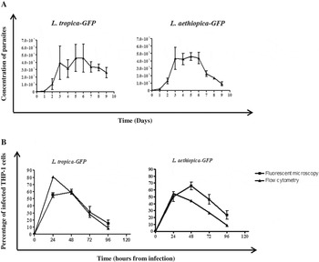
Promastigotes of transgenic GFP-expressing species were monitored for growth to determine infective metacyclic stationary phase. The growth cycle of transgenic species are comparable to each other in that stationary phase is reached at day 5 for all species (Fig. 5A and supplementary Fig. 3A – supplementary material available online only).
Extracellular and intracellular growth of GFP-expressing parasites. (A) Growth curve of GFP-expressing L. tropica and L. aethiopica promastigotes carried over 8 days. Results are the mean (±s.e.m.) of three independent experiments. (B) Time course of infection in THP-1 cells of GFP-expressing Leishmania species over a period of 96 h, determined by fluorescence microscopy (results are the mean [±s.e.m.] of three independent experiments for GFP-expressing L. tropica and four independent experiments for GFP-expressing L. aethiopica) and flow cytometry (results are the mean [±s.e.m.] of three independent experiments; where not shown, error bars fall within the symbols).

The metacyclic form of all four stable GFP-expressing species, isolated from late stationary phase promastigotes were able to infect terminally differentiated THP-1 cells. Infection was monitored at 24, 48, 72 and 96 h through both fluorescent microscopy and flow cytometry (Fig. 5B and supplementary Fig. 5B).
As previously reported (Singh et al. Reference Singh, Gupta, Jaiswal, Sundar and Dube2009), flow cytometry analysis enabled distinct separation of uninfected macrophage from an infected macrophage population based on differences in fluorescence levels between the two populations (Fig. 2B and supplementary Fig. 2B – supplementary material available online only). The percentage of infection in THP-1 cells calculated by either method followed a similar trend; percentage of infected cells peaked between 24 and 48 h and a significant decrease in infection ratio was detected after 48 h during infection with all four species (Fig. 5B and supplementary Fig. 5B). These results suggested that 24 h following infection is the best time point for assessing antileishmanial activity of reference drugs in this model.
GFP-expressing Leishmania species are susceptible to standard antileishmanial drugs
The evaluation of standard antileishmanial drugs, amphotericin B and miltefosine was carried out on both promastigote and intracellular amastigote stages of all four Leishmania species.
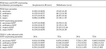
Half-maximal inhibitory constants were obtained for both amphotericin B and miltefosine following MTS assay on promastigotes. The IC50 values of transgenic GFP-expressing Leishmania spp. are comparable to those obtained for wild-type promastigotes (Table 1), suggesting introduction of GFP in all four Leishmania spp. retained their sensitivity to the standard antileishmanial drugs at the promastigote stage level. There were no significant differences in IC50 values between wild-type and transgenic parasites for all species (P>0·1). Specifically, wild-type L. aethiopica IC50 following treatment with miltefosine appears to be 1·24-fold different to the GFP-expressing strain, nevertheless this difference was not statistically significant (P = 0·2). Likewise, IC50 values obtained for wild-type L. major when tested with amphotericin B or miltefosine demonstrated a 1·62- and 2·18-fold difference in sensitivity, respectively, compared with GFP-expressing strain. However, these differences were not statistically significant (P = 0·2 and P = 0·1 for amphotericin B and miltefosine-treated parasites, respectively).
IC50 values of antileishmanial reference drugs against extracellular promastigote stage of L. mexicana-GFP, L. aethiopica-GFP, L. tropica-GFP and L. major-GFP and the correspondent wild type. Unpaired t-test coupled with Mann–Whitney U test of the IC50 between each of the wild type parasites and their transgenic counterpart showed no significant difference (P>0·1). Lower part, IC50 values of antileishmanial reference drugs against intracellular amastigotes stage of L. mexicana-GFP, L. aethiopica-GFP, L. tropica-GFP and L. major-GFP. Extracellular parasites were treated for 24 h; Intracellular parasites were treated for 24 and 72 h post infection. Results are the mean (±s.e.m.) of three independent experiments

Intracellular GFP-expressing amastigote forms of L. mexicana, L. major, L. aethiopica and L. tropica resident in THP-1 macrophages were susceptible to inhibition by amphotericin B and miltefosine over a 24 and 72 h treatment period following 48 and 96 h post-infection, respectively. IC50 data are summarized in Table 1. Susceptibility was comparable with published data (Escobar et al. Reference Escobar, Matu, Marques and Croft2002; Utaile et al. Reference Utaile, Kassahun, Abebe and Hailu2013), confirming that introduction of GFP in all four species did not alter sensitivity to drugs during the intracellular stage of the parasite's life cycle.
DISCUSSION
Development of genetically modified Leishmania species transfected with the reporter gene GFP has proved a powerful tool in Leishmania research.
Stable and constitutive expression of GFP in Leishmania species has been shown by targeted integration of exogenous genes in the transcriptionally active 18S ribosomal locus through homologous recombination (Mißlitz et al. Reference Mißlitz, Mottram, Overath and Aebischer2000). The presence of several copies of the 18S rRNA gene tandemly arrayed in the Leishmania parasite genome is advantageous in that introduction of transgenes into the chromosome has no detrimental effects on parasite survival (Mißlitz et al. Reference Mißlitz, Mottram, Overath and Aebischer2000). This strategy enabled uniform and high-level constitutive expression with no need for selective pressure and can offer a possibility for in vivo work, ideal for testing in intracellular amastigote forms. Therefore the 18S rRNA locus was chosen as the targeted integration site for transgene expression of GFP in this report. Homologous recombination and recombination rate in the Leishmania genome is dependent on several parameters such as length of the homologous region, level of sequence homology between donor and acceptor regions and copy number of the target genes (Papadopoulou and Dumas, Reference Papadopoulou and Dumas1997; Pulido et al. Reference Pulido, Muñoz, Restrepo, Mesa, Alzate, Vélez and Robledo2012). Based on the above criteria, the current study used pRib1.2αNEOαGFP as the vector for transfection of four Leishmania species. This construct was successfully used for stable integration of GFP in the 18S rRNA locus of the Leishmania donovani genome and no demonstrable effect of GFP expression on parasite morphology and infectivity was reported (Singh et al. Reference Singh, Gupta, Jaiswal, Sundar and Dube2009). Similarly, the current study has shown no effects of the GFP transgene on morphology and infectivity in L. mexicana, L. aethiopica, L. tropica and L. major. Transgenic parasites exhibited bright green fluorescence throughout the cytoplasm (Fig. 1A and supplementary Fig. 1A – supplementary material available online only) and were highly motile suggesting transgene expression did not affect motility. Furthermore, relative fluorescence measurements via flow cytometry data confirmed high level and homogeneous expression of GFP throughout parasite populations in all species (Fig. 2A and supplementary Fig. 2A) as well as a clear quantitative separation between transfected and wild type parasites with a percentage of GFP-expressing parasites ranging from 94·4% (L. major, supplementary Fig. 2A) to 99·1 (L. tropica, Fig. 2A). This mode of constitutive expression is under controlled regulation and not dependent on continuous drug selection pressure for high level of fluorescence expression (Pulido et al. Reference Pulido, Muñoz, Restrepo, Mesa, Alzate, Vélez and Robledo2012).
Suitability of L. mexicana, L. major, L. aethiopica and L. tropica GFP-expressing parasites as tools for drug screening and infection studies was confirmed via a number of tests. All transfectant clones expressed high levels of fluorescence and could be easily visualized intra- and extra-cellularly under the microscope (Fig. 1) and via flow cytometry (Fig. 2). All four parasite species were able to infect retinoic acid-treated THP-1 cells in vitro and infected cells could be detected up to 96 h post infection (Fig. 5B). These transgenic species proved ideal for setting up of flow cytometry-based assays in intracellular parasites, as the population of infected cells was clearly quantifiable over a period of 4 days. Data obtained from flow cytometry-based counting in at least four repeated experiments were extremely accurate as shown by the very small standard error (Fig. 5B). This observation is in line with published data that underline the high sensitivity and accuracy of the flow cytometry method compared with manual fluorescent microscopy counting (Pulido et al. Reference Pulido, Muñoz, Restrepo, Mesa, Alzate, Vélez and Robledo2012). Transgenic promastigotes were assessed for sensitivity to amphotericin B and miltefosine in a concentration-dependent manner and showed IC50 values comparable to those of the wild types (Table 1). This suggests that introduction of the GFP gene in the Leishmania genome of all four species did not alter their sensitivity profiles to the reference drugs and supports the use of such parasites for drug screening. The IC50s obtained via the semi-automatized medium throughput flow cytometry assay of THP-1 infected cells (Table 1) were also consistent with those obtained from previous studies.
Several molecular approaches have been used to express GFP in a variety of Leishmania strains. Antibiotic-induced expression of GFP was achieved in L. major, Leishmania amazonensis, L. donovani and Leishmania infantum by the transfection approach involving expression of GFP as an episomal transgene (Sean Ha et al. 1996; Kamau et al. Reference Kamau, Grimm and Hehl2001; Chan et al. Reference Chan, Bulinski, Chang and Fong2003; Okuno et al. Reference Okuno, Goto, Matsumoto, Otsuka and Matsumoto2003; Singh and Dube, Reference Singh and Dube2004). These fluorescent Leishmania species were amenable to in vitro drug testing assays in the promastigote stage. Leishmania donovani could also be used for testing drugs in the intracellular amastigote stage; however, due to the high background noise automated screening was impossible (Dube et al. Reference Dube, Gupta and Singh2009). Moreover episomal expression of GFP in Leishmania species is transient, resulting in a heterogeneous population of fluorescent promastigotes (Chan et al. Reference Chan, Bulinski, Chang and Fong2003; Varela et al. Reference Varela, Lorena Muñoz, Robledo, Kolli, Dutta, Chang and Muskus2009). Finally, continuous drug pressure is required for parasites to retain and express the GFP gene, as this cannot be maintained during infection; the parasite loses the gene during the intracellular stages of its life cycle both in vitro and in vivo.
Recent efforts have focused on the generation of stable recombinant Leishmania species constitutively expressing GFP through integration of the transgene in the Leishmania genome of various species. Integration of GFP has been stably carried out in various locations: in 18 s rRNA locus of L. donovani, L. mexicana, L. panamensis, L. braziliensis, L. amazonensis and L. infantum (Boucher et al. Reference Boucher, McNicoll, Dumas and Papadopoulou2002; Dube et al. Reference Dube, Gupta and Singh2009; Singh et al. Reference Singh, Gupta, Jaiswal, Sundar and Dube2009; Pulido et al. Reference Pulido, Muñoz, Restrepo, Mesa, Alzate, Vélez and Robledo2012); in the ssu locus of L. major, L. infantum and L. tarentolae (Bolhassani et al. Reference Bolhassani, Taheri, Taslimi, Zamanilui, Zahedifard, Seyed, Torkashvand, Vaziri and Rafati2011). Based on the above studies, all of the Leishmania species displayed homogeneous and constitutive expression of GFP and fluorescence persisted for long periods of time. These parasite species were able to infect macrophages in vitro and were able to maintain fluorescence during the intracellular stages. Furthermore, parasites were infective in vivo and fluorescence could be detected a month after infection (Singh et al. Reference Singh, Gupta, Jaiswal, Sundar and Dube2009; Bolhassani et al. Reference Bolhassani, Taheri, Taslimi, Zamanilui, Zahedifard, Seyed, Torkashvand, Vaziri and Rafati2011). On the basis of such promising results our efforts have been focusing towards the development of L. mexicana, L. major, L. aethiopica and L. tropica transgenic parasites in order to produce a larger repertoire of GFP-expressing Leishmania species covering the whole range of disease manifestations. Production of stable GFP transfectants of both L. mexicana and L. major has been previously reported and we have replicated it as controls and also validated such transfectants for flow cytometry-based drug assays. Clones of L. tropica and L. aethiopica constitutively expressing GFP have been produced for the first time. Leishmania tropica is mainly responsible for cutaneous manifestation but it is also known to spread to internal organs and cause visceral leishmaniasis. Leishmania aethiopica causes a spectrum of disease manifestations ranging from localized, self-healing lesions (local cutaneous leishmaniasis; LCL) to disfiguring mucocutaneous lesions (mucocutaneous leishmaniasis; MCL) to diffuse, persistent lesions (diffuse cutaneous leishmaniasis; DCL). Leishmania aethiopica also shows a high degree of resistance to first-line treatments such as antimonials which adds to its importance as model for drug discovery.
Current chemotherapeutic options are limited by toxicity, long treatment regimes and cost as well as intrinsic (in the case of L. aethiopica) and emerging parasite resistance (Zucca and Savoia, Reference Zucca and Savoia2011; Ejazi and Ali, Reference Ejazi and Ali2013). Research towards new therapeutic approaches for leishmaniasis is therefore imperative. Development of new treatments based on rational drug design of identified novel molecular targets is a widely used tool in the drug discovery process (Alam et al. Reference Alam, Goyal, Iqbal, Pal, Dey, Bindu, Maity and Bandyopadhyay2009). Nevertheless its application to leishmaniasis drug discovery is limited by the incomplete understanding of the complexities of the process of Leishmania survival. Therefore detection of new classes of compounds through screening of compound libraries in whole organisms remains the most exhaustive approach for selection of active compounds. An ideal screening procedure should involve a range of species and should be based on the intracellular stage of the parasite life cycle as this is the medically relevant one. Such screening should also be transferable to in vivo studies which are necessary as part of the drug development process. Green fluorescent protein is a reporter gene product renowned for its intrinsic fluorescent property which makes it ideal for both imaging and quantification by in vitro cell viability assays (Tsien, Reference Tsien1998; Kain, Reference Kain1999).
Our findings support the use of genomic integration approaches to generate transgenic GFP-expressing Leishmania species suitable for investigation of the infection process as well as for flow cytometry-based drug assay. Such an assay is faster and cheaper than those currently available; moreover it is based on the medically relevant stages of the parasite life cycle. Flow cytometry analysis proved to be fast, highly sensitive and accurate in obtaining IC50 data of reference drugs tested on the intracellular stage compared with the microscopy-based method of determination. Moreover, such parasites have the potential to be used for in vivo infection as a model for drug testing as well as for increasing our understanding of disease pathogenesis.
The above study succeeded in generating, to our knowledge for the first time, transgenic L. aethiopica and L. tropica parasites constitutively expressing GFP. Such strains as well as transgenic L. major and L. mexicana were successfully validated as representing the same characteristic as their wild type counterparts. Specifically, because such parasites maintain infectivity and drug susceptibility comparable to their wild type correspondents they are an ideal model both for infection study and drug assays.
SUPPLEMENTARY MATERIAL
To view supplementary material for this article, please visit http://dx.doi.org/S0031182013001777.
ACKNOWLEDGEMENTS
The authors would like to acknowledge Dr Simon Richardson for his valuable suggestions during sample preparation and fluorescence microscopy, Rajeev Raj for assistance in producing the images of GFP-expressing parasites and Sam Ingram for technical assistance.
FINANCIAL SUPPORT
This work received financial support from the University of Greenwich and Greenwich Research & Enterprise (GRE).