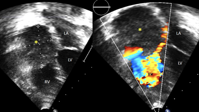
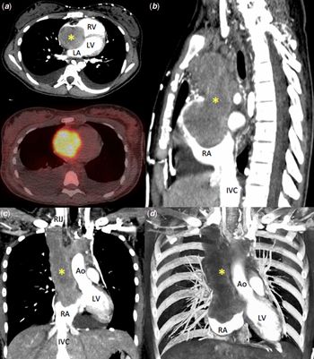
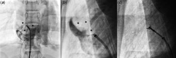
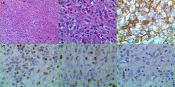
Primary pediatric cardiac tumors are extremely rare.Reference Davis, Allan and Perez1 We report a pediatric case of primary cardiac Hodgkin lymphoma with initial multi-modality imaging presentation. A 14-year-old female with 3-week history of dyspnea underwent echocardiography. A low-echogenic homogeneous mass (4.6 cm diameter) was found in the right atrium partially obstructing the tricuspid valve inflow, with no flow in the superior caval vein or innominate vein (Fig 1). Computed tomography (CT) demonstrated a large right atrial mass extending upward and occluding the superior caval vein and left innominate vein, with venous return from the head and neck vessels passing via the azygos/hemiazygos system and paravertebral venous collaterals (Fig 2). Due to extensive venous collaterals, she did not exhibit superior caval vein syndrome. Transcatheter tumor biopsy result was inconclusive (Fig 3). Histopathological findings from CT-guided core biopsy were consistent with classical Hodgkin lymphoma, demonstrating scattered large lymphocytes with Hodgkin cell-like morphology. Immunohistochemical staining showed positive CD30, PAX5, and vimentin biomarkers and negative CD45 biomarker (Fig 4). There was no immunosuppressive baseline status with negative human immunodeficiency virus testing. Cross-sectional imaging showed no evidence of separate nodal lesions except intracardiac/intravascular tumor and adjacent mildly enlarged anterior superior mediastinal lymph nodes. Disease isolated above the diaphragm and absence of B symptoms, classified her as stage IIAE. She was enrolled in Children’s Oncology Group study AHOD0031 and was treated with Doxorubicin, Bleomycin, Vincristine, Etoposide, Cyclophosphamide, and Prednisone. Follow-up CT showed an interval decrease in the size of right atrial mass, and positron-emission tomography scan showed no fluorodeoxyglucose uptake.
Transthoracic echocardiography, four-chamber view. A large mass (*) occupies right atrium, with flow acceleration of tricuspid valve inflow on Color Doppler imaging.

CT of chest. (a) Coronary view. A large mass in the right atrium shows a strong fluorodeoxyglucose uptake on positron-emission tomography. (b) Sagittal view. (c) Coronal view. The right atrial tumor extends into the superior caval vein and innominate vein. (d) Three-dimensional volume rendering image (Ao=ascending aorta; IVC=inferior caval vein; LA=left atrium; LV=left ventricle; RA=right atrium; RIJ=right internal jugular vein; RV=right ventricle).

Transcatheter tumor biopsy. (a and b) Angiography shows a large mass in the right atrium (*) in the anterior–posterior and lateral views. (c) A bioptone is opposed to a mass in the right atrium.

(a) Low-power (10×) magnification of an H&E slide of a pericardial biopsy specimen demonstrates a diffuse polymorphous cellular infiltrate. (b) At high-power (40×) magnification, the infiltrate consists of histiocytes, small lymphocytes, few eosinophils, and large atypical cells varied cytomorphologic features, including mono and polylobated nuclei and some with prominent nucleoli. Immunohistochemical stains show the large cells to be positive for (c) CD30 and (d) PAX5 and negative for (e) CD15 and (f) CD45.

Acknowledgements
None.
Financial support
This research received no specific grant from any funding agency, commercial or not-for-profit sectors.
Conflicts of interest
None.