Aorto-pulmonary collateral arteries are vessels developed from the ventral-splanchnic system; with normal pulmonary artery development, they regress early in gestation. Reference Boshoff and Gewillig1 With impaired pulmonary blood flow, collaterals may persist, often associated with pulmonary atresia with a ventricular septal defect. Reference Norgaard, Alphonso, Cochrane, Menahem, Brizard and d’Udekem2 Collaterals are rarely associated with heterotaxy, usually found postnatally. Reference Ota, Fujimoto and Murata3 We report a prenatally diagnosed case of aorto-pulmonary collaterals associated with a single ventricle and total anomalous pulmonary veins associated with right atrial isomerism.
Case
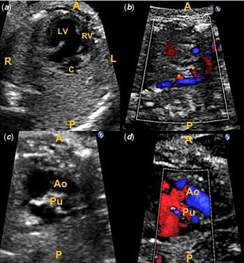
A 31-year-old G5P3T1 woman was referred for foetal echocardiography for the suspected cardiac anomaly. She had well-controlled hypertension and hypothyroidism. There was no family history of congenital heart disease. A 19-week foetal echocardiogram demonstrated dextrocardia, and an unbalanced atrioventricular septal defect with a dominant right-sided left ventricle. Both outlets arose from the single ventricle, with the aorta anterior to a smaller pulmonary artery. Severe sub-pulmonary stenosis and regurgitation were seen. Branch pulmonary arteries were confluent and hypoplastic. Inferior caval vein drained to the left-sided atrium while bilateral superior caval vein drained to their ipsilateral atrium. Pulmonary veins drained unobstructed through a confluence to the right-sided atrium. Ductus arteriosus was absent and two aorto-pulmonary collaterals from the thoracic aorta were identified (Fig 1, Supplementary videos 1 and 2). Ventricular function was normal and there was no atrioventricular valve regurgitation. The stomach was seen centrally and leftward, the liver was midline and there was asplenia; findings were in keeping with a right atrial isomerism. The patient was counselled about management. The challenge of small branch pulmonary arteries and collateral vessels in combination with anomalous pulmonary venous drainage and risk of pulmonary vein stenosis was reviewed. Pregnancy was continued and genetic testing declined. She was reassessed at 22-, 29-, and 36-weeks’ gestation.
Foetal echocardiogram at 29 weeks and 2 days’ gestation: ( a ) Four-chamber view showing single atrioventricular valve connection to a right-sided dominant left ventricle. A moderator band was not seen. A rudimentary right ventricle is seen leftward of the morphologic left ventricle. Pulmonary venous confluence was seen behind the larger right-sided atrium. ( b ) Sagittal imaging showing aortic arch with two large collaterals coursing towards the lungs. ( c and d ) Sagittal imaging showing larger and anterior aortic valve and posterior and smaller pulmonary valves with pulmonary regurgitation seen on colour Doppler. A = anterior; Ao = aorta; C = pulmonary venous confluence; L = left; LV = left ventricle; R = right; RV = right ventricle; P = posterior; Pu = pulmonary outflow; * = aorto-pulmonary collaterals.

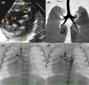
A term, 2.86 kg female neonate was born by caesarean section. Oxygen saturation ranged between 88 and 92% in room air. A postnatal echocardiogram confirmed prenatal diagnosis (Fig 2, Supplementary video 2). The abdominal ultrasound showed asplenia and midline liver without bowel malrotation. Chest computed tomography showed bilateral right bronchi (Fig 2). Ultrasound and computed tomography, in combination with an echocardiogram, confirmed right atrial isomerism. Genetic testing postnatally was declined. On day 3, she developed a bowel perforation and required partial colectomy and loop ileostomy. At 2 months, cardiac catheterization showed dual pulmonary perfusion by aorto-pulmonary collaterals and central pulmonary arteries (Fig 2, Supplementary videos 3 and 4). She underwent pulmonary outflow stenting and coil occlusion of collaterals. Pulmonary veins were not assessed. She developed respiratory distress from pulmonary vein stenosis. At 3 months, she received a 3.5-mm right Blaloch–Taussig shunt, stent removal, and suture-less pulmonary vein repair. She required extracorporeal membrane oxygenation and a delayed chest closure. Ongoing desaturation prompted repeat cardiac cross-sectional imaging; complete obstruction of the left upper pulmonary vein, severe stenosis of the right upper pulmonary vein, moderate stenosis of the right lower pulmonary vein, and minimal stenosis of the left lower pulmonary vein were found. She was deemed unsuitable for heart–lung transplantation. Given limited options, comfort care was initiated at 4 months and the patient demised.
Postnatal imaging confirming prenatal findings: ( a ) Postnatal two-dimensional echocardiogram in short axis showing a dominant, left ventricle that is rightward. A rudimentary right ventricle is seen leftward and anterior to the morphologic left ventricle. ( b ) CT chest showing symmetric bronchi in keeping with right atrial isomerism. ( c ) Angiogram showing one aorto-pulmonary collateral vessel arising from the aorta. ( d ) Angiogram showing a second aorto-pulmonary collateral arising from the aorta. A = anterior; L = left; LV = left ventricle; P = posterior; R = right; RV = right ventricle; * = aorto-pulmonary collateral.

Discussion
We present a case of antenatal diagnosis of aorto-pulmonary collaterals associated with right atrial isomerism, single ventricle, and total anomalous pulmonary venous drainage. To our knowledge, this is the first such case to be reported with a foetal diagnosis. Right atrial isomerism is often associated with complex cardiac malformations and commonly associated with pulmonary atresia and total anomalous pulmonary venous drainage; the presence of aorto-pulmonary collaterals with right atrial isomerism has rarely been reported postnatally. Reference Ota, Fujimoto and Murata3–Reference Reinhartz, Reddy and Petrossian5 Reinhart et al Reference Reinhartz, Reddy and Petrossian5 reported their experience with single ventricles with aorto-pulmonary collaterals from 1997 to 2005 and found that in a cohort of 14 patients, 7 patients received a Glenn but only 3 patients had Fontan completion. A postnatal cohort of 138 patients reported by Hsu et al Reference Hsu, Wang and Lin6 showed that aorto-pulmonary collaterals were uncommon in right atrial isomerism with pulmonary stenosis or atresia (6.5%; 9 patients). Although surgical mortality was similar in groups with or without aorto-pulmonary collaterals until Glenn connection, completion of total cavo-pulmonary connection was significantly lower in the collateral group; only one patient received Fontan completion. Mechanism underlying the development of collaterals remains unknown; a mouse model showed the combination of pulmonary atresia with an absent arterial duct can lead to aorto-pulmonary collaterals. Reference Rammeloo, DeRuiter, van den Akker, Wisse and Gittenberger-de Groot7 One could postulate that collaterals persisted due to the lack of antegrade pulmonary blood flow related to the combination of sub-pulmonary obstruction and small branch pulmonary arteries.
While there is reported poor prognosis related to collaterals in the context of single-ventricle lesions, total anomalous pulmonary venous drainage was also present which carries a risk of obstruction. There is a risk of re-stenosis of pulmonary veins post-intervention which should be reviewed during counselling. Pulmonary vein stenosis is thought to be associated with intimal proliferation of myofibroblasts. Reference Sadr, Tan, Kieran and Jenkins8 Risk factors for pulmonary vein stenosis, in this case, include heterotaxy, presence of single ventricle additional procedures at the time of suture-less repair, and pre-operative stenosis. Reference White, Ho and Faerber9 Mortality associated with pulmonary stenosis is signficant, Reference Backes, Nealon and Armstrong10 particularly with three or four stenosed pulmonary veins. Reference Backes, Nealon and Armstrong10 It is unclear how the aorto-pulmonary collaterals affected the development of pulmonary veins stenosis or caused re-stenosis post-repair.
Diagnosis of right atrial isomerism and pulmonary atresia can be reliably made prenatally; eliciting the presence of aorto-pulmonary collaterals and anomalous pulmonary veins is important to guide prenatal counselling. Association of collaterals in combination with anomalous pulmonary veins carries a poor prognosis. This case highlights the need for thorough foetal cardiac imaging to identify all known and rare associations with heterotaxy and counsel families accordingly.
Conclusion
This is the first reported foetal diagnosis of aorto-pulmonary collateral vessels in the setting of right atrial isomerism, a single ventricle, and total anomalous pulmonary veins. Foetal cardiologists should complete an accurate anatomic assessment to provide families with realistic expectations of postnatal outcomes.
Supplementary material
To view supplementary material for this article, please visit https://doi.org/10.1017/S1047951120003649.
Acknowledgements
Thank you to the patient who provided permission to review fetal case for educational purposes.
Financial support
This research received no specific grant from any funding agency, commercial or not-for-profit sectors.
Conflicts of interest
None.
Ethical standards
Written consent was obtained from the patient prior to reporting this case.