Pulmonary arteriovenous malformation is the presence of a direct relation between the pulmonary artery and pulmonary vein without a capillary structure. It is a rarely encountered disease and has an incidence of 3 out of 1500 in autopsies. The lesions are characterised by thin-walled vessels that are most frequently found in the lower lobes of lungs. Reference Enon, Penny, Shaddy, Penny, Feltes, Cetta and Mital1 Systemic venous blood skips the lungs flowing into the left atrium, thereby resulting in right-to-left shunt and arterial desaturation. The arteriovenous fistulae found in this disease can be microvascular and multiple while they can be in the form of a large arteriovenous connection. They may emerge congenitally or due to several acquisition-related reasons. Reference Guvenc, Saygi, Demir and Odemis2 Although aetiology is not exactly known, it can be seen more often after cavopulmonary shunt surgeries and in patients with hepatic failure. Reference Enon, Penny, Shaddy, Penny, Feltes, Cetta and Mital1
Extracorporeal membrane oxygenation is used to support the patient haemodynamically and respirationally in medical treatment-resistant, severe and revocable cardiac or pulmonary insufficiency cases. Reference Abraham, Gilliam, Kim, Wolf, Vincent and Petit3 The most frequent ECMO indication in children is CHD. Extracorporeal membrane oxygenation support is needed in several situations such as post-cardiotomy, myocarditis, cardiomyopathies, pulmonary haemorrhage, sepsis, pneumonia, asthma attack, and diseases such as resistant arrhythmias and pulmonary hypertension. In a report published in 2016 by the paediatric extracorporeal life support organisation, the survival and discharge ratios among neonatal and children receiving extracorporeal membrane oxygenation support were reported in the range of 44–54%. Reference Taka, Kotani, Kuroko, Iwadou, Iwasaki and Kasahara4 Cardiac catheterisation is used for diagnosis and treatment in children receiving extracorporeal membrane oxygenation support. This paper presents an infant with pulmonary arteriovenous malformation who was connected extracorporeal membrane oxygenation support and received a successful transcatheter fistula embolisation since haemodynamic stability could not be ensured with mechanical ventilator support.
Case report
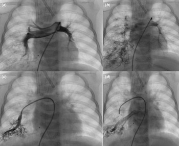
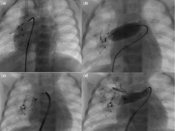
A four-month-old male patient, who had no previously known diseases, had a CT scan due to low saturation at an external centre, and multiple and dilated vascular structures in the right lung consistent with arteriovenous fistula were detected. The patient had significantly low saturation, overall poor condition and hypotension and was admitted to the ICU. The patient’s circulation was poor with a blood pressure of 52/30 mm Hg, heart rate 115 beats/min, saturation 25% under 100% FiO2 support while his capillary filling time was 6 seconds, and peripheral pulses could not be palpated. The patient, who developed bradycardia followed by cardiopulmonary arrest, received cardiopulmonary resuscitation for two minutes. The patient’s cardiac activity was restored. Despite adequate fluid support and infusions of dopamine, adrenaline, noradrenaline, and milrinone infusions, the patient’s progression was bradycardic and hypotensive. The blood gas exam showed severe respiratory and metabolic lactic acidosis. After the patient was admitted to the ICU, he had arrest two times. After cardiopulmonary resuscitation for two minutes, the heart rate became 100 beats/min. The pulmonary X-ray did not show pneumothorax or any other pathological signs. The patient’s severe hypoxia did not regress; his saturation was around 25%, and metabolic lactic acidosis gradually increased. He underwent a right jugular incision and was started on veno-arterial extracorporeal membrane oxygenation support with right carotid artery and internal jugular vein cannulation. Afterwards, his blood pressure retreated to 75/55 mmHg, saturation level increased to 96%, and metabolic acidosis was reduced. The patient’s urinary output was normal, his pupils were isochoric, and light reflex was bilaterally positive. The patient’s blood parameters were improved, and vital signs were within normal limits. His inotropes were gradually reduced. On day four of his hospitalisation, he underwent angiography in the catheterisation laboratory under extracorporeal membrane oxygenation support. After the injection of selective contrast media into the right pulmonary artery, multiple pulmonary arteriovenous fistulae were detected in the right lung. Occlusion was applied on the two fistulae on the lower right side using an Amplatzer® vascular plug 4 (8 and 7 mm; Abbott Vascular, USA) and a Lifetech® vascular plug (10 mm; Lifetech Medical Corporation, China) and on the wide fistula in the right middle lobe using an Amplatzer® vascular plug 4 (7 mm) (Fig 1). The patient successfully left after the extracorporeal membrane oxygenation support in the evening of the day of procedure. The patient’s saturation value was around 85%, and he was admitted to the catheter laboratory for the second time as intubated two days later. The wide vascular structure carrying blood to the right lung middle and lower lobes was occluded with an Amplatzer® vascular plug 1 (16 mm) while the fistula in the right lung upper lobe with an Amplatzer® vascular plug 4 (8 mm) (Fig 2). The patient’s saturation rose to 95% after the procedure. The patient was started on aspirin treatment at an antiaggregating dose, and extubation was done one day later. The patient’s vital signs and neurological exam result were normal. He was discharged on day 11 of his hospitalisation. The patient has been under regular outpatient follow-up for two years, and his saturation ranged between 92 and 94%. The patient’s family provided informed consent for the use of the images of the patient’s clinical signs, laboratory, and imaging results.
(a) Pulmonary artery and right and left pulmonary artery anatomy can be seen in contrast agent injection into the main pulmonary artery. ( b) Multiple pulmonary AV fistula is seen in the right lung. ( c) Selective contrast agent injection into the peripheral pulmonary artery located in the lower zone of the right lung is observed. ( d) An associated pulmonary AV fistula is seen in contrast agent injection into the peripheral pulmonary artery located in the lower zone of the right lung.

(a) After the first angiography performed with extracorporeal membrane oxygenation support, the devices used to close the fistulas are monitored. ( b) In the second angiography procedure performed on the patient who left extracorporeal membrane oxygenation support, it is seen that test occlusion was performed before fistula closure. ( c) In the second angiography procedure, the device used to close the fistula associated with the middle and lower lobes of the right lung is seen. (d) In the second angiography procedure, it is observed that the device used to close the fistula associated with the right lung upper lobe and there is no distal flow in the contrast agent injection.

Discussion
Pulmonary arteriovenous fistula is a rare reason for cyanosis. If a cyanosis patient does not have pulmonary parenchymal pathology and an intercardiac shunt was not detected in the echocardiographic exam, this disease should be considered. The diagnosis of extracardiac shunt can be confirmed via contrast echocardiographic evaluation using agitated saline, and the diagnosis can be made using CT angiography or conventional pulmonary angiography. Reference Khanra, Razi, Tiwari, Soni and Thakur5 Pulmonary angiography is considered the golden standard in diagnosis. Reference Enon, Penny, Shaddy, Penny, Feltes, Cetta and Mital1 Hereditary haemorrhagic telangiectasis (Osler-Weber-Rendu syndrome) can be part of the disease or emerge as an isolated abnormality. Reference Enon, Penny, Shaddy, Penny, Feltes, Cetta and Mital1 Depending on the fistula size and amount of shunt, the patient asymptomatic while signs such as respiratory trouble, severe cyanosis, finger clubbing, chest pain, and haemoptysis may develop. In untreated patients, heart failure, chronic cyanotic complications, neurological complications due to paradoxical embolism, endocarditis, and death due to aneurysmal fistula rupture can be seen. Reference Enon, Penny, Shaddy, Penny, Feltes, Cetta and Mital1,Reference Guvenc, Saygi, Demir and Odemis2,Reference Khanra, Razi, Tiwari, Soni and Thakur5 With the advancements in transcatheter devices and increased experience in this matter, fistula embolisation procedures can be successfully performed as an alternative to surgical treatment. Even large fistulae can be closed using one or multiple devices. Reference Khanra, Razi, Tiwari, Soni and Thakur5 Today, the procedure of transcatheter embolisation using coils or vascular plugs for lesions ≥3 mm is accepted as a treatment of priority. Reference Enon, Penny, Shaddy, Penny, Feltes, Cetta and Mital1 In our patient, fistulae were detected under CT imaging and pulmonary angiography. Since our patient did not have family history, epistaxis, telangiectasia in the skin or mucous membranes and visceral organ involvement, the hereditary haemorrhagic telangiectasis disease was not considered. The case presented with treatment-resistant severe hypoxia and cyanosis. The patient was intubated, and his clinical signs and blood gas parameters did not improve despite mechanical ventilator treatment; therefore, he was placed under extracorporeal membrane oxygenation support. The pulmonary arteriovenous fistulae identified in the patient were successfully closed in two sessions using five different devices with a transcatheter method.
Among children, publications and patient-related results related to cardiac catheterisation under extracorporeal membrane oxygenation support are inadequate. In one study, 75 catheterisation procedures performed during extracorporeal membrane oxygenation support due to CHD or cardiomyopathy in children between 2003 and 2013 were investigated. Nearly three-fourths of catheterisation procedures are invasive interventions. In this study, the conclusion was that cardiac catheterisation in children under extracorporeal membrane oxygenation support was a rare situation even in large-volume centres and that cardiac catheterisation and transcatheter invasive procedures could be performed in children safely and with low complication rates. Reference Abraham, Gilliam, Kim, Wolf, Vincent and Petit3
In the literature, no patients were reported who received transcatheter fistula occlusion under extracorporeal membrane oxygenation support with a diagnosis of pAVM. In a case report, it was reported that a 19-year-old patient who had massive haemoptysis due to bronchial arteriovenous malformation, severe hypoxia, and haemodynamic instability was put under extracorporeal membrane oxygenation support and underwent surgical right bilobectomy as endovascular intervention was unsuccessful. Reference Shepherd, Kotkar, Bhalla, Rostambeigi, Kreisel and Puri6 The case that we present indicates a successful and multidisciplinary approach to the treatment of a rarely seen disease. The infant pulmonary arteriovenous malformation patient with haemodynamic instability due to severe hypoxia received extracorporeal membrane oxygenation support under emergency conditions, and the transcatheter fistula embolisation procedure was successfully performed.
In conclusion, pulmonary arteriovenous malformations, which are a rare cause of cyanosis among children, can be closed using the transcatheter method. In patients with an overall poor condition who are haemodynamically unstable, fistula embolisation can be performed after ensuring extracorporeal membrane oxygenation support.
Acknowledgement
None.
Author contribution
Study conception and design: OG, MS, MAO.
Data collection: OG, MS.
Analysis and interpretation of results: MS, EO.
Draft manuscript preparation: OG, MS, MAO, EO.
All authors reviewed the results and approved the final version of the manuscript.
Financial support
This research received no specific grant from any funding agency, commercial, or not-for-profit sectors.
Conflict of interest
None.
Ethical standards
This case description does not contain any study with animal performed by any of the authors. All applicable and international, national, and/or institutional guidelines for the case were followed. The procedure performed in the case description was in accordance with the ethical standards of the institutional and/or research committee and with the declaration and with the 1964 Helsinki declaration and its later amendments or comparable ethical standards.
Consent was obtained from the parents of the individual included in the study.