Introduction
Alzheimer's disease (AD), dementia with Lewy bodies (DLB) and vascular dementia (VaD) are the most common causes of dementia (Stevens et al., Reference Stevens, Livingston, Kitchen, Manela, Walker and Katona2002). In addition to cognitive decline, accompanying motor dysfunctions such as slowness, rigidity and impaired balance and gait are frequently reported in patients with these disorders (Kurlan et al., Reference Kurlan, Richard, Papka and Marshall2000; Prehogan and Cohen, Reference Prehogan and Cohen2004). The development of motor disturbances has been shown to vary within and between the types of dementia (Román et al., Reference Román1993; O'Keeffe et al., Reference O'Keeffe, Kazeem, Philpott, Playfer, Gosney and Lye1996).
Paratonia is a form of hypertonia that is defined as an active unintentional resistance against passive movement. Paratonia is a motor problem that is frequently observed in individuals with dementia and results in a loss of mobility and the development of contractures (Souren et al., Reference Souren, Franssen and Reisberg1997; Hobbelen et al., Reference Hobbelen, Koopmans, Verhey, van Peppen and de Bie2006). The prevalence of paratonia increases with the progression of dementia and is estimated to reach 100% in patients with later stages of dementia (Souren et al., Reference Souren, Franssen and Reisberg1997). The effect of paratonia on a patient's quality of life is devastating, and the caregiver's burden increases substantially with time (Souren et al., Reference Souren, Franssen and Reisberg1997).
In early-stage dementia, paratonia has been associated with the development of apraxia, and in late-stage dementia, it has been associated with the reappearance of neonatal reflexes that are known as frontal release symptoms (Franssen et al., Reference Franssen, Kluger, Torossian and Reisberg1993; Beversdorf and Heilman, Reference Beversdorf and Heilman1998; Vahia et al., Reference Vahia, Cohen, Prehogan and Memon2007).
The pathogenesis of paratonia is not well understood. The increased prevalence in patients with neurodegenerative disorders suggests a central cerebral pathology, possibly in the substantia nigra (Risse et al., Reference Risse1990; O'Keeffe et al., Reference O'Keeffe, Kazeem, Philpott, Playfer, Gosney and Lye1996). However, positron emission tomography (PET) scan analyses and autopsy studies have provided no evidence for dopaminergic nigrostriatal dysfunction in AD patients with paratonia (Tyrrell et al., Reference Tyrrell1990). Alternatively, peripheral biomechanical changes have been hypothesized (Hobbelen et al., Reference Hobbelen, de Bie and van Rossum2003). Previous research has reported that diabetes mellitus (DM) and general multimorbidity are risk factors for muscle rigidity in non-dementia patients (Arvanitakis et al., Reference Arvanitakis, Wilson, Schneider, Bienias, Evans and Bennett2004; Mourey et al., Reference Mourey, Manckoundia, Martin-Arveux, Tavernier-Vidal and Pfitzenmeyer2004). In addition, studies have shown that antipsychotic medication may induce paratonia-like rigidity in dementia patients (Risse et al., Reference Risse1990; Soininen et al., Reference Soininen, Laulumaa, Helkala, Hartikainen and Riekkinen1992).
Most previous studies on paratonia have focused on patients in the late stages of dementia with severe paratonia, making it difficult to investigate retrospectively the factors that contributed to the development of paratonia. To date, no longitudinal studies exist that examine the contributing factors to the development of paratonia.
Recently, a consensus definition was established, and a new, valid and reliable assessment instrument for paratonia became available, stimulating new research in this field (Hobbelen et al., Reference Hobbelen, Koopmans, Verhey, van Peppen and de Bie2006; Reference Hobbelen, Koopmans, Verhey, Habraken and de Bie2008).
In the present study, we performed a one-year longitudinal study among dementia day-care patients to explore the possible contributing factors to the development of paratonia in dementia patients. The specific research questions included the following: (1) Do differences exist in the profiles of the participants with and without paratonia? (2) During what stage of dementia does paratonia develop? (3) Do differences exist in the course and the development of paratonia between the different types of dementia? (4) Is decreased functional mobility an early indicator of the development of paratonia? (5) Does diabetes mellitus, multimorbidity or the use of antipsychotic medication contribute to the development of paratonia?
Methods
Design
We performed a multi-center, longitudinal, one-year follow-up cohort study in which the participants were visited at their center every three months.
Study population
Dementia day-care centers in nursing homes and residential homes with dementia special care units (DCUs) in the regions of Eindhoven, Helmond and Tilburg in the Netherlands were randomly selected as recruitment facilities. The participants were considered to be eligible for inclusion in the present study when they met the following criteria: (1) an established diagnosis of dementia according to DSM-IV criteria (American Psychiatric Association, 1994); (2) a score of stage 6 or lower on the Global Deterioration Scale (GDS; Reisberg et al., Reference Reisberg, Ferris, de Leon and Crook1982); and (3) the ability to walk at least 10 meters without assistance (a walking aid was allowed), which was necessary for the assessment of functional mobility. Participants were excluded from the study if they had unstable health owing to an interfering comorbidity.
The staff of the participating day-care centers and DCUs were asked to identify eligible participants, after which an information brochure about the study and a written consent application form was sent to the eligible participants and their legal representatives. A participant was only included in the present study if written informed consent was received from their representative. The patients’ general practitioners (GPs) were notified about the study and were mandated by the participants to provide information from their records. Those participants who initially agreed to participate but refused further collaboration during the study were excluded. The local ethical committee of the Arnhem/Nijmegen region approved the study.
Assessments
The primary outcome measure in the present study was the Paratonia Assessment Instrument (PAI) (Hobbelen et al., Reference Hobbelen, Koopmans, Verhey, Habraken and de Bie2008). The PAI is a dichotomous assessment instrument with which an examiner can establish the presence (or absence) of paratonia by successively moving all four limbs passively in flexion and extension while the participant is in a sitting position (Hobbelen et al., Reference Hobbelen, Koopmans, Verhey, Habraken and de Bie2008). The examiner starts with a slow movement of the limb, after which the movement is accelerated. The PAI is a construct of five criteria that allow for a categorical diagnosis of paratonia, i.e. paratonia can only be diagnosed when the following five criteria are all present: (1) an involuntary variable resistance; (2) a degree of resistance that varies depending on the speed of the movement (e.g. a low resistance to slow movements and a high resistance to fast movement); (3) resistance to passive movement in any direction; (4) no clasp-knife phenomenon; and (5) resistance in two movement directions in one limb or in two different limbs.
Baseline variables
Each patient's functional mobility was assessed using the Timed Up and Go (TUG) test (Podsiadlo and Richardson, Reference Podsiadlo and Richardson1991; Ries et al., Reference Ries, Echternach, Nof and Gagnon Blodgett2009). The TUG measures the time in seconds that it takes a patient to stand up from a chair (with an approximate height of 46 cm), walk 3 meters, turn around a cone, walk back to the chair and sit down. A score of 20 seconds or more is associated with a higher risk of falling.
Each patient's quality of life was assessed with the Qualidem (Ettema et al., Reference Ettema, Droes, de Lange, Mellenbergh and Ribbe2007). The Qualidem is a 40-item, caregiver-rated assessment scale that was developed for dementia patients in residential care. The maximum score is 120, which indicates a high quality of life.
Each patient's severity of dementia was classified using the 7-point Global Deterioration Scale (GDS; Reisberg et al., Reference Reisberg, Ferris, de Leon and Crook1982). The GDS rates cognitive deterioration in dementia patients, from normal cognition (stage 1) to a very severe cognitive decline (stage 7). We considered GDS 3 and 4 as mild dementia, GDS 5 as moderate dementia and GDS 6 as severe dementia.
Each patient's cognitive function was tested using the Mini-mental State Examination (MMSE) (Folstein et al., Reference Folstein, Folstein and McHugh1975). The MMSE is an 11-item questionnaire with a maximum score of 30, which indicates no cognitive decline, and a minimum score of 0, which indicates a very severe cognitive decline.
The diagnosis of dementia and its subtypes (AD, VaD and DLB) was made using regular guidelines (McKhann et al., Reference McKhann, Drachman, Folstein, Katzman, Price and Stadlan1984; Román et al., Reference Román1993; McKeith et al., Reference McKeith1996). The presence of comorbidities and the use of medication were retrieved from the participants’ medical records and the GP files. The comorbidities were classified according to the Dutch classification of diseases in nursing home care. The medications were classified according to the international Anatomic Therapeutic Chemical (ATC) classification system (Skrbo et al., Reference Skrbo, Begović and Skrbo2004).
One experienced and well-trained assessor administered the PAI, TUG and MMSE and assessed all of the patients at every time point. Trained personnel at the participating day-care centers assessed the participants using the Qualidem and the GDS. The PAI was assessed at baseline and every three months after baseline. All the other variables were assessed at baseline and after 12 months.
Statistical analysis
The data were analyzed using SPSS 16.0 for Macintosh. With an expected paratonia prevalence of 25% in this population, an estimated one-year incidence of 25% and the presence of six factors that may influence the development of paratonia (e.g. age, medication type, severity of dementia, type of dementia, functional mobility and comorbidities), the sample size calculations indicated that 240 participants would be needed to study the development of paratonia (with 10 participants per factor). These prevalence estimates were derived from our development of the PAI combined with the estimates of Souren et al., who used an equivalent definition of paratonia (Hobbelen et al., Reference Hobbelen, Koopmans, Verhey, van Peppen and de Bie2006; Souren et al., Reference Souren, Franssen and Reisberg1997).
We first analyzed the baseline characteristics and the different determinants of paratonia in the baseline cohort with independent sample t-tests or cross-tabulation chi-squared tests. Second, the, at baseline paratonia-free cohort, was studied to establish the hazard ratio between the different GDS stages and the different types of dementia. For these analyses, we performed a Cox regression with the PAI as dependent variable in time intervals of three months, the total number of days in the study as the time variable, and the GDS and the type of dementia as independent variables, in two separate analyses. Furthermore, we analyzed the characteristics of those participants who developed paratonia in one year's time at baseline and after one year. Lastly, we analyzed the risk factors for paratonia using a logistic regression with the PAI after 12 months as dependent variable and with age, gender, MMSE score, TUG score, number of co-morbidities, diabetes mellitus, stroke/TIA score, type of medication, use of antipsychotic medications and the type and severity of dementia as the independent variables. In this logistic regression analysis, we also discarded data from those participants who exhibited paratonia at baseline.
For all analyses, we considered p-values <0.05 to be statistically significant.
Results
Of the 366 eligible participants, 210 (57.4%) agreed to participate. Four participants were excluded due to severe illness, resulting in 206 participants in the present study. Over one year, 59 participants were lost to follow-up: two before baseline measures due to the withdrawal of informed consent, 41 died (cause of death not noted), and 16 were transferred to unknown addresses or became severely ill (Figure 1).
Study flow chart.

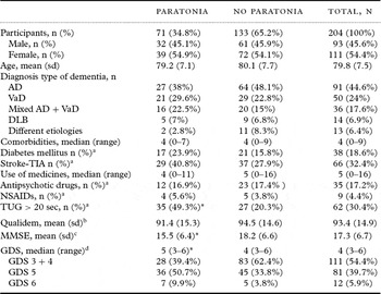
Baseline measures were assessed in 204 participants – 111 (54.4%) females and 93 (45.6%) males, with a mean age of 79.8 years (range 56–97) (Table 1). We obtained medical files for all participants and received GP files for 54% of the participants (n = 110). Most participants had AD (44.6%, n = 91), VaD (24.5%, n = 50) or mixed dementia (VaD and AD) (17.6%, n = 36).
Baseline characteristics

*p-value < 0.01.
aThe variables diabetes mellitus, stroke/TIA, antipsychotic drugs, NSAIDS and Timed Up and Go (TUG) > 20 seconds are dichotomous.
bQualidem score range 0–120; higher score indicates a higher quality of life.
cMMSE cognitive decline score range 0–30:, score 24–28 indicates very mild; 19–23: mild; 14–18 moderately severe; <14 severe.
dGDS stages range from 1 (normal cognition) to 7 (very severe cognitive decline).
AD = Alzheimer's disease; VaD = vascular dementia; DLB = dementia with Lewy bodies; TIA = transient ischemic attack; NSAIDs = non-steroidal anti-inflammatory drugs; TUG = Timed Up and Go; MMSE = Mini-mental State Examination; GDS = Global Deterioration Scale.
Baseline characteristics
Paratonia was diagnosed in 71 patients (34.8%) at baseline. The prevalence of paratonia was highest in the patients with mixed dementia (44.4%, 16 out of 36) and in the VaD group (42%, 21 out of 50). However, when compared to the prevalence of paratonia in the AD group (29.7%, 27 out of 91), the prevalence in the VaD group was not significantly different. The prevalence of paratonia increased significantly as the severity of dementia increased: 25.2% (28 out of 111) of patients with mild dementia (GDS 3 and 4), 44.4% (36 out of 81) of patients with moderate dementia (GDS 5) and 58.3% (7 out of 12) of patients with severe dementia (GDS 6) (cross-tabulation χ2, p < 0.01).
Patients with paratonia generally had a higher GDS rating (χ2, p < 0.01), a lower MMSE score (scores: 15.4 versus 18.2; independent sample t-test, p <0.01) and a longer time on the TUG (cross-tabulation χ2, p<0.01).
Longitudinal data
One hundred and forty-seven participants completed the study. Of the 71 diagnosed with paratonia at baseline, 27 were lost to follow-up. The remaining 44 participants were diagnosed again with paratonia after one year.
Of the 133 participants who did not have paratonia at baseline, 51 participants (38.3%) were diagnosed with paratonia over the course of one year, of which 39 completed the study, and 12 were lost to follow-up. Of the remaining 82 participants who were not diagnosed with paratonia, 64 completed the study, and 18 were lost to follow-up. The patients without a complete follow-up did not differ from those who completed the study with regard to age, gender or severity of dementia.
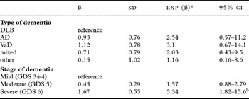
For the different types of dementia, VaD participants had the highest hazard ratio for developing paratonia (HR = 3.07, 95% CI 0.7–14.1, NS), with DLB as a reference. The hazard ratio between the different stages of dementia, with mild dementia as a reference, was significant, indicating that the incidence of paratonia increases with increasing severity of dementia (severe versus mild dementia: HR = 5.34, 95% CI 1.82–15.6). Table 2 shows the hazard ratio of the different types and stages of dementia.
Cox regression with the PAI as dependent variable and the type and stage of dementia as the independent variables (analyzed separately)

aExp (ß) = hazard ratio.
bp-value <0.05.
PAI = Paratonia Assessment Instrument; DLB = dementia with Lewy bodies; AD = Alzheimer's disease; VaD = vascular dementia; GDS = Global Deterioration Scale.
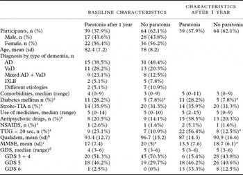
The baseline characteristics of the 39 cases of paratonia in comparison with the baseline characteristics of the 64 participants who did not develop paratonia and completed the study are outlined in Table 3. The baseline scores were lower and the one-year rates of decline in both the MMSE and the TUG were higher in the patients who developed paratonia compared with those who did not (MMSE: a mean decline of 3.5 points in the paratonia group and 1.3 in the non-paratonia group, p<0.01).
Characteristics of participants who go on to develop paratonia after one year compared with those who remain paratonia-free

*p-value <0.01.
aThe variables diabetes mellitus, stroke/TIA, antipsychotic drugs, NSAIDS and Timed Up and Go (TUG) >20 seconds are dichotomous.
bQualidem score range 0–120; higher score indicates a higher quality of life.
cMMSE cognitive decline score range 0–30:, score 24–28 indicates very mild; 19–23: mild; 14–18 moderately severe; <14 severe.
dGDS stages range from 1 (normal cognition) to 7 (very severe cognitive decline).
AD = Alzheimer's disease; VaD = vascular dementia; DLB = dementia with Lewy bodies; TIA = transient ischemic attack; NSAIDs = non-steroidal anti-inflammatory drugs; TUG = Timed Up and Go; MMSE = Mini-mental State Examination; GDS = Global Deterioration Scale.
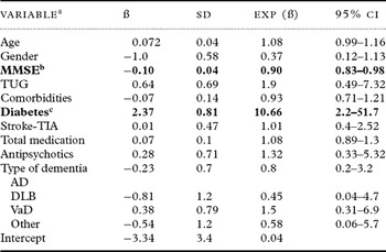
The logistic regression on the longitudinal data indicates that a decrease in the MMSE score was a significant risk factor for developing paratonia in one year (OR = 0.90, indicating that a one-point decrease on the MMSE increases the risk of paratonia by 10%; 95% CI = 0.83–0.98). Furthermore, we found that the participants with DM (n = 39) had an almost eleven-fold higher risk of developing paratonia in one year than those without DM (OR = 10.7; 95% CI = 2.2–51.7). Other comorbidities and the use of antipsychotics or other medications were not related to an increased prevalence or incidence of paratonia (Table 4).
logistic regression with baseline data of those participants with no paratonia at baseline

aDependent variable: Paratonia Assessment Instrument (PAI); the presence of paratonia "No/Yes" after one year.
Model: (Intercept), Age, Gender, Mini-mental State Examination (MMSE), Timed Up and Go (TUG), total amount of co-morbidities, Diabetes Mellitus, Stroke-TIA, total amount of medication, antipsychotics, Type of dementia (AD+VaD reference group).
bp-value <0.05.
cp-value <0.01.
MMSE = Mini-mental State Examination; TIA = transient ischemic attack; AD = Alzheimer's disease; DLB = dementia with Lewy bodies; VaD = vascular dementia.
Discussion
The profile of a typical participant with paratonia includes having a higher GDS rating, a lower MMSE score and lower functional mobility. The present study confirmed that the risk of developing paratonia increases with the progression of dementia and a decrease in cognitive abilities (Souren et al., Reference Souren, Franssen and Reisberg1997).
In general, the prevalence (34.8%) and incidence (38.3%) of paratonia in this cohort was higher than we expected at the start of the study; however, these higher rates enhanced the power of the present study.
Our most striking result was the finding that participants with DM had an almost eleven-fold higher risk of developing paratonia. DM was not a significant variable in our cross-sectional analyses at baseline. This difference can be explained by the obvious disadvantage of the cross-sectional analyses that included patients who are likely to develop paratonia in the data, masking the importance of this particular factor. The 95% CI was very broad, indicating that the uncertainty of the true OR was high, which was probably caused by the relatively small group of 39 participants with paratonia in this analysis.
DM appeared to be a factor of importance in the development of paratonia. It has already been shown that DM is associated with a variety of complications and an increased risk of dementia (Araki and Ito, Reference Araki and Ito2009). Recent findings have suggested that different patterns of cerebral injury in dementia exist with or without DM, including microvascular infarcts and the activation of neuroinflammation in individuals with dementia and DM (Sonnen et al., Reference Sonnen2009). Moreover, research by Arvanitakis et al. (Reference Arvanitakis, Wilson, Bienias and Bennett2007) has shown that DM causes rigidity and gait disturbances in older persons without dementia. These authors suggested that, aside from possible damage to the nigrostriatal system and/or white matter changes, DM also causes damage to the peripheral nervous system. Furthermore, it is known that high levels of glucose cause nonenzymatic glycation with advanced glycation endproducts (AGE), forming cross-links in collagen. This process causes a stiffening of all tissues, a process normally seen in aging that is accelerated by DM (Ulrich and Cerami, Reference Ulrich and Cerami2001). We must acknowledge the possibility that the PAI was not able to distinguish between this stiffness and mild paratonia, resulting in an overestimation of the importance of DM as a risk factor. Further longitudinal research with a longer follow-up period is necessary to identify the contribution of DM to the development of paratonia. This topic is especially interesting because it is clear that the negative long-term effects of DM can be influenced by various preventive interventions; in particular, an increase in physical activity has been shown to be very effective (Sigal et al., Reference Sigal, Kenny, Wasserman, Castaneda-Sceppa and White2006; Chodzko-Zajko et al., Reference Chodzko-Zajko2009).
Paratonia is seen in all types of dementia patients. The higher prevalence of paratonia and the higher hazard ratio in the VaD and mixed group of AD and VaD are an indication that vascular damage, in conjunction with dementia, most likely plays an important role in the development of paratonia. It is interesting to note that DM is a known cause of vascular damage and that both VaD and DM are likely important factors in the development of paratonia. We may be observing a common pathway in both conditions that enhances the development of paratonia. However, the risk of paratonia associated with DM did not appear to be more pronounced in participants with VaD compared to other dementias. It should be noted that our analysis did not indicate collinearity between VaD and DM and stroke. Further fundamental research is recommended to reveal the pathways through which vascular damage may contribute to the development of paratonia.
A decline in functional mobility appears to be a good indicator of the presence of paratonia in patients with dementia. However, the results of the present study do not indicate that a decline in functional mobility can predict the development of paratonia in one year. Therefore, the hypothesis that paratonia is enhanced by changes in the biomechanical properties that are equivalent to those seen in stroke and cerebral palsy patients is therefore not supported. Paratonia may be a cause of the decline in functional mobility. However, because this finding is the result of a cross-sectional analysis, it should be interpreted with caution.
Limitations
A limitation of the present study is that we included fewer early-stage dementia patients than we expected, influencing the expected prevalence and incidence of paratonia in this cohort. In addition, the participants in the selected day-care centers were generally at a more advanced stage of deterioration than we anticipated. This level of deterioration is one possible explanation for the relatively high attrition rate of 28% of the participants (n = 57).
A further limitation of this study was the difficulty we encountered in retrieving the patients’ medical records from their GPs. Although all of the participants provided written (proxy) consent, the GPs were very reluctant to share information, and in some cases (46%, n = 94), we only received information from the records that were available at the DCUs. It is possible that this difficulty caused some bias in the analysis of the comorbidities and medication.
Although the ad hoc power calculation indicated that we attained enough power for our analyses, the level of power was not enough for a proper analysis of the effect of medication on paratonia. Our study sample used such a wide variety of medication that the clustering of medication types was not sufficient for the analysis of 204 participants. We therefore only separated the use of antipsychotics as a variable in the analysis, as use of this type of medication can induce paratonia-like rigidity. As shown in Tables 1 and 3, the use of antipsychotic medication was not a factor that influenced the development of paratonia.
Another limitation was that behavioral symptoms were not quantifiably measured in this study. This type of analysis could have been relevant, as anxiety and other behavioral disorders have been associated with an increased prevalence of paratonia (Hobbelen et al., Reference Hobbelen, Koopmans, Verhey, van Peppen and de Bie2006). The measurement of behavioral symptoms is a subject for further study with a larger patient sample.
The cohort in the present study was heterogeneous, with participants ranging from mild to severe dementia and having different types of dementia. Some selection bias may have also occurred, especially in the mild dementia cases, because we recruited participants for this study in DCUs. It can be hypothesized that only mild dementia patients who have a more problematic disease course are offered DCU treatment. In addition, we realize that the MMSE is more of a cognitive screening tool than a reliable measure of cognitive function; nevertheless, this scale is now used worldwide to determine the degree of overall cognitive impairment.
Finally, a follow-up time of one year is brief, as most types of dementia progress over 5 to 10 years. Further longitudinal research over a longer period of time with larger cohorts is therefore recommended to verify our conclusions.
Clinical implications
For daily clinical practice, the early detection of patients with dementia who are at risk of developing paratonia is important. Our findings suggest that patients with DM and vascular risk factors experience higher risks of developing paratonia. Furthermore, it is important to diagnose paratonia in early-stage dementia patients when investigating risk factors that enhance the development of paratonia. Our findings may be helpful in the development of a more focused care program.
Conclusion
A decline of functional mobility is a good indicator of the presence of paratonia in patients with dementia. DM and likely vascular damage are risk factors for the development of paratonia. This finding enables us to look further into the possible pathogenesis of paratonia. Furthermore, it is a pretext for preventive intervention.
Conflict of interest declaration
The first author, J. Hobbelen, was a part-time employee at the Vitalis WoonZorg Groep (a non-profit foundation) in Eindhoven, which supported this study with a grant. The sponsor had no role in the design, methods, subject recruitment, data collection, analysis, or preparation of the manuscript.
Description of authors’ roles
J. Hobbelen designed the study, coordinated the study, analyzed and interpreted the data and prepared the paper. F. Tan performed the statistical analyses and helped write the paper. F. Verhey, R. Koopmans and R. de Bie supervised the design of the study and the data collection, interpreted the results of the analysis and helped write the paper.
Acknowledgments
This study was supported by a grant from the Vitalis WoonZorg Groep, Eindhoven. We thank all the participants and their relatives for their cooperation in this study, as well as the participating DCUs and day-care centers from “de Zorgboog” Helmond, “SVVE” Eindhoven, “de Wever” Tilburg and “Vitalis WoonZorg Groep” Eindhoven. We are grateful for the help of the GPs and personnel who actively contributed to this study. We thank Sytse Zuidema, a senior scientist in the Department of Primary and Community Care, Center for Family Medicine, Geriatric Care and Public Health, Radboud University Nijmegen Medical Center, Nijmegen, the Netherlands, for his enthusiastic and insightful contribution to the analyses of this study. Lastly, we thank Bob Wilkinson from the Maastricht University Translation and Editing Department.