The best surgical technique to repair an interrupted aortic arch remains disputed. Although a direct anastomosis after sufficient mobilisation of both the ascending and the descending aorta is thought to provide satisfactory patency and growth, the distance between them is problematic in terms of anastomotic tension and angulation, which are related to recoarctation or bronchial compression. At present, direct anastomosis with patch augmentation is the most preferred procedure for interrupted aortic arch repair and has a low incidence of complications.Reference Mishra 1 We present here two cases of aortic arch reconstruction using a pulmonary artery autograft patch combined with a reversed left subclavian artery flap in patients with an interrupted aortic arch type B complex.
Case reports
Case 1
An 8-day-old female infant was referred to our institution because of cardiac murmur, respiratory distress, and oliguria. She had been born with a birth weight of 2735 g and gestational age of 38 weeks. Echocardiography and three-dimensional computed tomographic angiography revealed an interrupted aortic arch type B with a perimembranous ventricular septal defect and left ventricular outflow tract obstruction; the aortic annulus was 4.0 mm (z-score, −3.57) and the subaortic area was 3.6 mm in diameter. After administration of prostaglandin E1, bilateral pulmonary artery banding was performed at 9 days of life. As the left ventricular outflow tract obstruction was moderated, with an aortic annulus of 4.6 mm (z-score, −3.17) and a subaortic area of 4.0 mm in diameter after 3 weeks, biventricular repair was performed at 34 days of life.
Case 2
A female infant was delivered with a birth weight of 3070 g and gestational age of 38 weeks at our institution. Examinations revealed an interrupted aortic arch type B with a perimembranous ventricular septal defect and left ventricular outflow tract obstruction; the aortic annulus and subaortic area was 4.3 mm (z-score, −3.52) and 4.0 mm in diameter, respectively. Bilateral pulmonary artery banding was performed at 5 days of life after prostaglandin E1 administration. Biventricular repair was performed at 48 days of life because of an improvement of the left ventricular outflow tract obstruction with an aortic annulus of 5.0 mm (z-score, −2.98) and subaortic area of 4.5 mm in diameter.
Operation
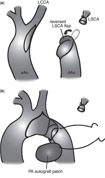
After median sternotomy, cardiopulmonary bypass was established using arterial cannulation in the brachiocephalic artery and descending aorta and bicaval venous cannulation with moderate hypothermia. Cardiac arrest was achieved with antegrade blood cardioplegia. The bilateral pulmonary artery branches were debanded, and the ventricular septal defect was closed with a Dacron patch after infundibular wedge resection. The patent ductus arteriosus was divided and excised completely. Because the ductal tissue was extended to the base of the left subclavian artery, we did not reconstruct the left subclavian artery. After transection of the left subclavian artery as distally as possible, the left subclavian artery flap was created and turned up to the descending aorta with a continuous 7-0 polypropylene suture. The lesser curvature of the aortic arch was incised longitudinally from the base of the left common carotid artery, proximally to the ascending aorta. A direct end-to-side anastomosis was performed between the arch and descending aorta, except for its anterior area, using 7-0 polypropylene sutures. After harvesting the pulmonary artery autograft patch from the main pulmonary artery, the anterior area of the neo-aortic arch was augmented with it. The defect of main pulmonary artery was corrected with a fresh autopericardial patch using 7-0 polypropylene sutures (Fig 1).
Schematic illustration of the operative procedures. (a) Reversed LSCA flap and (b) PA autograft patch for aortic arch augmentation. aAo = ascending aorta; dAo = descending aorta; LSCA = left subclavian artery; LCCA = left common carotid artery; PA = pulmonary artery.

Post-operative outcomes
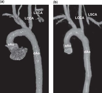
The post-operative course during the 1-year follow-up was uneventful in both patients. Three-dimensional computed tomographic angiography showed that the reconstructed aortic arch had no significant stenosis at the repair site and created a sufficient aortopulmonary space with a wide arch angle (Fig 2). Echocardiography showed that the pressure gradient across the repair site was <10 mmHg and that the pulmonary valvular function was well preserved without main pulmonary artery stenosis.
Post-operative three-dimensional computed tomographic angiography (anterolateral view, extracted aorta). ( a ) Case 1. ( b ) Case 2. aAo = ascending aorta; dAo = descending aorta; LSCA = left subclavian artery; LCCA = left common carotid artery.

Discussion
Interrupted aortic arch type B is one of the incremental risk factors for reinterventions for recurrent arch obstruction, and this morphological subtype is consistently associated with an increased mortality.Reference McCrindle, Tchervenkov and Konstantinov 2 The interrupted gap between the left common carotid artery and left subclavian artery carries the risk of recoarctation or bronchial compression due to tension and angulation at the anastomosis site in interrupted aortic arch repair.
Patch augmentation of the aortic arch has often been performed in interrupted aortic arch repair. Various patch materials have been used, including fresh or glutaraldehyde-treated autopericardia, xenopericardia, homografts, and prostheses. The pulmonary artery autograft patch has previously been reported in single-stage repair for an interrupted aortic arch complex.Reference Cetin, Tireli, Ozkara and Oztunc 3 – Reference Tateishi and Kawada 7 The rate of recoarctation and bronchial compression after aortic arch reconstruction with a pulmonary artery autograft patch was significantly lower than that with a homograft or pericardial patch.Reference Roussin, Belli and Lacour-Gayet 5 The reversed left subclavian artery flap is another surgical technique for aortic arch reconstruction in interrupted aortic arch repair.Reference Dietl and Torres 8 It is non-circumferential with a better growth potential; however, the use of this technique alone could not give rise to an adequate lumen because of its limited size.
In the present cases, a combination of a pulmonary artery autograft patch with a reversed left subclavian artery flap has made it possible to enlarge the neo-aortic arch with a tension-free anastomosis and to generate a sufficient aortopulmonary space with a wide arch angle. The pulmonary artery autograft patch enabled augmentation of the anterior area of the neo-aortic arch, whereas the reversed left subclavian artery flap provided additional augmentation to its posterior area. No post-operative complications of recoarctation or bronchial compression have been observed in the two patients to date. Although the reversed left subclavian artery flap has the potential disadvantage of an impaired blood supply to the left arm, both infants did not experience any symptoms of left upper extremity ischaemia. The autologous tissues have growth potential, non-antigenicity, and antithrombogenicity. In comparison with an autopericardium, the theoretical advantage of a pulmonary artery autograft is the uniform embryological origin with the aortic wall, possibly leading to a better compatibility in case of aortic arch reconstruction.Reference Malyshev, Gladyshev, Safuanov, Siniukov, Borovikov and Rostovykh 9
Association of cardiac anomalies presents a surgical challenge in definitive repair of the interrupted aortic arch complex. In the present cases of interrupted aortic arch type B with a ventricular septal defect and left ventricular outflow tract obstruction, bilateral pulmonary artery banding was performed as an initial palliative surgery to control excessive pulmonary blood flow for haemodynamic stability. This approach has been extended to infants with borderline development of left heart structures. Recently, Ballard et alReference Ballard, Tibby and Miller 10 reported that bilateral pulmonary artery banding for borderline hypoplastic left heart allows time for growth of left heart structures and may lead to achieving a systemic circulation for a biventricular repair, possibly because of an increased blood flow across the aortic annulus. Biventricular repair with a left ventricular outflow tract bypass procedure such as a staged Yasui operation would be recommended if the aortic annulus is less than the patient's weight plus 1.0 mm, as advocated by a previous report.Reference Hirata, Quaegebeur, Mosca, Takayama and Chen 11 In the present cases, the growth of the aortic annulus and subaortic area after bilateral pulmonary artery banding allowed us to establish a regular biventricular repair with ventricular septal defect closure. Fortunately, the main pulmonary artery was ∼2.5 times larger than the ascending aorta in both cases, which proved to be advantageous in attaining adequate pulmonary artery autograft patch. At this moment, there has been no problematic lesion in the patched sites of the neo-aortic arch and main pulmonary artery, but further careful follow-up of these sites should be necessary.
In summary, we report two successful cases of an aortic arch reconstruction using a pulmonary artery autograft patch and a reversed left subclavian artery flap in patients with an interrupted aortic arch type B ventricular septal defect and left ventricular outflow tract obstruction. This surgical technique can be one of the procedural options for an interrupted aortic arch type B complex repair with autologous materials alone.
Acknowledgements
None.
Financial Support
This research received no specific grant from any funding agency, commercial or not-for-profit sectors.
Conflicts of Interest
None.