Introduction
Rhinoscleroma of the nose is a disease frequently reported in the literature.
Rosai–Dorfman disease, although previously a rare entity in the differential diagnosis of cervical lymphadenopathy, is now being increasingly considered. The aetiopathogenesis of Rosai–Dorfman disease still remains obscure, although newer studies postulate viral or chronic granulomatous infections as a likely cause, but this is not yet proven.
The coexistence of rhinoscleroma in the nose or nasopharynx together with Rosai–Dorfman disease in the neck nodes has also been reported.
In the current case report, both rhinoscleroma and Rosai–Dorfman disease were seen coexisting in the same nasal lesion, as proved by histopathological examination. This strongly supports an aetiological relationship between rhinoscleroma and Rosai–Dorfman disease, as already postulated. A literature review did not reveal any previous report of the coexistence of both conditions in the same lesion. This, along with the histological evidence, makes this case unique.
Case report
A 42-year-old woman attended our out-patient department in November 2005 due to nasal obstruction and neck swelling, more pronounced on the right side.
On examination, the nasal cavities had firm, non-bleeding, non-sensitive, nodular masses, mostly on the right side and with a few on the left side at depth. The cervical nodes were multiple, non-tender, matted, firm and involved the upper and mid-cervical groups on both sides.
All results of investigations were within normal limits, apart from a raised erythrocyte sedimentation rate of 60 mm/h.
An excision biopsy of the nasal masses was performed, and the histopathology report indicated rhinoscleroma.
The results of a fine needle aspiration of the neck nodes were interpreted as Rosai–Dorfman disease.
Thus, this patient constituted a case of rhinoscleroma of the nasal cavities together with Rosai–Dorfman disease of the cervical nodes.
She was treated with injected streptomycin and oral tetracycline. Due to severe vertigo, the streptomycin was discontinued. The neck disease was not addressed as the patient was lost to follow up.
She returned with a recurrence two years later. The nasal masses and neck swelling were now larger than in the previous presentation.
Computed tomography showed polypoidal masses filling the right paranasal sinuses, nasal cavity and nasopharynx and completely obstructing the airway.
Incision biopsy again confirmed the nasal mass as rhinoscleroma, but with a suspicion of Rosai–Dorfman disease. Fine needle aspiration of the neck nodes again revealed Rosai–Dorfman disease.
Surgery via a Caldwell–Luc approach revealed an antrum filled with friable, polypoidal, fleshy masses eroding the medial wall and extending through the meati to the choanae and nasopharynx and into the left deep nasal cavity. After removal of the lesion in its entirety, it was sent for histopathological examination at two centres, for cross-confirmation.
The histopathology report was as follows.
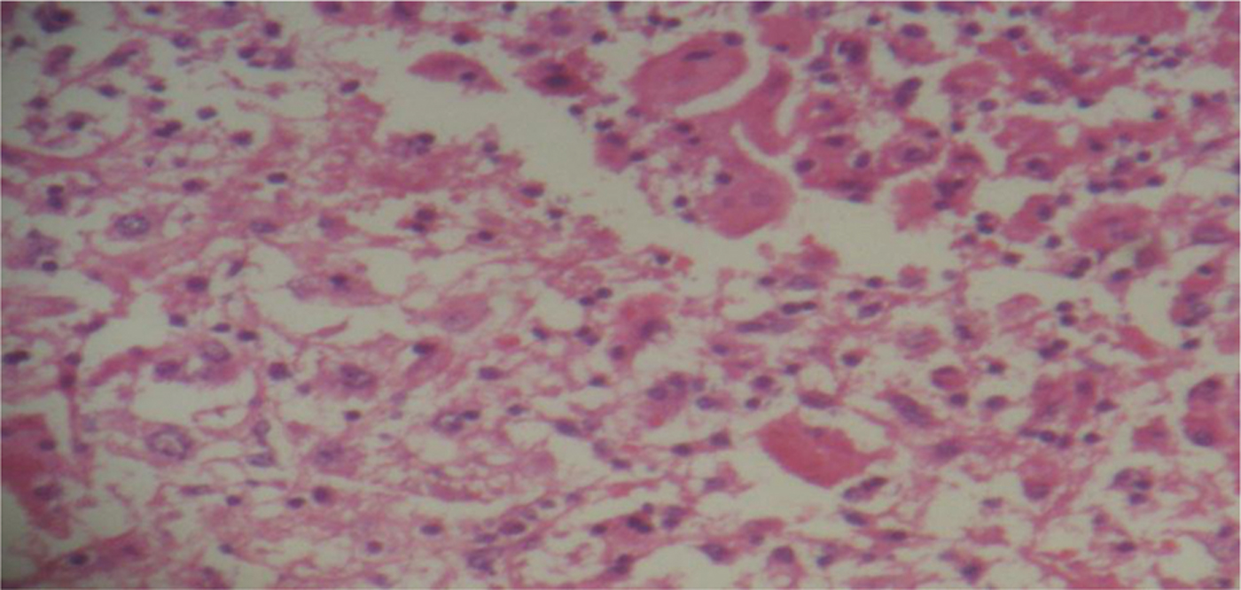
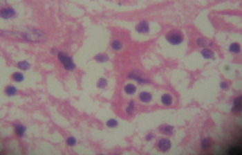
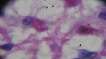
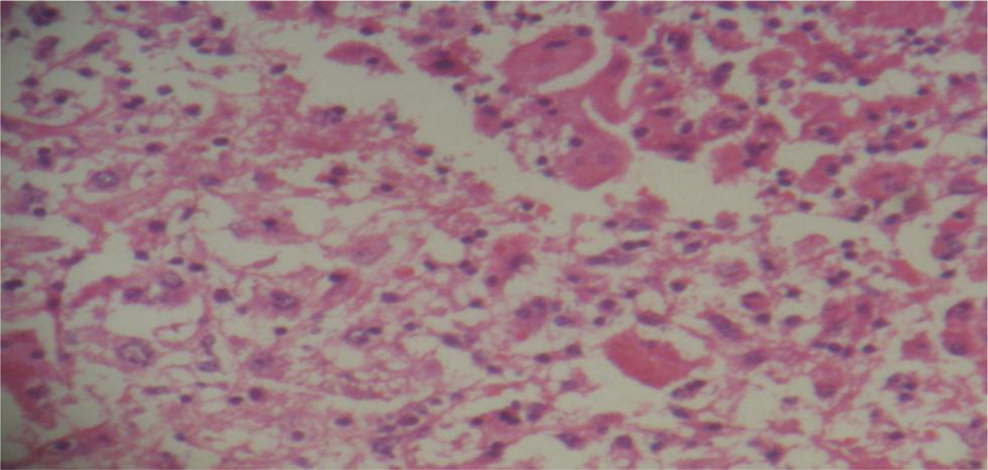
… [T]issue showing numerous foamy cells and macrophages admixed with lymphocytes and plasma cells. Many macrophages show lymphocytes in the cytoplasm (emperipolesis) [See Figures 1 and 2]. Odd Russell body is present. Gram staining shows some rod-like Gram-negative bacteria in the foamy cells and some in between the cells? Klebsiella rhinoscleromatis. [Periodic-Acid Schiff] staining shows no fungal bodies, but the bacterial rods show faint staining [Figure 3]. Immunostaining done at Wellspring Laboratory, Bombay, showed S-100 positivity of the macrophage cells. Impression: Rosai–Dorfman disease with evidence of Klebsiella rhinoscleromatis in the tissue examined suggesting associated rhinoscleroma.
Photomicrograph showing macrophages with emperipolesis (top right section of figure) and several foamy macrophages (Mikulicz cells; lower left half of figure). (H&E; ×40)
High-power photomicrograph of macrophage with engulfed lymphocytes (emperipolesis). (H&E; ×100)

High-power photomicrograph of a Mikulicz cell, showing bacilli (arrows) taking up Periodic-Acid Schiff stain within vacuolated cytoplasm. (×100)

From the histopathology reports, a final diagnosis of rhinoscleroma with coexisting extranodal Rosai–Dorfman disease of the nasal cavity and Rosai–Dorfman disease of the neck was arrived at.
After treatment with high dose ofloxacin (400 mg twice daily) for one month, the patient developed joint swellings and pain but continued the treatment. This was followed by oral prednisolone in tapering doses for one month, for the Rosai–Dorfman disease. The patient's neck nodes subsided considerably.
At the time of writing, the patient had been reviewed twice more, with minor recurrences in the nasal cavities alone, managed endoscopically. There had been no enlargement of the cervical lymph nodes, and she had been treated with high-dose ofloxacin as before.
Discussion
Rhinoscleroma is a progressive granulomatous disease starting in the nose and later extending into the nasopharynx and oropharynx, larynx, and, sometimes, the trachea and bronchi. It is more common in women and the middle-aged.
The disease is known to progress through three stages: (1) atrophic, (2) granulomatous and (3) cicatrising. Lymphadenopathy is not an associated feature.
Rhinoscleroma is caused by the frisch bacillus, a Gram-negative, rod-like organism. Histologically, affected tissues show granulomatous tissue infiltrates in the submucosa characterised by the presence of plasma cells, lymphocytes and eosinophils. In addition, there are scattered large foam cells (Mikulicz cells) which have a central nucleus and vacuolated cytoplasm, with the vacuoles containing rod-like bacilli and Russell bodies. Mikulicz cells are transformed macrophages with engulfed bacilli which persist intracellularly. Russell bodies are globules of immunoglobulins arranged in a crystalline array in the cytoplasm of plasma cells in areas of chronic inflammation. Organisms are easily visible on haematoxylin and eosin (H&E) staining, but are more easily seen with Periodic-Acid Schiff and silver impregnation techniques. Gram and Periodic-Acid Schiff staining were used in the current case, although the organisms were seen easily with just H&E staining.
The disease follows a self-limiting course, ending in the cicatrising stage, and the organism can be extremely difficult to eradicate with antimicrobials. Treatment involves surgical debridement and prolonged antimicrobial therapy but has a high relapse rate. The organism is sensitive to a variety of antimicrobial agents. However, prolonged (one to three months) treatment with fluoroquinolones such as ciprofloxacin or ofloxacin has produced the best results, with low relapse rates.Reference Hart1 In our patient, high-dose ofloxacin gave good results.
Rosai–Dorfman disease, also known as sinus histiocytosis with massive lymphadenopathy, is a benign, idiopathic histiocytic proliferation affecting lymph nodes and extranodal sites in all age groups. The predominant clinical manifestation is massive cervical lymphadenopathy. Extranodal disease is observed in 28–43 per cent of cases, with a preference for the head and neck region. The concomitant involvement of one or more sites in the same individual is seen in 44.7 per cent of cases. The current case showed Rosai–Dorfman disease of the neck nodes and a naso-antral mass.
The aetiology of Rosai–Dorfman disease remains obscure, but two theories exist. The first associates the disease with a specific infectious process, the second with a disturbance of the immune system. However, no laboratory evidence pointing to an aetiological agent, and no other clinicopathological signs suggesting a deficient humoral or cellular immunity, have been reported.Reference Foucar, Rosai and Dorfman2
First described in 1969 by Rosai and Dorfman in the lymph nodes, the characteristic histopathological feature of the disease is a proliferation of distinct histiocytic cells that demonstrate emperipolesis (engulfment of lymphocytes and plasma cells by the histiocytes) against a background of inflammatory infiltrate, abundant plasma cells and lymphocytes.Reference Layfield3 Immunohistochemical studies show that histiocytes are S-100-positive.
Although rhinoscleroma and Rosai–Dorfman disease have been reported in the same patient in several instances in the literature, they have always been at separate sites. The presence of the two diseases in the same lesion, anywhere in the body, has not previously been reported. The coexistence of rhinoscleroma with Rosai–Dorfman disease in the mass removed from the naso-antrum of the current patient was proved by the presence of typical bacilli in Mikulicz cells, which also stained positive with Periodic-Acid Schiff, and also the lymphoplasmacytic infiltrate occurring alongside large histiocytes clearly showing emperipolesis; these histiocytes were also S-100-positive. The histology slides clearly showed, in the same field, the distinctive histology of both diseases (Figure 1) already confirmed previously in this patient.
• Rhinoscleroma is a chronic granulomatous disease affecting the respiratory passages
• Slides show rod-like, Gram-negative bacilli within Mikulicz cells
• Rosai–Dorfman disease causes nodal and extranodal lesions
• Slides show emperipolesis within large histiocytes
• The two diseases have not previously been shown to coexist within the same lesion
• Such coexistence is reported here, suggesting an aetiological relationship
Fine needle aspiration cytology of the neck nodes of this patient also showed features confirming Rosai–Dorfman disease (also termed sinus histiocytosis with massive lymphadenopathy).
Lampert and Lennert studied 15 cases of this disease, and found elevated antibody titres to klebsiella antigens in the serum on repeated occasions.Reference Lampert and Lennert4 Typing with different klebsiella-O antigens showed that Klebsiella rhinoscleromatis and K ozaenae always gave the strongest reactions. In rhinoscleroma, the diagnosis is usually established from the pathognomonic histological findings of masses of plasma cells interspersed with Mikulicz cells, although, in many instances, klebsiella organisms are directly detected in the lesions.Reference Lampert and Lennert4 This same histology may be seen in Rosai–Dorfman disease, but without the organism being detected and with the presence of emperipolesis. Furthermore, Lampert and Lennert also suggested that Rosai–Dorfman disease might be caused in some instances by a rare klebsiella organism.
Conclusion
Although rhinoscleroma and Rosai–Dorfman disease have been reported worldwide, and have been described to coexist in the same patient at different sites, never have the two pathologies been demonstrated within the same lesion at one site, anywhere in the body. The aetiology of Rosai–Dorfman disease could not previously be proved. The current report presents a case of a middle-aged woman with recurrent nasal lesions which showed histological evidence of both rhinoscleroma and Rosai–Dorfman disease in the same lesion. The author believes that this provides strong evidence supporting the hypothesis of an aetiological relationship between rhinoscleroma and Rosai–Dorfman disease.