Introduction
Perforation of the tympanic membrane is a sequela of otitis media and primarily results from middle-ear infection, trauma or iatrogenic causes.Reference Zhang, Huang, Zheng, Sun, Chen and Si1 Since the earliest attempts at tympanic membrane perforation closure, various graft materials have been used for reconstruction. Temporalis fascia remains the most frequently used graft material. However, in the presence of advanced middle-ear pathology, retraction pockets and atelectatic ears, temporalis fascia tends to undergo post-operative atrophy, regardless of the placement technique.Reference Gamra, Mbarek, Khammassi, Methlouthi, Ouni and Hariga2, Reference Khan and Parab3 The use of cartilage for tympanic membrane reconstruction is not a recent idea; many tympanoplasty techniques using cartilage have been described.Reference Tos4
The greatest advantage of cartilage–perichondrium composite grafts was initially thought to be their very low metabolic rate. However, this tissue can receive nutrition by diffusion; it is easy to work with because it is pliable and can resist deformation by pressure variations.Reference Levinson5 The major advantage of cartilage–perichondrium composite grafts is their stiffness and bradytrophic metabolism, as summarised in detail by Zhang et al., Tos and Yung.Reference Zhang, Huang, Zheng, Sun, Chen and Si1, Reference Tos4, Reference Yung6 In our department, temporalis fascia grafts were previously used to reconstruct the tympanic membrane. However, the long-term outcome was not ideal, especially for large perforations. Hence, we developed a new technique for tympanic membrane reconstruction using a sliced tragal cartilage–perichondrium composite graft.Reference Khan and Parab3 To avoid the possibility of a thick cartilage graft interfering with sound conduction,Reference Zahnert, Huttenbrink, Murbe and Bornitz7, Reference Overbosch8 we prefer to use sliced cartilage.Reference Khan and Parab3 Most previous studies compared temporalis fascia grafts with whole thickness cartilage grafts in tympanoplasty.Reference Kazikdas, Onal, Boyraz and Karabulut9, Reference Albirmawy10 However, it is unknown whether cartilage–perichondrium composite grafts have an advantage over other graft materials for repairing small perforations.Reference Zhang, Huang, Zheng, Sun, Chen and Si1
This study aimed to compare the anatomical and audiological outcomes of using a sliced tragal cartilage–perichondrium composite graft versus a temporalis fascia graft for type I tympanoplasty in our otological practice.
Materials and methods
A retrospective study of all type I tympanoplasties performed from May 2005 to January 2008 using sliced tragal cartilage–perichondrium composite graftsReference Khan and Parab3 and temporalis fascia grafts was carried out in the Maharashtra Institute of Medical Education and Research Medical College and The Sushrut ENT Hospital, Talegaon-Dabhade, Pune, India. The results for type I tympanoplasty using a sliced tragal cartilage–perichondrium composite graft have already been published.Reference Khan and Parab3
Study population
A total of 390 ears (226 from males and 164 from females) with tympanic membrane perforations were operated on in our department and our hospital from May 2005 to January 2008 with a minimum follow up of 4 years. Patient age ranged from 11 to 57 years. Only primary cases in which the ossicular chain was intact and no mastoid surgery was performed were included. All patients had dry ears and presented with an air–bone gap (ABG) of 30.68 ± 4.77 dB and 32.91 ± 4.88 dB upon audiometry (0.5, 1, 2 and 4 kHz) in the cartilage and fascia groups, respectively. The patients were grouped into those with large and those with small perforations, and were randomly allocated to surgery using temporalis fascia or sliced tragal cartilage grafts by both authors. The choice of graft type to be used in tympanoplasty was explained to all patients, and their consent was obtained. A total of 268 ears were operated on using sliced tragal cartilage–perichondrium composite graft and 223 were available for follow up.Reference Khan and Parab3 A total of 203 ears were operated on using temporalis fascia grafts and 167 were available for follow up. The institutional review board of our hospital approved the study.
Anaesthesia
All patients were operated on under local anaesthesia with adequate sedation, except for children who were operated on under general anaesthesia. Pre-medication included an intravenous injection of 0.30–0.50 mg/kg (maximum 30 mg) pentazocine lactate and 0.08–0.10 mg/kg (maximum 5 mg) midazolam under the care of an anaesthetist. A 2 per cent lidocaine solution containing 5 µg/ml adrenaline was used for infiltration.
Procedure
All procedures were performed via endaural incision. Patients were randomly assigned to tympanoplasty with either temporal fascia or sliced tragal cartilage–perichondrium composite grafts. All patients were operated on a day care surgery basis.
Tragal cartilage–perichondrium composite graft harvest and preparation was previously described in detail.Reference Khan and Parab3 Briefly, a Lempert endaural incision was made and a tragal cartilage graft (measuring approximately 15 mm × 15 mm) was harvested under aseptic conditions. The tragal cartilage was then cut into slices using a Precise Cartilage Knife (Heinz Kurz, GmbH Medizintechnik, Dusslingen, Germany; Figure 1).
Photograph showing the tragal cartilage sliced using a precise cartilage Knife.

A temporalis fascia graft was harvested via the same type of incision (Figure 2). The edges of the perforation in the pars tensa were neatened using a sickle knife. The tympanomeatal flap was elevated after making incisions at the 6 o'clock and 12 o'clock positions. This could be extended up to the 2 o'clock position on the anterior canal wall (for the right ear), leaving 6 mm of canal skin laterally from the annulus tympanicus. The whole elevated tympanomeatal flap was parked superiorly in the attic area, and then ossicular mobility and continuity was assessed. The handle of the malleus was denuded, and an underlay technique was used to place the 0.5-mm thick sliced cartilage–perichondrium composite shield graft or temporalis fascia over the entire tympanic membrane including the perforation (Figure 3). The tympanomeatal flap was then repositioned, Gelfoam® was placed over the graft, a meatal pack was added, the endaural incision was sutured and a mastoid bandage applied.
Photograph showing temporalis fascia harvested via an endaural incision.
Photograph showing a sliced tragal cartilage–perichondrium composite graft used as an underlay graft.
Follow up
The meatal pack and mastoid bandage were removed after 48 hours. Patients were then advised on ear care and the use of topical antibiotics (steroid ear drops, comprising neomycin, polymyxin B and hydrocortisone). Subsequent post-operative visits took place at weekly intervals for one month, and monthly thereafter for six months. After three months, pure tone audiometry was performed to evaluate ABG closure. Functional evaluations also took place after six months and after one, two, three and four years. Post-operative otoendoscopy recordings were made for all patients.
Results
From May 2005 to January 2008, 223 ears were operated on using a sliced tragal cartilage–perichondrium composite graft and 167 ears were operated on using a temporalis fascia graft, with a minimum follow up of 4 years. Table I shows the age and sex distribution of both patient groups. There were 129 males and 94 females in the cartilage group, and 97 males and 70 females in the temporalis fascia study group.
Age and sex distribution for the tragal cartilage and temporalis fascia groups
y = years
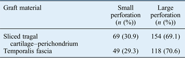
A Student's t-test found no significant difference in pre-operative ABG for patients in the cartilage and temporalis fascia groups. Table II shows patient numbers by perforation size and graft material. Perforations affecting more than 50 per cent of the tympanic membrane area were defined as large.Reference Zhang, Huang, Zheng, Sun, Chen and Si1, Reference Yung6
Patient numbers by perforation size and graft material
Post-operative anatomical results
There were no recurrent tympanic membrane defects in any of the 118 patients with small perforations after surgery using either graft material after 4 years; the graft acceptance rate was 100 per cent for this group. Anatomical results for the 272 patients with large perforations are shown in Tables III and IV.
Age and gender distribution of perforations in the cartilage group after four years
M = male; F = female
Post-operative morphological data in patients with large perforations
res = residual; rec = recurrent
Table III shows the age and gender distribution of post-operative perforations for patients in the cartilage and the temporalis fascia groups. Table IV shows the occurrence of re-perforations after 6 months, 12 months, 2 years, 3 years and 4 years. In the cartilage–perichondrium composite graft group, two ears had residual and two had recurrent perforations (Figure 4) at the two-year follow up,Reference Khan and Parab3 and two patients had residual and three had recurrent perforations at the four-year follow up. In the temporalis fascia graft group, 2 ears had residual perforations and 19 had recurrent perforations at 2 years, and 27 ears had recurrent perforations after 4 years (Figure 5).
Photograph showing an example of recurrent perforation following sliced cartilage type I tympanoplasty.
Photograph showing an example of recurrent perforation following temporalis fascia type I tympanoplasty.
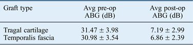
Table V shows the pre- and post-operative ABGs for the total cartilage and temporalis fascia study groups. The post-operative ABG after two years was 7.06 ± 3.39 dB in the cartilage–perichondrium composite graft group and 7.14 ± 2.25 dB in the temporalis fascia graft group.Reference Khan and Parab3 At the four-year follow up, the post-operative ABGs for these groups were 7.10 ± 3.01 dB and 8.05 ± 3.22 dB, respectively. The slightly higher post-operative ABG in the temporalis fascia tympanoplasty group can be attributed to the higher number of recurrent perforations.
Pre- and post-operative air–bone gaps for the total cartilage and temporalis fascia groups*
* Data are presented as the mean ± standard deviation. Avg = average; pre-op = pre-operative; post-op = post-operative; ABG = air–bone gap
Table VI shows pre- and post-operative ABGs (after four years) for ears closed successfully by cartilage and temporalis fascia. The Student's t-test found no significant difference in the post-operative ABG between the sliced cartilage and temporalis fascia groups (p < 0.05). This result shows that tympanoplasty using sliced tragal cartilage achieves an acoustic benefit comparable with that obtained after temporalis fascia tympanoplasty.
Pre- and post-operative air–bone gaps for intact ears in the cartilage and temporalis fascia groups after four years*
* Data are presented as the mean ± standard deviation. Avg = average; pre-op = pre-operative; post-op = post-operative; ABG = air–bone gap
Discussion
Since Berthold first adopted the full-thickness skin graft as an autologous substitute in 1878, many kinds of autologous substitutes have gradually come to be used in myringoplasty.Reference Zhang, Huang, Zheng, Sun, Chen and Si1 Temporalis fascia remains the most frequently used graft material, with tympanic membrane closure rates of 70–90 per cent for primary tympanoplasties in different hands.Reference Khan and Parab3 Typically, autologous substitutes only provide stent effects and do not increase the thickness of the tympanic membrane. Thus, newly formed tympanic membranes often lack an intermediate layer of elastic fibres, which serves to resist the negative pressure of the tympanum (Figure 6).Reference Zhang, Huang, Zheng, Sun, Chen and Si1 Therefore, repaired tympanic membranes often re-perforate, particularly when the initial perforation is large and the eustachian tube is dysfunctional. Furthermore, the blood supply to the central part of the newly formed tympanic membrane is usually so poor that re-perforation occurs easily.Reference Zhang, Huang, Zheng, Sun, Chen and Si1
Photograph showing a thin neotympanum four years after type I tympanoplasty using a temporalis fascia graft.
The use of cartilage in middle-ear reconstruction is not a new concept.Reference Yung6 Advantages of the cartilage graft include its very low metabolic rate and ability to receive nutrients by diffusion. It is also very easy to work with because it is pliable, resists deformation from pressure variations and incorporates well into the tympanic membrane.Reference Khan and Parab3, Reference Levinson5 Human and animal studies have found that although some softening occurs over time, the cartilage matrix remains intact but with empty lacunae showing chondrocyte degeneration.Reference Khan and Parab3, Reference Yamamoto, Iwanaga and Fukumoto11, Reference Hamed, Samir and El Bigermy12 Cartilage retains its rigidity and resists retraction, resorption and re-perforation, even with continuous eustachian tube dysfunction (Figure 7).Reference Khan and Parab3, Reference Dornhoffer13
Well-functioning sliced tragal cartilage four years after surgery.
Zahnert et al. reported that a tympanic membrane with a large perforation repaired using a thick cartilage fragment had poor conductivity in response to low-frequency sounds; this could be improved by reducing the cartilage thickness.Reference Zahnert, Huttenbrink, Murbe and Bornitz7 They also showed that, compared with normal tympanic membrane, 0.5-mm thick cartilage maintained mechanical stability and reduced sound energy loss during sound transmission. Hence, we prefer to slice the tragal cartilage with a precise mathematical slicer to reliably obtain a 0.5-mm thick tragal cartilage–perichondrium graft.Reference Khan and Parab3
This is the first study to compare the results of type I tympanoplasty using the sliced tragal cartilage–perichondrium composite graft and the temporalis fascia graft. All previous studies have compared tympanoplasty outcomes using temporalis fascia with whole thickness cartilage.Reference Zhang, Huang, Zheng, Sun, Chen and Si1, Reference Kazikdas, Onal, Boyraz and Karabulut9, Reference Albirmawy10, Reference Ozbek, Ciftçi, Tuna, Yazkan and Ozdem14, Reference Onal, Arslanoglu, Songu, Demiray and Demirpehlivan15 A total of 223 ears underwent type I tympanoplasty using a sliced tragal cartilage–perichondrium composite graft and 167 underwent temporalis fascia tympanoplasty, with a minimum follow up of 4 years.Reference Khan and Parab3 Success was defined as anatomical intactness of the tympanic membrane and hearing improvement by post-operative ABG closure.
Tympanic membrane intactness
In the anatomical evaluation of the tympanic membrane, the criterion for success is an absence of re-perforation. In our study, the graft take-up rate was 98.20 per cent in the sliced cartilage group and 87.42 per cent in the temporalis fascia group. There were 2 residual perforations in each group, 2 recurrent perforations in the cartilage group and 19 recurrent perforations in the temporalis fascia group at the 2-year follow up.Reference Khan and Parab3 At the 4-year follow up, graft take-up was 97.75 per cent and 82.63 per cent in the cartilage and temporalis fascia groups, respectively, with 3 recurrent perforations in the cartilage group and 27 in the temporalis fascia group.
Air–bone gap closure
The ABG closure was 7.10 ± 3.01 dB in the cartilage group and 7.99 ± 4.22 dB in the temporalis fascia group. These values were not significantly different (p < 0.05), which suggests that slicing the tragal cartilage prevents it from interfering with sound conduction. The higher mean ABG noted in the temporalis fascia group can be attributed to the greater number of re-perforations.
In a study of type I tympanoplasty, Ozbek et al. concluded that tympanoplasty using the palisade cartilage technique resulted in a significantly higher graft acceptance rate (100 per cent) compared with the fascia technique (70.2 per cent; p = 0.008).Reference Ozbek, Ciftçi, Tuna, Yazkan and Ozdem14 Speech reception threshold levels, pure tone average and ABGs improved significantly after surgery in both the palisade and fascia groups (p < 0.001). There was no significant between-group difference in audiological results (p > 0.05).Reference Ozbek, Ciftçi, Tuna, Yazkan and Ozdem14
In a study on palisade cartilage tympanoplasty by Kazikdas et al., graft take was achieved in 22 patients (95.7 per cent) in the palisade cartilage group and 21 patients (75 per cent) in the temporalis fascia group (p = 0.059). For both techniques, there were no significant changes in the mean speech reception threshold, ABG and pure tone average scores (p > 0.05). However, functional success (as indicated by improvements in mean ABG and speech reception threshold) was achieved with the palisade cartilage technique.Reference Kazikdas, Onal, Boyraz and Karabulut9
In a study by Albirmawy, a significantly greater graft acceptance rate was achieved for type I tympanoplasty using a ring graft technique (95 per cent) compared with a temporalis fascia graft (76.2 per cent; p < 0.01).Reference Albirmawy10 Pure tone average ABG and speech reception threshold levels improved significantly in both the ring graft and fascia groups (p < 0.001). There was no significant difference in audiometric results between the two groups (p > 0.05), although there was a trend towards better post-operative results in the ring group.Reference Albirmawy10
Graft success rates of 65.9 per cent for the fascia group and 92.3 per cent for the cartilage group were reported by Onal et al.Reference Onal, Arslanoglu, Songu, Demiray and Demirpehlivan15 Post-operatively, the mean ± standard deviation air conduction threshold was 28.54 ± 14.20 dB for the fascia group and 22.97 ± 8.37 dB for the cartilage group, while the mean ± standard deviation bone conduction threshold was 11.71 ± 8.50 dB for the fascia group and 7.15 ± 5.56 dB for the cartilage group.
In a study of 1000 patients by Dornhoffer the average pre- and post-operative ABGs were 21.7 ± 13.5 dB and 11.9 ± 9.3 dB (p < 0.05) in 215 cases of high-risk perforation. Complications included recurrent perforation (4.2 per cent), conductive hearing loss requiring revision (1.9 per cent), and post- and intra-operative tube insertion in four (1.9 per cent) and six ears (2.8 per cent), respectively.Reference Dornhoffer16
• Temporalis fascia is the most commonly used graft material for tympanic membrane reconstruction
• Sliced tragal cartilage is routinely used for tympanoplasty in our otological practice
• This study compared the four-year success rate for temporalis fascia and sliced tragal cartilage graft tympanoplasty
• Primary (sliced) cartilage tympanoplasty has a better success rate than temporalis fascia grafting for large perforations
• Poor outcomes associated with tragal cartilage thickness can be overcome by slicing
Aidonis et al., reported a graft take-up rate for cartilage shield tympanoplasty of 98.4 per cent, with no post-operative complications. The average pre- and post-operative pure tone average ABGs were 32.4 ± 14.1 dB and 24 ± 13 .7 dB, respectively (p < 0.05).Reference Aidonis, Robertson and Sismanis17
The current study suggests that a cartilage graft is better than a temporalis fascia graft for tympanic membrane perforation closure. The disadvantage of using thicker cartilage, thought to interfere with hearing and sound conduction, was overcome by slicing to achieve a thickness comparable with that of the temporalis fascia. This process also retains cartilage rigidity and so prevents recurrence, re-perforation and retraction. The comparable post-operative ABG closure with both the sliced tragal cartilage–perichondrium composite graft and temporalis fascia graft (p > 0.05) indicates that slicing the tragal cartilage does not interfere with sound conduction, thus providing an advantage over the use of thick unsliced cartilage.
Conclusion
The overall success rate is better for primary cartilage tympanoplasty (using sliced cartilage) than for temporalis fascia grafting for large perforations.
We conclude that, after a four-year follow up, tympanoplasty using the tragal cartilage–perichondrium composite graft gave a better therapeutic outcome regarding tympanic membrane intactness and hearing improvement compared with temporalis fascia. The disadvantage of using thick tragal cartilage, which is thought to interfere with sound conduction, can be overcome by slicing, as indicated by post-operative ABG closure.
Acknowledgements
We wish to acknowledge Prof Swati Raje, Department of Preventive and Social Medicine, MIMER Medical College, Pune, India, for performing the statistical analyses.