




No CrossRef data available.
Article contents
- Abstract
- INTRODUCTION
- TICK CELL CULTURE: A MODEL FOR THE STUDY OF TICK–A. MARGINALE INTERACTIONS
- ROLE OF MSP1A AND 1B IN INFECTION AND TRANSMISSION OF A. MARGINALE
- EVOLUTION AND PHYLOGEOGRAPHY OF A. MARGINALE
- PROSPECTS FOR CONTROL OF A. MARGINALE BY USE OF PATHOGEN- AND TICK-DERIVED ANTIGENS
- ACKNOWLEDGEMENTS
- References
Anaplasma marginale (Rickettsiales: Anaplasmataceae): recent advances in defining host–pathogen adaptations of a tick-borne rickettsia
Published online by Cambridge University Press: 19 April 2005
Article contents
- Abstract
- INTRODUCTION
- TICK CELL CULTURE: A MODEL FOR THE STUDY OF TICK–A. MARGINALE INTERACTIONS
- ROLE OF MSP1A AND 1B IN INFECTION AND TRANSMISSION OF A. MARGINALE
- EVOLUTION AND PHYLOGEOGRAPHY OF A. MARGINALE
- PROSPECTS FOR CONTROL OF A. MARGINALE BY USE OF PATHOGEN- AND TICK-DERIVED ANTIGENS
- ACKNOWLEDGEMENTS
- References
Abstract
The tick-borne intracellular pathogen Anaplasma marginale (Rickettsiales: Anaplasmataceae) develops persistent infections in cattle and tick hosts. While erythrocytes appear to be the only site of infection in cattle, A. marginale undergoes a complex developmental cycle in ticks and transmission occurs via salivary glands during feeding. Many geographic isolates occur that vary in genotype, antigenic composition, morphology and infectivity for ticks. In this chapter we review recent research on the host–vector–pathogen interactions of A. marginale. Major surface proteins (MSPs) play a crucial role in the interaction of A. marginale with host cells. The MSP1a protein, which is an adhesin for bovine erythrocytes and tick cells, is differentially regulated and affects infection and transmission of A. marginale by Dermacentor spp. ticks. MSP2 undergoes antigenic variation and selection in cattle and ticks, and contributes to the maintenance of persistent infections. Phylogenetic studies of A. marginale geographic isolates using msp4 and msp1α provide information about the biogeography and evolution of A. marginale: msp1α genotypes evolve under positive selection pressure. Isolates of A. marginale are maintained by independent transmission events and a mechanism of infection exclusion in cattle and ticks allows for only the infection of one isolate per animal. Prospects for development of control strategies by use of pathogen and tick-derived antigens are discussed. The A. marginale/vector/host studies described herein could serve as a model for research on other tick-borne rickettsiae.
Information
- Type
- Research Article
- Information
- Copyright
- © 2004 Cambridge University Press
INTRODUCTION
Anaplasma marginale is a tick-borne pathogen that causes the disease anaplasmosis in cattle (Ristic, 1968; Bram, 1975). This pathogen is classified within the Order Rickettsiales which was recently reorganized into two families, Anaplasmataceae and Rickettsiaceae, based on genetic analyses of 16S rRNA, groELS and surface protein genes (Dumler et al. 2001) (Table 1). Organisms of the family Anaplasmataceae are obligate intracellular organisms that are found exclusively within membrane-bound vacuoles in the host cell cytoplasm. Within the family Anaplasmataceae, phylogenetic analyses consistently supported the formation of four distinct genetic groups of the organisms: (1) Anaplasma with a 96·1% similarity; (2) Ehrlichia (97·7% similarity); (3) Wolbachia with a minimum of 95·6% similarity; and (4) Neorickettsia with a minimum of 94·9% similarity (Dumler et al. 2001). The genus Anaplasma currently includes the three pathogens of ruminants, A. marginale, A. centrale which was originally described by Theiler (1911) as a subspecies of A. marginale and A. ovis, as well as the addition of A. bovis (formerly Ehrlichia bovis), A. phagocytophilum (formerly E. phagocytophilum, E. equi and the HGE agent), and A. platys (formerly E. platys). Aegyptianella, also included in this genus, was retained as a genus incertae sedis due to lack of sequence information.

A. marginale is distributed worldwide in tropical and subtropical regions of the New World, Europe, Africa, Asia and Australia. Several geographic isolates of A. marginale have been identified in North and South America, which differ in morphology, protein sequence, antigenic characteristics and their ability to be transmitted by ticks (Smith et al. 1986; Wickwire et al. 1987; Allred et al. 1990; Rodriguez et al. 2000; de la Fuente et al. 2001a,c, 2003a; Palmer, Rurangirwa & McElwain, 2001).
A. marginale develops persistent infections in mammalian and tick hosts, both of which serve as reservoirs for infection of susceptible hosts. The only known site of replication of A. marginale in cattle is bovine erythrocytes (Richey, 1981) (Fig. 1). Within these cells membrane-bound inclusion bodies contain from 4–8 rickettsiae, and as many as 70% or more of the erythrocytes may become infected during acute infection and disease. Removal of infected cells by the bovine reticuloendothelial system results in mild to severe anaemia and icterus (Richey, 1981). The acute phase of the disease is characterized by weight loss, fever, abortion, lowered milk production and often death.

Fig. 1. Bovine erythrocytes infected with Anaplasma marginale. (A) Inclusion bodies are located at the periphery of the erythrocyte in a stained blood film. (B) An electron micrograph of an A. marginale inclusion that contains five organisms. A, bar=10 μm; B, bar=0·5 μm.
Mechanical transmission of A. marginale occurs when infected blood is transferred to susceptible cattle via blood-contaminated fomites or mouthparts of biting flies (Ewing, 1981). Biological transmission of A. marginale is effected by ticks and approximately 20 species of ticks have been incriminated as vectors worldwide (Dikmans, 1950; as reviewed by Ewing, 1981). An updated listing of tick species tested as vectors of A. marginale (Table 2) lists conflicting reports on transmission of A. marginale by some tick species. These inconsistencies may have resulted because some A. marginale isolates (i.e. those from Illinois, Florida and California) have subsequently proven not to be transmissible by Dermacentor spp. ticks (Smith et al. 1986; Wickwire et al. 1987; de la Fuente et al. 2001a, 2003a). In general, tick vectors of A. marginale include Boophilus spp., selected Dermacentor spp., Ixodes ricinus and Rhipicephalus spp., while Amblyomma spp. do not appear to transmit A. marginale. Although transovarial transmission has been reported for D. andersoni (Howell, Stiles & Moe, 1941), others have not demonstrated this mode of transmission of A. marginale. However, transovarial transmission of A. marginale by the one-host Boophilus ticks has not been thoroughly investigated and may warrant further studies.

Intrastadial transmission of A. marginale is effected by male ticks. Recent studies have demonstrated that male Dermacentor ticks may play an important role in the biological transmission of A. marginale because they become persistently infected with A. marginale and can transmit A. marginale repeatedly when they transfer among cattle (Kocan et al. 1992a,b). Male ticks, therefore, also serve as a reservoir of A. marginale, along with persistently infected cattle (Kocan et al. 1992a,b; Ge et al. 1996; Kocan, Blouin & Barbet, 2000).
The developmental cycle of A. marginale was described in persistently infected male ticks infected as adults and this cycle is complex and coordinated with the tick feeding cycle (Kocan, 1986; Kocan et al. 1992a,b). Infected erythrocytes taken into ticks with the blood-meal provide the source of A. marginale infection for tick gut cells (Fig. 2A & B). After development of A. marginale in tick gut cells, many other tick tissues become infected, including the salivary glands (Fig. 2C & D) from where the rickettsiae are transmitted to vertebrates during feeding (Kocan, 1986; Kocan et al. 1992a,b; Ge et al. 1996). At each site of infection in ticks, A. marginale develops within membrane-bound vacuoles or colonies. The first form of A. marginale seen within the colony is the reticulated (vegetative) form that divides by binary fission (Fig. 3) forming large colonies that may contain hundreds of organisms. The reticulated form then changes into the dense form (Fig. 3) which is the infective form and can survive outside of cells. Cattle become infected with A. marginale when the dense form is transmitted during tick feeding via the salivary glands.

Fig. 2. Micrographs of colonies of Anaplasma marginale in tick gut and salivary gland cells. (A) A light micrograph of a large colony (arrow) in a tick gut cell. (B) An electron micrograph of a colony (C) in a tick gut cell. (C) A light micrograph of many colonies (arrow) in a salivary gland cell. (D) An electron micrograph of a tick salivary gland cell (arrow) that contains several A. marginale colonies. A, bar=5 μm; B, bar=5 μm; C, bar=10 μm; D, bar=5 μm.

Fig. 3. Electron micrograph of the two developmental stages of Anaplasma marginale within colonies in tick cells. Reticulated forms within a colony on the left divide by binary fission and dense forms are within a second colony in a cultured tick cell. Bar=1 μm.
Six major surface proteins (MSPs) have been identified on A. marginale derived from bovine erythrocytes and found to be conserved on tick- and cell culture-derived organisms (as reviewed by Kocan, Blouin & Barbet, 2000). Three of these MSPs, namely MSP1a, MSP4 and MSP5, are from single genes and do not vary antigenically during the multiplication of the bacterium (Barbet et al. 1987; Allred et al. 1990; Bowie et al. 2002), while the other three, MSP1b, MSP2 and MSP3, are from multigene families and may vary antigenically, most notably in persistently infected cattle (French et al. 1998; French, Brown & Palmer, 1999; Barbet et al. 2000, 2001; Brayton et al. 2001, 2002; Meeus & Barbet, 2001; Bowie et al. 2002). However, recent results demonstrated selection of msp2 sequence variants in persistently infected ticks (Rurangirwa et al. 1999; Rurangirwa, Stiller & Palmer, 2000; de la Fuente & Kocan, 2001).
MSP1 is a heterodimer composed of two structurally unrelated polypeptides: MSP1a and MSP1b. MSP1a is encoded by msp1α (Allred et al. 1990) and is involved in adhesion to bovine erythrocytes and tick cells, and transmission of A. marginale by Dermacentor spp. (de la Fuente et al. 2001a,b; McGarey & Allred, 1994; McGarey et al. 1994). MSP1b is encoded by at least two genes, msp1β1 and msp1β2 (Barbet et al. 1987; Camacho-Nuez et al. 2000; Viseshakul et al. 2000; Bowie et al. 2002) and has been suggested to be an adhesin for bovine erythrocytes but proved not to be an adhesin for tick cells (McGarey et al. 1994; McGarey & Allred, 1994; de la Fuente et al. 2001b). In addition, MSP1 has a neutralization-sensitive epitope (Palmer et al. 1987). The molecular weight of MSP1a varies in size among strains of A. marginale because of different numbers of tandemly repeated 28–29 amino acid peptides (Allred et al. 1990; de la Fuente, Van Den Bussche & Kocan, 2001; de la Fuente et al. 2001c, 2002c, 2003a,b,c). msp1α is a stable genetic marker for identification of A. marginale strains in individual animals during acute and chronic phases of infection and before, during and after tick transmission (Bowie et al. 2002; Palmer, Rurangirwa & McElwain, 2001).
TICK CELL CULTURE: A MODEL FOR THE STUDY OF TICK–A. MARGINALE INTERACTIONS
Development of the A. marginale/tick cell culture system
A major impediment in anaplasmosis research has been the lack of a laboratory model or a cell culture system. Although cell culture systems were investigated using bovine erythrocytes and other arthropod cell lines (Marble & Hanks, 1972; Hildago, 1975; Mazzola, Amerault & Roby, 1976, 1979; Kessler & Ristic, 1979; Kessler et al. 1979; Mazzola & Kuttler, 1980; Samish, Pipano & Hana, 1988), none of these cell culture systems sustained the replication and continuous propagation of A. marginale. Recently our laboratory, in collaboration with Drs U. G. Munderloh and T. J. Kurtti at the University of Minnesota, developed methods for culturing A. marginale in an Ixodes scapularis tick cell culture which provides new research opportunities for studying the interaction of A. marginale and tick cells (Munderloh et al. 1996a; Blouin & Kocan, 1998; Blouin et al. 1999). A series of continuous tick cell lines were developed by Drs Munderloh and Kurtti (Munderloh & Kurtti, 1989; Munderloh et al. 1994). All cell lines have been characterized extensively by karyotyping and isoenzyme analysis (Munderloh et al. 1994). These analyses have also confirmed the species identity of the cell lines.
Bovine erythrocytes infected with A. marginale were used to inoculate monolayers of the tick cell line, IDE8, that was originally derived from embryos of Ixodes scapularis. We documented infection and transformation of A. marginale in tick cells from the erythrocyte inclusion body stage to development of large colonies in the cultured tick cells (Munderloh et al. 1996a; Blouin & Kocan, 1998; Blouin et al. 1999). The developmental cycle of A. marginale in cultured tick cells was similar to that described previously in naturally infected ticks (Blouin & Kocan, 1998) (Fig. 4). Cultured A. marginale retained its antigenic composition and infectivity for cattle after successive passages (Blouin & Kocan, 1998; Barbet et al. 1999). Infection and development of A. marginale in cell culture was predictable, and cell monolayers became completely infected by 10–12 days post exposure (PE), after which cultures developed a cytopathic effect as cells began to detach (referred to as ‘terminal cultures’). The cell culture-derived organisms proved to be infective for cattle and Dermacentor spp. ticks which fed on those cattle. The six major surface proteins described on erythrocytic A. marginale were conserved on cell culture-derived organisms and the cell culture-derived A. marginale isolates retained their antigenic identity as determined by the molecular weight and sequence of MSP1a (Barbet et al. 1999). At present three geographic isolates originally derived from Virginia, Oklahoma and Oregon have been propagated in cell culture (Blouin & Kocan, 1998; Blouin et al. 2002a). A. marginale derived from cell culture proved to be a potent antigen for use in vaccine preparations (Kocan et al. 2001; de la Fuente et al. 2002b) and serologic tests (Saliki et al. 1998). Subsequently, this same IDE8 cell line was used in other laboratories to successfully propagate A. phagocytophila (Munderloh et al. 1996b, 1999), E. canis (Kocan, Munderloh & Ewing, 1998) and E. (Cowdria) ruminantium (Bell-Sakyi et al. 2000).

Fig. 4. Electron micrographs of A. marginale in cultured IDE8 tick cells. (A) A large colony of A. marginale adjacent to the nucleus (Nu) within a cultured tick cell. (B) An A. marginale adhered to the cell membrane of a cultured tick cell (TC). (C) A reticulated form of A. marginale within a membrane-bound colony. (D) A colony that contains reticulated forms of A. marginale that are dividing by binary fission (small arrows). (E) A colony that contains dense forms of A. marginale. (F) A. marginale being released (arrow) from a cultured tick cell. A, bar=5 μm; B, bar=0·5 μm; C, bar=0·5 μm; D, bar=5 μm; E, bar=5 μm; F, bar=1 μm.
The cultured tick cells were adapted for use in experimental studies and assays (as reviewed by Blouin et al. 2002a). Infected cell cultures were frozen and stored in liquid nitrogen, and were then recovered and used to inoculate uninfected cell monolayers. Several pools of cell culture-derived A. marginale were frozen in liquid nitrogen to serve as seed stocks and as a permanent sample of the original isolate in the event of antigenic variation during subsequent passages. The A. marginale/tick cell culture system was adapted for short-term growth in 24-well and 96-well plate formats for use in the development of various assays. A competitive ELISA using a monoclonal antibody (MAb) directed against MSP5 and cell culture-derived A. marginale was developed in our laboratory for determination of serologic responses of cattle in vaccine trials (Saliki et al. 1998) similar to a cELISA developed for serodiagnosis of A. marginale in cattle (Visser et al. 1992; Knowles et al. 1996). A second indirect ELISA was developed in our laboratory for detection and quantification of A. marginale in cell cultures (Kocan et al. 2001).
Infection and development of A. marginale in cultured tick cells
The developmental cycle of A. marginale in tick cell culture has been described, including the entry and exit of the rickettsia from the IDE8 cells (Blouin & Kocan, 1998) (Fig. 4). Uninfected monolayers of IDE8 cells were inoculated with A. marginale culture inoculum at a high multiplicity of infection and a short exposure period allowed for synchronization of development in the initial rickettsial cycles. Development of A. marginale in the cultured tick cells was documented using light and electron microscopy. Infection of IDE8 cells occurred within 15 minutes post-exposure (PE). Host cell invasion was initiated by the adhesion of the dense form of A. marginale (diameter 0·5 μm to 0·8 μm) to the host cell membrane. Distinct projections of the host cell plasmalemma adhered along the outer membrane of A. marginale. The adhesion between the rickettsiae and tick cell membrane increased along a continuous section forming a depression in the host cell membrane. A. marginale was subsequently enclosed by the host cell membrane and internalized within a vacuole A. marginale transformed into the reticulated (vegetative) form that divides by binary fission. Repeated division resulted in the formation of colonies by 48 hr PE that contained hundreds of rickettsiae. The reticulated forms of A. marginale subsequently transformed by day 3 PE into the infective or dense forms that were rounded and contained a denser and more uniform distribution of ribosomes and in which DNA fibrils were not readily apparent. On day 4 PE colony membranes were fused with the host cell plasmalemma, followed by rupture of the membrane complex. A flap opened in the fused cell membranes that allowed for release of the dense forms from the parasitophorous vacuole without loss of host cell cytoplasm. The released rickettsiae then initiated a new series of infections resulting in host cells containing 5 or more colonies per cell (Blouin & Kocan, 1998). Tick cell death occurred after most of the cells became infected, resulting in detachment of tick cell monolayers and the cytopathic effect was then apparent with light microscopy. The mechanism of A. marginale entry into tick cells by endocytosis and exit by a process involving the fusion of the colony and host cell membranes, described herein for the first time, appears to be a mechanism controlled both by the cell and the parasite (Blouin & Kocan, 1998). The adherence of rickettsiae to the tick cell membrane prior to infection suggested the presence of adhesion molecules on the surface of A. marginale that were recognized by tick cell receptors. The sequence of infection and development of A. marginale in tick cell culture was predictable and easily followed, and the cell cultures provide a controlled system in which to study the effects of various agents on the infection and development of A. marginale in the cultured tick cells.
Infectivity of A. marginale isolates for tick cells
Many of the geographic isolates of A. marginale described in the US are infective and transmissible by ticks (i.e. Virginia, Oklahoma, Idaho and Oregon) while other isolates (i.e. Florida, Okeechobee, Illinois and California) do not appear to be infective for, or transmitted by, ticks (Smith et al. 1986; Wickwire et al. 1987; de la Fuente et al. 2001a, 2003a). Furthermore, the infectivity of A. marginale isolates appears to be directly related to the adhesive properties of the isolate MSP1a expressed in recombinant E. coli (de la Fuente et al. 2001a, 2003a).
We have used uniform techniques in an attempt to isolate and propagate several A. marginale isolates in the IDE8 tick cell line, including ones from Virginia, Oklahoma, Florida, Okeechobee, California, and Oregon. Splenectomized calves were experimentally-infected with each of the isolates and blood was collected aseptically at peak parasitaemia from each of the calves and processed as described previously (Munderloh et al. 1996a; Blouin et al. 1999). Three A. marginale isolates (Virginia, Oklahoma and Oregon), also shown to be infective for ticks, proved to be infective for the cultured tick cells, while other isolates (Florida, Okeechobee and California), which were not infective for ticks, did not prove after repeated attempts to be infective for cultured tick cells (Blouin et al. 2002a). We have observed from these many studies that A. marginale isolates that are not infective for ticks are also not infective for cultured tick cells and, therefore, cannot be propagated in this cell culture system.
Infection exclusion of A. marginale in cattle, ticks and tick cell culture
Recent studies (de la Fuente et al. 2003c; Palmer et al. 2001) on A. marginale-infected cattle in endemic areas demonstrated that multiple msp1α genotypes were present, but that only one genotype was found per individual bovine. These findings suggested that infection of cattle with other genotypes was excluded.
Studies were undertaken to confirm infection exclusion of A. marginale genotypes in infected bovine erythrocytes, cultured tick cells and naturally infected ticks (de la Fuente et al. 2002a; de la Fuente, Blouin & Kocan, 2003). Two tick transmissible isolates of A. marginale, Virginia and Oklahoma, were used for these studies. In two separate trials, cattle inoculated with equal doses of the two isolates developed infection with only one genotype. Tick cell cultures inoculated with equal doses of the two isolates became infected only with the Virginia isolate. When cultures were inoculated with different ratios of the Oklahoma and Virginia isolates, the isolate inoculated in the higher ratio became established and excluded the other. When cultures with established infections of one isolate were subsequently infected with the other, only the established isolate was detected. Infection exclusion during initial infection in cell culture was documented by labeling each isolate with a different fluorescent dye. After 2 days in culture, only a single isolate was detected per cell by fluorescent microscopy. Finally, when A. ovis infections were established in cultures that were subsequently inoculated with the Virginia or Oklahoma isolates, A. marginale was excluded. These studies confirmed that infection exclusion occurs with A. marginale in bovine erythrocytes and tick cells, resulting in establishment of only one genotype. The phenomenon of infection exclusion was also recently demonstrated in ticks (de la Fuente, Blouin & Kocan, 2003). When ticks were allowed to feed on two cattle, first one infected with a Virginia isolate and the second infected with an Oklahoma isolate, PCR studies on the tick salivary glands demonstrated that only the Virginia isolate became established in the ticks and apparently excluded the Oklahoma isolate.
These results point to the existence of some mechanisms common to tick cells and mammalian host cells during the multiplication of Anaplasma spp. The characterization of the mechanism of infection exclusion in A. marginale resulting in one genotype per animal would contribute to our understanding of anaplasmosis epidemiology. If cattle infected with a single A. marginale genotype were introduced into an endemic herd, these genotypes would be maintained and most likely also become endemic, if they are transmitted to susceptible cattle. Both persistently infected cattle and ticks could serve as reservoirs of the introduced genotype. The phenomenon of infection exclusion, which results in one genotype infection per animal, may constrain the mobility and establishment of multiple A. marginale isolates per geographic area and supports the need for novel vaccine preparations that are cross-protective against multiple genotypes that may be introduced from other areas via shipment of cattle.
The tick cell culture as a biological assay system
The development of the cell culture system for the propagation of A. marginale provided many new opportunities for research on this economically important pathogen of cattle (as reviewed by Blouin et al. 2002a). The A. marginale/tick cell culture system has been used to develop biological assays to determine the adhesive properties of selected A. marginale MSPs, the inhibition of A. marginale infection of tick cells by anti-A. marginale antibodies, the efficacy of drugs screened for affecting the multiplication of A. marginale in tick cells, and the effect of exogenous compounds in infection and transmission of A. marginale.
The role of MSP1a in adhesion to tick cells was confirmed using various adhesion assays, as well as by the demonstration of inhibition of infection by antibodies against the individual proteins (MSP1a and MSP1b) (de la Fuente et al. 2001a,b, 2003a; Blouin et al. 2003). The effect of tetracycline on A. marginale in cell culture demonstrated the utility of the tick cell culture system as a method of screening drugs (Blouin et al. 2002b). Additionally, tick saliva and bee venom phospholipase A2 enhanced infection rates in ticks, supporting the concept that the vector itself contributes exogenous factors, which enhance infection rates of tick cells (Blouin et al. 2002a).
The ultimate goal of A. marginale research is to define host–pathogen relationships in order to discover a way to not only control disease but to prevent infection. The tick cell culture system has proved to be a source of antigens for the development of anti-A. marginale vaccines and a useful tool for conducting research on the relationship between A. marginale and tick cells, and the information gained from these studies will most likely be applicable to other tick-borne pathogens.
ROLE OF MSP1A AND 1B IN INFECTION AND TRANSMISSION OF A. MARGINALE
MSP1a and MSP1b form the MSP1 complex. MSP1a is variable in molecular weight among geographic isolates because of different numbers of tandem 28 or 29 amino acid repeats located in the amino terminal portion of the protein (Allred et al. 1990; de la Fuente et al. 2001c,d, 2002c, 2003a,b,c). Because of the variation in the repeated portion of the msp1α, this gene has been used as a stable genetic marker for identification of A. marginale geographic isolates (Barbet et al. 1987, 1999; de la Fuente, Van Den Bussche & Kocan, 2001; de la Fuente et al. 2001c). The sequence of msp1α is conserved during the multiplication of the rickettsia in cattle and ticks (Palmer et al. 2001; Bowie et al. 2002). A neutralization-sensitive epitope was demonstrated on the MSP1a tandem repeats (Palmer et al. 1987; Oberle et al. 1988) and was found to be conserved among A. marginale isolates (Palmer et al. 1987; de la Fuente, Van Den Bussche & Kocan, 2001; de la Fuente et al. 2001c, 2002c, 2003a,b,c). MSP1a was shown to be an adhesin for bovine erythrocytes and both native and cultured tick cells using recombinant E. coli expressing MSP1a in microtitre haemagglutination and adhesion recovery assays and by microscopy (McGarey & Allred, 1994; McGarey et al. 1994; de la Fuente et al. 2001b). The portion of MSP1a with the tandem repeats was necessary and sufficient to effect adhesion to bovine erythrocytes and tick cells (de la Fuente et al. 2003a; as reviewed by Kocan et al. 2003b). MSP1a was shown to be involved in infection and transmission of A. marginale by Dermacentor spp. ticks (de la Fuente et al. 2001a) and to be involved in immunity to A. marginale infection in cattle (Palmer et al. 1987; Brown et al. 2001). Furthermore, a correlation was established between the adhesive properties of recombinant MSP1a and the transmissibility of the isolate by Dermacentor spp. ticks. Additional studies demonstrated the critical role of the 20th amino acid of the repeat in the interaction of MSP1a with host cell receptors (de la Fuente et al. 2003a). However, the analysis of tandemly repeated MSP1a peptides of several geographic isolates of A. marginale revealed a complex relationship between the msp1α genotype and the tick-transmissible phenotype of the isolate and suggested that both the sequence and conformation of the repeated peptides influenced the adhesive properties of MSP1a (de la Fuente et al. 2003a).
MSP1b is polymorphic among geographic isolates of A. marginale (Barbet et al. 1987; Camacho-Nuez et al. 2000; Viseshakul et al. 2000; Bowie et al. 2002). Although from a multigene family, only small variations in protein sequences were observed in MSP1b1 and MSP1b2 during the life cycle in cattle and ticks (Bowie et al. 2002). This protein, which forms a complex with MSP1a, has been shown to be an adhesin for bovine erythrocytes (McGarey & Allred, 1994; McGarey et al. 1994). However, MSP1b was recently demonstrated to be an adhesin only for bovine erythrocytes and did not prove to be an adhesin for tick cells (de la Fuente et al. 2001b).
Recent analyses conducted in our laboratory demonstrated that MSP1a and MSP1b are glycosylated. A similar observation was made by McBride, Xue-Jie Yu & Walker (2000) for E. chaffeensis and E. canis major surface proteins. The observed molecular weights of the two MSP1 proteins (90 kDa for MSP1a and 100 kDa for MSP1b) were greater than the predicted molecular weights (63 kDa and 79 kDa, respectively) (Garcia-Garcia et al. 2002a). The analysis of amino acid sequence showed that while MSP1b (Oklahoma isolate) contains only one potential O-glycosylation site and seven potential N-glycosylation sites, MSP1a (Oklahoma isolate) contains only one potential N-glycosylation site and 25 potential O-glycosylation sites, particularly in the N-terminal repeated peptides region where Ser/Thr accounts for 32% of the amino acids. Recombinant MSP1a and MSP1b proteins had the same molecular weights as the native proteins, suggesting that the post-translational modifications were similar in E. coli. Carbohydrates were found on both recombinant MSP1a and MSP1b using periodate oxidation and biotin–hydrazide conjugation. Only three sugar residues (glucose, galactose and xylose) were identified by gas chromatography, demonstrating the absence of conserved sugar moieties usually found in O-linked and N-linked oligosaccharides. Therefore, enzymes that recognize carbohydrate sequences containing amino sugars, such as PNGase F, NANase II, GALase III, HEXase I and O-glycosidase DS, were unable to deglycosylate these proteins. Although glycoproteins from both MSP1a and MSP1b contained the same sugars, the molar ratio varied between them. MSP1a had a higher content of glucose and only residual contents of xylose and galactose, while MSP1b had a higher content of xylose and galactose. These differences probably reflect variation in the nature of the glycosylation, since the oligosaccharides should be O-linked in MSP1a and N-linked in MSP1b. The molecular weights of the N-terminal portion of MSP1a containing the adhesive repeated peptides and the conserved C-terminal region of MSP1a were also larger than predicted. Carbohydrates were detected in both regions, although the degree of modification in the N-terminal region was higher. The significance and biological function of MSP1a and MSP1b glycosylation is unknown but the carbohydrate residues may contribute to the adhesive properties of the MSPs. Glycosylation of membrane proteins may also function in protection of the protein against proteases during development, and could contribute to the generation of antigenic and phenotypic variation of the pathogen.
The expression of MSP1a is differentially regulated in erythrocytic and tick stages of A. marginale. Cattle immunized with A. marginale from tick cells or bovine erythrocytes produced antibodies against the A. marginale MSP5 but a differential antibody response to MSP1a and MSP1b was observed (Kocan et al. 2001; de la Fuente et al. 2002b; Garcia-Garcia et al. 2002b). Cattle immunized with erythrocyte-derived A. marginale elicited an antibody response mainly against MSP1a, while animals immunized with cell culture-derived antigen produced predominantly antibodies to MSP1b. The molecular bases of this differential antibody response were then studied by comparing the levels of MSP1a, MSP1b and MSP5 on A. marginale harvested from the two host cells. The amount of MSP1b and MSP5 was similar on A. marginale from both host cells, but the amount of MSP1a was higher in the erythrocyte-derived A. marginale. These differences were also found when RNA transcripts were analyzed by RT-PCR, demonstrating that the differential expression of MSP1a and MSP1b was regulated at the transcriptional level. Since MSP1a is an A. marginale adhesin for tick cells, biological transmission of the pathogen could be enhanced by increased levels of this surface protein. Differences in the level of surface exposed molecules may also contribute to phenotypic and antigenic variation of the pathogen.
EVOLUTION AND PHYLOGEOGRAPHY OF A. MARGINALE
The phylogenetic relationship and evolution of A. marginale isolates is important for understanding the biology and the possibilities for control of anaplasmosis. We chose two A. marginale MSPs, MSP1a and MSP4, for phylogenetic analysis. MSPs are involved in interactions with both vertebrate and invertebrate hosts (McGarey & Allred, 1994; McGarey et al. 1994; de la Fuente et al. 2001b,c) and, therefore, are also likely to evolve more rapidly than other nuclear genes because they are subjected to selective pressures exerted by host immune systems. For instance, the repeated sequence motifs in A. marginale MSP1a are important in host–pathogen interactions by serving as adhesins required for invasion and transmission of A. marginale. The role of MSP1a repeats is relevant to tick–pathogen interactions. Therefore, the function of MSP1a may have resulted from distinct evolutionary pressures, specifically from those exerted by ligand–receptor and host–pathogen interactions. Both msp1α and msp4 have been found to be stable genetic markers during the multiplication of A. marginale (de la Fuente et al. 2001c; Bowie et al. 2002).
Phylogenetic analysis of A. marginale geographic isolates from the US was performed using msp1α and msp4 gene and derived protein sequences (de la Fuente, Van Den Bussche & Kocan, 2001). Results of these analyses strongly supported a southeastern clade of A. marginale comprised of Virginia and Florida isolates. Furthermore, analysis of 16S rDNA fragment sequences from the tick vector of A. marginale, D. variabilis, from various areas of the US was performed and suggested co-evolution of the vector and pathogen (de la Fuente, Van Den Bussche & Kocan, 2001).
Phylogenetic studies were also executed using New World isolates of A. marginale from United States, Mexico, Brazil and Argentina. Seventeen isolates of A. marginale, plus two outgroup taxa (A. centrale and A. ovis), were included for the analysis of MSP4 sequences (de la Fuente et al. 2002c). Maximum-parsimony analysis of MSP4 sequences provided phylogenetic information on the evolution of A. marginale isolates. Strong bootstrap support was detected for a Latin American clade of A. marginale isolates. Moreover, within this Latin American clade, strong bootstrap support was detected for Mexican and South American clades. Isolates of A. marginale from the United States also grouped into two clades, a southern clade consisting of isolates from Florida, Mississippi, and Virginia and a west-central clade consisting of isolates from California, Idaho, Illinois, Oklahoma and Texas. Although little phylogeographic resolution was detected within any of these higher clades, msp4 sequences appear to be good genetic markers for inferring phylogeographic patterns of isolates of A. marginale on a broad geographic scale. In contrast to the phylogeographic resolution provided by MSP4, DNA and protein sequence variation from MSP1a representing 20 New World isolates of A. marginale failed to provide phylogeographic resolution (de la Fuente et al. 2002c). Most variation in MSP1a sequences appeared unique to a given isolate. In fact, similar DNA sequence variation in msp1a was detected within isolates from Idaho and Florida and from Idaho and Argentina. These results suggested that msp1a sequences may be rapidly evolving and questioned the use of msp1a sequences for defining geographic isolates of A. marginale. Nevertheless, it was necessary to address the possibility that msp1a may provide phylogeographic information when numerous strains from a given area are included in the analysis.
Eleven A. marginale isolates from Oklahoma isolated from cattle with anaplasmosis during 2001, plus two previous isolates from Wetumka (Oklahoma isolate in de la Fuente, Van Den Bussche & Kocan, 2001; de la Fuente et al. 2001a,b,c, 2002c, 2003c) and Pawhuska identified in 1997 and 1960s, respectively, were then analyzed for msp1a and msp4 gene and protein sequences. Maximum parsimony and maximum likelihood (ML) phylogenies of msp4 sequences of 13 strains from Oklahoma and in comparison with 7 Latin American and 12 strains from the US using as outgroups A. centrale and A. ovis sequences provided strong bootstrap support for a Latin American clade; within this clade, support was detected for Mexican and South American clades. Isolates of A. marginale from the US also grouped into two clades from the southern (isolates from Florida, Mississippi, and Virginia) and west-central (isolates from California, Idaho, Illinois, Oregon, Missouri, and Texas) states. Both clades contained strains from Oklahoma, suggesting extensive cattle movement in the past. Within Oklahoma, ML analysis of msp4 sequences provided bootstrap support for east-central and north-central clades, both including strains from Stillwater, Oklahoma. ML analysis of codon and amino acid changes over the MSP4 phylogeny evidenced that msp4 is not under positive selection pressure. In contrast, ML phylogeny of msp1a DNA and protein sequences of 13 strains from Oklahoma and in comparison with 7 Latin American and 13 strains from the US demonstrated no geographic clustering and showed that the gene is under positive selection pressure (de la Fuente et al. 2003c).
These results indicate that msp1a is not a marker for the characterization of geographic isolates of A. marginale and suggest that the genetic heterogeneity observed among strains of A. marginale within Oklahoma, and probably within other endemic regions, could be explained by cattle movement and maintenance of different genotypes by independent transmission events. Therefore, if cattle infected with a new A. marginale genotype were imported into a region, the new isolate could become established by mechanical and/or biological transmission to susceptible cattle. In regions with few cattle introductions, like Australia, little genotypic variation is found within A. marginale isolates (Bock & de Vos, 2001). In regions with extensive cattle movement, like Oklahoma, a highly heterogeneous A. marginale population would be expected. msp4 gene sequences appear to be good genetic markers for evolutionary studies within the genus Anaplasma and for inferring phylogeographic patterns of A. marginale isolates.
PROSPECTS FOR CONTROL OF A. MARGINALE BY USE OF PATHOGEN- AND TICK-DERIVED ANTIGENS
The characterization of antigens identified on A. marginale and the vaccine preparations developed for the control of infections with A. marginale were recently reviewed by Kocan et al. (2003b) and will not be covered in this review. The recent report of vaccine preparations with antigens derived from A. marginale grown in cultured IDE8 cells has opened new possibilities for the development of safe, reproducible vaccine formulations against a broad spectrum of A. marginale strains (Kocan et al. 2001; de la Fuente et al. 2002b). However, these vaccine formulations have only provided partial protection against infections with A. marginale, diminishing the severity and duration of the clinical signs.
These results stress the need for more efficacious vaccines against infections with A. marginale. The use of recombinant antigens for the development of subunit vaccines against anaplasmosis has provided promising results (Kocan et al. 2000, 2003b; reviewed by Palmer, 1989). However, the incorporation of recombinant antigens into vaccines for control of A. marginale infections will require a better understanding of the mechanisms governing bacterial infection and transmission and the nature of the protective response developed in persistently infected cattle. Furthermore, the differences in protein expression and structure between rickettsia grown in tick cells and bovine erythrocytes are crucial for the development of vaccine formulations capable of preventing infection and transmission of the pathogen, the ultimate goal of the vaccine against A. marginale. Current results suggest that differences exist between A. marginale in tick cells and bovine erythrocytes (Kocan et al. 2003a,b). Further research is needed to define the adaptations of A. marginale to cattle and ticks.
Finally, the inclusion of tick antigens in A. marginale-specific vaccines could enhance their protective effect and increase efficacy (Nuttall, 1999). This transmission-blocking approach is supported by evidence that host resistance to ticks provides some protection against tick-borne transmission of viruses and B. burgdorferi (Wikel et al. 1997). Furthermore, vaccination against B. microplus has been demonstrated to contribute to the control of tick-borne diseases (reviewed by de la Fuente & Kocan, 2003; de la Fuente et al. 1998). Therefore, the combination of anti-tick antigens with A. marginale-derived proteins may provide a means to control A. marginale infection and transmission through immunization of cattle.
ACKNOWLEDGEMENTS
This research was supported by the project No. 1669 of the Oklahoma Agricultural Experiment Station, the Endowed Chair for Food Animal Research (K. M. Kocan, College of Veterinary Medicine, Oklahoma State University), NIH Centers for Biomedical Research Excellence through a subcontract to J. de la Fuente from the Oklahoma Medical Research Foundation, and the Oklahoma Center for the Advancement of Science and Technology, Applied Research Grant, AR00(1)-001, and a Grant-in-Aid of Research from the National Academy of Sciences through Sigma Xi to J. C. Garcia-Garcia. J. C. Garcia-Garcia is supported by a Howard Hughes Medical Institute Predoctoral Fellowship in Biological Sciences. Dollie Clawson and Joy Yoshioka (Department of Veterinary Pathobiology, Oklahoma State University) are acknowledged for technical assistance and review of the manuscript, respectively. Sue Ann Hudiburg and Janet J. Rogers (Core Sequencing Facility, Department of Biochemistry and Molecular Biology, Noble Research Center, Oklahoma State University) are acknowledged for oligonucleotide synthesis and DNA sequencing, respectively. The collaboration of Drs Anthony F. Barbet (University of Florida, Gainesville, FL, USA), Jeremiah T. Saliki and Ronald A. Van Den Bussche (Oklahoma State University, Stillwater, OK, USA), Guy Palmer (Washington State University, Pullman, WA, USA), Sergio D. Rodríguez and Miguel A. García (CENID-Parasitología Veterinaria, Instituto Nacional de Investigaciones Forestales, Agrícolas y Pecuarias, SAGARPA. Apdo. Postal 206, CIVAC 62550, Jiutepec, Morelos, México), Alberto A. Guglielmone and Atilio J. Mangold (Instituto Nacional de Tecnología Agropecuaria, Estación Experimental Agropecuaria Rafaela, Santa Fe, Argentina), Lygia M. Friche Passos (Escola de Veterinaria, Universidade Federal de Minas Gerais, Belo Horizonte, Brazil), and Mucio F. Barbosa Ribeiro (Instituto de Ciencias Biologicas, Universidade Federal de Minas Gerais, Belo Horizonte, Brazil) is greatly appreciated.
References
REFERENCES
ALLRED, D. R., McGUIRE, T. C., PALMER, G. H., LEIB, S. R., HARKINS, T. M., McELWAIN, T. F. &
BARBET, A. F. (
1990).
Molecular basis for surface antigen size polymorphisms and conservation of a neutralization-sensitive epitope in Anaplasma marginale.
Proceedings of the National Academy of Sciences, USA
87, 3220–3224.CrossRefGoogle Scholar
ANTHONY, D. W. &
ROBY, T. O. (
1962).
Anaplasmosis transmission studies with Dermacentor variabilis (Say) and Dermacentor andersoni (Stiles) (=D. venustus Marx) as experimental vectors. In
Proceedings of the 4th National Anaplasmosis Research Conference, pp.
78–81.
Reno, Nevada.
ANTHONY, D. W. &
ROBY, T. O. (
1966).
The experimental transmission of bovine anaplasmosis by three species of North American ticks.
American Journal of Veterinary Research
27, 191–198.Google Scholar
BARBET, A. F., BLENTLINGER, R., JOOYOUNG YI, LUNDGREN, A. M., BLOUIN, E. F. &
KOCAN, K. M. (
1999).
Comparison of surface proteins of Anaplasma marginale grown in tick cell culture, tick salivary glands, and cattle.
Infection and Immunity
67, 102–107.Google Scholar
BARBET, A. F., JOOYOUNG YI, LUNDGREN, A., McEWEN, B. R., BLOUIN, E. F. &
KOCAN, K. M. (
2001).
Antigenic variation of Anaplasma marginale: Major surface protein 2 diversity during cyclic transmission between ticks and cattle.
Infection and Immunity
69, 3057–3066.CrossRefGoogle Scholar
BARBET, A. F., LUNDGREN, A., JOOYOUNG YI, RURANGIRWA, F. R. &
PALMER, G. H. (
2000).
Antigenic variation of Anaplasma marginale by expression of MSP2 mosaics.
Infection and Immunity
68, 6133–6138.CrossRefGoogle Scholar
BARBET, A. F., PALMER, G. H., MYLER, P. J. &
McGUIRE, T. C. (
1987).
Characterization of an immunoprotective protein complex of Anaplasma marginale by cloning and expression of the gene coding for polypeptide Am105L.
Infection and Immunity
55, 2428–2435.Google Scholar
BELL-SAKYI, L. M., PAXTON, E. A., MUNDERLOH, U. G. &
SUMPTION, K. J. (
2000).
Growth of Cowdria ruminantium, the causative agent of heartwater, in a tick cell line.
Journal of Clinical Microbiology
38, 1238–1240.Google Scholar
BLOUIN, E. F., BARBET, A. F., JOOYOUNG YI, KOCAN, K. M. &
SALIKI, J. T. (
1999).
Establishment and characterization of an Oklahoma isolate of Anaplasma marginale in cultured Ixodes scapularis cells.
Veterinary Parasitology
87, 301–313.Google Scholar
BLOUIN, E. F., DE LA FUENTE, J., GARCIA-GARCIA, J. C., SAUER, J. R., SALIKI, J. T. &
KOCAN, K. M. (
2002 a).
Applications of a cell culture system for studying the interaction of Anaplasma marginale with tick cells.
Animal Health Research Reviews
3, 57–68.Google Scholar
BLOUIN, E. F. &
KOCAN, K. M. (
1998).
Morphology and development of Anaplasma marginale (Rickettsiales: Anaplasmataceae) in cultured Ixodes scapularis (Acari: Ixodidae) cells.
Journal of Medical Entomology
35, 788–797.CrossRefGoogle Scholar
BLOUIN, E. F., KOCAN, K. M., DE LA FUENTE, J. &
SALIKI, J. T. (
2002 b).
Effect of tetracycline on development of Anaplasma marginale in cultured Ixodes scapularis cells.
Veterinary Parasitology
107, 115–126.Google Scholar
BLOUIN, E. F., SALIKI, J. T., DE LA FUENTE, J., GARCIA-GARCIA, J. C. &
KOCAN, K. M. (
2003).
Antibodies to Anaplasma marginale Major Surface Protein 1a and 1b inhibit infectivity for cultured tick cells.
Veterinary Parasitology
111, 247–260.CrossRefGoogle Scholar
BOCK, R. E. &
DE VOS, A. J. (
1999).
Effect of cattle on innate resistance to infection with Anaplasma marginale transmitted by Boophilus microplus.
Australian Veterinary Journal
77, 748–751.CrossRefGoogle Scholar
BOCK, R. E. &
DE VOS, A. J. (
2001).
Immunity following use of Australian tick fever vaccine: a review of the evidence.
Australian Veterinary Journal
79, 832–839.CrossRefGoogle Scholar
BOWIE, J. V., DE LA FUENTE, J., KOCAN, K. M., BLOUIN, E. F. &
BARBET, A. F. (
2002).
Conservation of major surface protein 1 genes of the ehrlichial pathogen Anaplasma marginale during cyclic transmission between ticks and cattle.
Gene
282, 95–102.Google Scholar
BOYNTON, W. H., HERMES, W. B., HOWELL, D. E. &
WOODS, G. M. (
1936).
Anaplasmosis transmission by three species of ticks in California.
Journal of the American Veterinary Medical Association
88, 500–502.Google Scholar
BRAM, R. A. (
1975).
Tick-borne livestock diseases and their vectors. 1. The global problem.
World Animal Review
6, 1–5.Google Scholar
BRAYTON, K. A., KNOWLES, D. P., McGUIRE, T. C. &
PALMER, G. H. (
2001).
Efficient use of a small genome to generate antigenic diversity in tick-borne ehrlichial pathogens.
Proceedings of the National Academy of Sciences, USA
98, 4130–4135.CrossRefGoogle Scholar
BRAYTON, K. A., PALMER, G. H., LUNDGREN, A., JOOYOUNG YI &
BARBET, A. F. (
2002).
Antigenic variation of Anaplasma marginale msp2 occurs by combinatorial gene conversion.
Molecular Microbiology
43, 1151–1159.CrossRefGoogle Scholar
BROWN, W. C., PALMER, G. H., LEWIN, H. A. &
McGUIRE, T. C. (
2001).
CD4(+) T lymphocytes from calves immunized with Anaplasma marginale major surface protein 1 (MSP1), a heteromeric complex of MSP1a and MSP1b, preferentially recognize the MSP1a carboxyl terminus that is conserved among strains.
Infection and Immunity
69, 6853–6862.CrossRefGoogle Scholar
BRUMPT, E. (
1931).
Transmission d'Anaplasma marginale par Rhipicephalus bursa et par margraopus.
Annuals de Parasitologie
9, 4–9.Google Scholar
CAMACHO-NUEZ, J., DE LOURDES MUÑOZ, M., SUAREZ, C. E., McGUIRE, T. C., BROWN, W. C. &
PALMER, G. H. (
2000).
Expression of polymorphic msp1β genes during acute Anaplasma marginale rickettsemia.
Infection and Immunity
68, 1946–1952.CrossRefGoogle Scholar
CHRISTENSEN, J. F. &
HOWARD, J. A. (
1966).
Anaplasmosis transmission by Dermacentor occidentalis taken from cattle in Santa Barbara County, CA.
American Journal of Veterinary Research
27, 1473–1475.Google Scholar
DE LA FUENTE, J., BLOUIN, E. F. &
KOCAN, K. M. (
2003).
Infection of ticks with the intracellular rickettsia Anaplasma marginale excludes infection with other genotypes.
Clinical and Diagnostic Laboratory Immunology
10, 182–184.Google Scholar
DE LA FUENTE, J., GARCIA-GARCIA, J. C., BLOUIN, E. F. &
KOCAN, K. M. (
2001 a).
Major surface protein 1a effects tick infection and transmission of the ehrlichial pathogen Anaplasma marginale.
International Journal for Parasitology
31, 1705–1714.Google Scholar
DE LA FUENTE, J., GARCIA-GARCIA, J. C., BLOUIN, E. F. &
KOCAN, K. M. (
2001 b).
Differential adhesion of major surface proteins 1a and 1b of the ehrlichial cattle pathogen Anaplasma marginale to bovine erythrocytes and tick cells.
International Journal for Parasitology
31, 145–153.Google Scholar
DE LA FUENTE, J., GARCIA-GARCIA, J. C., BLOUIN, E. F. &
KOCAN, K. M. (
2003 a).
Characterization of the functional domain of major surface protein 1a involved in adhesion of the rickettsia Anaplasma marginale to host cells.
Veterinary Microbiology
91, 265–283.Google Scholar
DE LA FUENTE, J., GARCIA-GARCIA, J. C., BLOUIN, E. F., RODRIGUEZ, S. D., GARCIA, M. A. &
KOCAN, K. M. (
2001 c).
Evolution and function of tandem repeats in the major surface protein 1a of the ehrlichial pathogen Anaplasma marginale.
Animal Health Research Reviews
2, 163–173.Google Scholar
DE LA FUENTE, J., GARCIA-GARCIA, J. C., BLOUIN, E. F., SALIKI, J. T. &
KOCAN, K. M. (
2002 a).
Infection of tick cells and bovine erythrocytes with one genotype of the intracellular ehrlichia Anaplasma marginale excludes infection with other genotypes.
Diagnostic Laboratory Immunology
9, 658–668.Google Scholar
DE LA FUENTE, J., GOLSTEYN THOMAS, E. J., VAN DEN BUSSCHE, R. A., HAMILTON, R. G., TANAKA, E. E., DRUHAN, S. E. &
KOCAN, K. M. (
2003 b).
Characterization of Anaplasma marginale isolated form North American bison.
Applied and Environmental Microbiology
69, 5001–5005.Google Scholar
DE LA FUENTE, J. &
KOCAN, K. M. (
2001).
Expression of Anaplasma marginale major surface protein 2 variants in persistently infected ticks.
Infection and Immunity
69, 5151–5156.CrossRefGoogle Scholar
DE LA FUENTE, J. &
KOCAN, K. M. (
2003).
Advances in the identification and characterization of protective antigens for development of recombinant vaccines against tick infestations.
Expert Review of Vaccines
2, 583–593.CrossRefGoogle Scholar
DE LA FUENTE, J., KOCAN, K. M., GARCIA-GARCIA, J. C., BLOUIN, E. F., CLAYPOOL, P. L. &
SALIKI, J. T. (
2002 b).
Vaccination of cattle with Anaplasma marginale derived from tick cell culture and bovine erythrocytes followed by challenge-exposure by infected ticks.
Veterinary Microbiology
89, 239–251.Google Scholar
DE LA FUENTE, J., RODRIGUEZ, M., REDONDO, M., MONTERO, C., GARCIA-GARCIA, J. C., MENDEZ, L., SERRANO, E., VALDES, M., ENRIQUEZ, A., CANALES, M., RAMOS, E., BOUE, O., MACHADO, H., LLEONART, R., DE ARMAS, C. A., REY, S., RODRIGUEZ, J. L., ARTILES, M. &
GARCIA, L. (
1998).
Field studies and cost-effectiveness analysis of vaccination with Gavac against the cattle tick Boophilus microplus.
Vaccine
16, 366–373.Google Scholar
DE LA FUENTE, J., VAN DEN BUSSCHE, R. A., GARCIA-GARCIA, J. C., RODRÍGUEZ, S. D., GARCÍA, M. A., GUGLIELMONE, A. A., MANGOLD, A. J., PASSOS, L. M., BLOUIN, E. F. &
KOCAN, K. M. (
2002 c).
Phylogeography of New World isolates of Anaplasma marginale (Rickettsiaceae: Ehrlichieae) based on major surface protein sequences.
Veterinary Microbiology
88, 275–285.Google Scholar
DE LA FUENTE, J., VAN DEN BUSSCHE, R. A. &
KOCAN, K. M. (
2001).
Molecular phylogeny and biogeography of North American isolates of Anaplasma marginale (Rickettsiaceae: Ehrlichieae).
Veterinary Parasitology
97, 65–76.CrossRefGoogle Scholar
DE LA FUENTE, J., VAN DEN BUSSCHE, R. A., PRADO, T. &
KOCAN, K. M. (
2003 c).
Anaplasma marginale major surface protein 1α genotypes evolved under positive selection pressure but are not a marker for geographic isolates.
Journal of Clinical Microbiology
41, 1609–1616.Google Scholar
DIKMANS, G. (
1950).
The transmission of anaplasmosis.
American Journal of Veterinary Research
11, 5–16.Google Scholar
DUMLER, J. S., BARBET, A. F., BEKKER, C. P. J., DASCH, G. A., PALMER, G. H., RAY, S. C., RIKIHISA, Y. &
RURANGIRWA, F. R. (
2001).
Reorganization of the genera in the families Rickettsiaceae and Anaplasmataceae in the order Rickettsiales: unification of some species of Ehrlichia with Anaplasma, Cowdria with Ehrlichia and Ehrlichia with Neorickettsia, descriptions of six new species combinations and designation of Ehrlichia equi and ‘HGE agent’ as subjective synonyms of Ehrlichia phagocytophila.
International Journal of Systematic Evolutionary Microbiology
51, 2145–2165.CrossRefGoogle Scholar
ERIKS, I. S., STILLER, D. &
PALMER, G. H. (
1993).
Impact of persistent Anaplasma marginale rickettsemia on tick infection and transmission.
Journal of Clinical Microbiology
31, 2091–2096.Google Scholar
EWING, S. A. (
1981).
Transmission of Anaplasma marginale by arthropods. In
Proceedings of the 7th National Anaplasmosis Conference, pp.
395–423.
Mississippi State University, MS, USA.
EWING, S. A., PANCIERA, R. J., KOCAN, K. M., GE, N. L., WELSH, R. D., OLSON, R. W., BARKER, R. &
RICE, L. E. (
1997).
A winter outbreak of anaplasmosis in a non-endemic area of Oklahoma: a possible role for Dermacentor albipictus.
Journal of Veterinary Diagnostic Investigation
9, 206–208.CrossRefGoogle Scholar
FRENCH, D. M., BROWN, W. C. &
PALMER, G. H. (
1999).
Emergence of Anaplasma marginale antigenic variants during persistent rickettsemia.
Infection and Immunity
67, 5834–5840.Google Scholar
FRENCH, D. M., McELWAIN, T. F., McGUIRE, T. C. &
PALMER, G. H. (
1998).
Expression of Anaplasma marginale major surface protein 2 variants during persistent cyclic rickettsemia [published erratum appears in Infection and Immunity 1998, May, 66, 2400].
Infection and Immunity
66, 1200–1207.Google Scholar
GARCIA-GARCIA, J. C., DE LA FUENTE, J., BELL, G., BLOUIN, E. F. &
KOCAN, K. M. (
2002 a).
Characterization of the glycosylation of Anaplasma marginale major surface proteins 1a and 1b.
83rd Conference of Research Workers in Animal Diseases, St. Louis, MO. Abstract 200.
GARCIA-GARCIA, J. C., DE LA FUENTE, J., BLOUIN, E. F., SALIKI, J. T. &
KOCAN, K. M. (
2002 b).
Differential expression of major surface protein 1a by the cattle pathogen Anaplasma marginale in bovine erythrocytes and tick cells. In
Proceedings of the 83rd Conference of Research Workers in Animal Diseases, St. Louis, MO. Abstract 199.
GE, N. L., KOCAN, K. M., BLOUIN, E. F. &
MURPHY, G. L. (
1996).
Developmental studies of Anaplasma marginale (Rickettsiales[ratio ]Anaplasmataceae) in male Dermacentor andersoni (Acari[ratio ]Ixodidae) infected as adults by using non-radioactive in situ hybridization and microscopy.
Journal of Medical Entomology
33, 911–920.CrossRefGoogle Scholar
HELM, R. (
1924).
Beitrag zum Anaplasmen-Problem.
Zeitschrift für Infektionskr
25, 199–226.Google Scholar
HILDAGO, R. J. (
1975).
Propagation of Anaplasma marginale in bovine lymph node cell culture.
American Journal of Veterinary Research
36, 635–640.Google Scholar
HOWARTH, J. A. &
HOKAMA, Y. (
1973).
Tick transmission of anaplasmosis under laboratory conditions. In
Proceedings of the 6th National Anaplasmosis Research Conference, pp.
117–120.
Las Vegas, Nevada.
HOWARTH, J. A. &
ROBY, T. O. (
1972).
Transmission of anaplasmosis by field collections of Dermacentor occidentalis Marx (Acarina: Ixodidae).
76th Meeting of the United States Animal Health Association, pp.
98–102.
HOWELL, D. E., STILES, G. W. &
MOE, L. H. (
1941).
The fowl tick (Argas persicus), a new vector of anaplasmosis.
American Journal of Veterinary Research
4, 73–75.Google Scholar
KESSLER, R. H. &
RISTIC, M. (
1979).
In vitro cultivation of Anaplasma marginale: invasion of and development of noninfected erythrocytes.
American Journal of Veterinary Research
40, 1744–1776.Google Scholar
KESSLER, R. J., RISTIC, M., SELLS, D. M. &
CARSON, C. A. (
1979).
In vitro cultivation of Anaplasma marginale: growth pattern and morphological appearance.
American Journal of Veterinary Research
40, 1767–1773.Google Scholar
KNOWLES, D. P., TORIONI DE ECHAIDE, S., PALMER, G. H., McGUIRE, T. C., STILLER, D. &
McELWAIN, T. F. (
1996).
Antibody against an Anaplasma marginale MSP5 epitope common to tick and erythrocyte stages identified persistently infected cattle.
Journal of Clinical Microbiology
34, 2225–2230.Google Scholar
KOCAN, K. M. (
1986).
Development of Anaplasma marginale in ixodid ticks: coordinated development of a rickettsial organism and its tick host. In
Morphology, Physiology, and Behavioral Ecology of Ticks (
ed. Sauer, J. R. & Hair, J. A.), pp.
472–505.
Chichester, Horwood, UK.
KOCAN, K. M., BLOUIN, E. F. &
BARBET, A. F. (
2000).
Anaplasmosis control: past, present and future.
Annals of the New York Academy of Science
916, 501–509.CrossRefGoogle Scholar
KOCAN, K. M., DE LA FUENTE, J., BLOUIN, E. F. &
GARCIA-GARCIA, J. C. (
2003 a).
Adaptation of the tick-borne pathogen, Anaplasma marginale, for survival in cattle and tick hosts. In
Proceedings of the 4th International Conference on Ticks and Tick-borne Pathogens, Experimental and Applied Acarology (
ed. Jongejan, F. & Kaufman, W. R.), Dordrecht, Boston, London, Kluwer Academic Publishers.
KOCAN, K. M., DE LA FUENTE, J., GUGLIELMONE, A. A. &
MELENDÉZ, R. D. (
2003 b).
Anaplasma marginale: Antigens and control alternatives for a rickettsial hemoparasite of cattle.
Clinical Microbiology Reviews, In press.Google Scholar
KOCAN, K. M., GOFF, W. L., STILLER, D., CLAYPOOL, P. L., EDWARDS, W., EWING, S. A., HAIR, J. A. &
BARRON, S. J. (
1992 a).
Persistence of Anaplasma marginale (Rickettsiales: Anaplasmataceae) in male Dermacentor andersoni (Acari: Ixodidae) transferred successively from infected to susceptible calves.
Journal of Medical Entomology
29, 657–668.Google Scholar
KOCAN, K. M., HAIR, J. A., EWING, S. A. &
STRATTON, J. G. (
1981).
Transmission of Anaplasma marginale Theiler by Dermacentor andersoni Stiles and Dermacentor variabilis (Say).
American Journal of Veterinary Research
42, 15–18.Google Scholar
KOCAN, K. M., HALBUR, T., BLOUIN, E. F., ONET, V., DE LA FUENTE, J., GARCIA-GARCIA, J. C. &
SALIKI, J. T. (
2001).
Immunization of cattle with Anaplasma marginale derived from tick cell culture.
Veterinary Parasitology
102, 151–161.CrossRefGoogle Scholar
KOCAN, K. M., MUNDERLOH, U. G. &
EWING, S. A. (
1998).
Development of the Ebony isolate of Ehrlichia canis in cultured Ixodes scapularis cells.
79th Conference of Research Workers in Animal Diseases, Chicago. Abstract 95.
KOCAN, K. M., STILLER, D., GOFF, W. L., CLAYPOOL, P. L., EDWARDS, W., EWING, S. A., McGUIRE, T. C., HAIR, J. A. &
BARRON, S. J. (
1992 b).
Development of Anaplasma marginale in male Dermacentor andersoni transferred from parasitemic to susceptible cattle.
American Journal of Veterinary Research
53, 499–507.Google Scholar
MARBLE, D. W. &
HANKS, M. A. (
1972).
A tissue culture method for Anaplasma marginale.
Cornell Veterinarian
62, 196–205.Google Scholar
MAZZOLA, V., AMERAULT, T. E. &
ROBY, T. O. (
1976).
Survival of Anaplasma marginale in Aedes albopictus cells.
American Journal of Veterinary Research
37, 987–989.Google Scholar
MAZZOLA, V., AMERAULT, T. E. &
ROBY, T. O. (
1979).
Electron microscope studies of Anaplasma marginale in an Aedes albopictus culture system.
American Journal of Veterinary Research
40, 1812–1815.Google Scholar
MAZZOLA, V. &
KUTTLER, K. L. (
1980).
Anaplasma marginale in bovine erythrocyte cultures.
American Journal of Veterinary Research
41, 2087–2088.Google Scholar
McBRIDE, J. W., XUE-JIE YU &
WALKER, D. H. (
2000).
Glycosylation of homologous immunodominant proteins of Ehrlichia chaffeensis and Ehrlichia canis.
Infection and Immunity
68, 13–18.CrossRefGoogle Scholar
McGAREY, D. J. &
ALLRED, D. R. (
1994).
Characterization of hemagglutinating components on the Anaplasma marginale initial body surface and identification of possible adhesins.
Infection and Immunity
62, 4587–4593.Google Scholar
McGAREY, D. J., BARBET, A. F., PALMER, G. H., McGUIRE, T. C. &
ALLRED, D. R. (
1994).
Putative adhesins of Anaplasma marginale: major surface polypeptides 1a and 1b.
Infection and Immunity
62, 4594–4601.Google Scholar
MEEUS, P. F. &
BARBET, A. F. (
2001).
Ingenious gene generation.
Trends in Microbiology
9, 353–355.CrossRefGoogle Scholar
MUNDERLOH, U. G., BLOUIN, E. F., KOCAN, K. M., GE, N. L., EDWARDS, W. &
KURTTI, T. J. (
1996 a).
Establishment of the tick (Acari: Ixodidae)-borne cattle pathogen Anaplasma marginale (Rickettsiales: Anaplasmataceae) in tick cell culture.
Journal of Medical Entomology
33, 656–664.Google Scholar
MUNDERLOH, U. G., JAURON, S. D., FINGERLE, V., LEITRITZ, L., HAYES, S. F., HAUTMAN, J. M., NELSON, C. M., HUBERTY, B. W., KURTTI, T. J., AHLSTRAND, G. G., GREIG, B., MELLENCAMP, M. A. &
GOODMAN, J. L. (
1999).
Invasion and intracellular development of the human granulocytic ehrlichiosis agent in tick cell culture.
Journal of Clinical Microbiology
37, 2518–2524.Google Scholar
MUNDERLOH, U. G. &
KURTTI, T. J. (
1989).
Formulation of medium for tick cell culture.
Experimental and Applied Acarology
7, 219–229.CrossRefGoogle Scholar
MUNDERLOH, U. G., MADIGAN, J. E., DUMLER, J. S., GOODMAN, J. L., HAYES, S. F., BARLOUGH, J. E., NELSON, C. M. &
KURTTI, T. J. (
1996 b).
Isolation of the equine granulocytic ehrlichiosis agent, Ehrlichia equi, in tick cell culture.
Journal of Clinical Microbiology
34, 664–670.Google Scholar
MUNDERLOH, U. G., WANG, Y. L. M., CHEN, C. &
KURTTI, T. J. (
1994).
Establishment, maintenance and description of cell lines from the tick Ixodes scapularis.
Journal of Parasitology
80, 533–543.CrossRefGoogle Scholar
NUTTALL, P. A. (
1999).
Pathogen–tick–host interactions: Borrelia burgdorferi and TBE virus.
Zentralblatt für Bakteriologie
289, 492–505.CrossRefGoogle Scholar
OBERLE, S. M., PALMER, G. H., BARBET, A. F. &
McGUIRE, T. C. (
1988).
Molecular size variations in an immunoprotective protein complex among isolates of Anaplasma marginale.
Infection and Immunity
56, 1567–1573.Google Scholar
PALMER, G. H. (
1989).
Anaplasmosis vaccines. In
Veterinary Protozoan and Hemoparasite Vaccines (
ed. Wright, I. G.), pp.
1–29.
CRC Press, Boca Raton, FL.
PALMER, G. H., RURANGIRWA, F. R. &
McELWAIN, T. F. (
2001).
Strain composition of the ehrlichia Anaplasma marginale within persistently infected cattle, a mammalian reservoir for tick transmission.
Journal of Clinical Microbiology
39, 631–635.CrossRefGoogle Scholar
PALMER, G. H., WAGHELA, S. D., BARBET, A. F., DAVIS, W. C. &
McGUIRE, T. C. (
1987).
Characterization of a neutralization sensitive epitope on the AM 105 surface protein of Anaplasma marginale.
Journal of Parasitology
17, 1279–1285.CrossRefGoogle Scholar
PARKER, R. J. (
1982).
The Australian brown dog tick Rhipicephalus sanguineus as an experimental parasite of cattle and vector of Anaplasma marginale.
Australian Veterinary Journal
58, 47–51.CrossRefGoogle Scholar
PETERSON, K. J., RALEIGH, R. J., STROUND, R. K. &
GOULDING, R. L. (
1977).
Bovine anaplasmosis transmission studies conducted under controlled natural exposure in a Dermacentor andersoni=(venustus) indigenous areas of eastern Oregon.
American Journal of Veterinary Research
38, 351–354.Google Scholar
PIERCY, P. L. (
1938).
Fifty-first Annual Report, Texas Agricultural Experiment Station, pp.
12.
PIERCY, P. L. &
SCHMIDT, H. (
1941).
Fifty-fourth Annual Report, Texas Agricultural Experiment Station, pp.
110–111.
POTGIETER, F. T. (
1979).
Epizootiology and control of anaplasmosis in South Africa.
Journal of the South African Veterinary Association
504, 367–372.Google Scholar
POTGIETER, F. T., KOCAN, K. M., McNEW, R. W. &
EWING, S. A. (
1983).
Demonstration of colonies of Anaplasma marginale in the midgut of Rhipicephalus simus.
American Journal of Veterinary Research
44, 2256–2261.Google Scholar
REES, C. W. (
1930).
Experimental transmission of anaplasmosis by Rhipicephalus sanguineus.
North American Veterinarian
11, 17–20.Google Scholar
REES, C. W. (
1932).
The experimental transmission of anaplasmosis by Dermacentor variabilis.
Science
75, 318–320.CrossRefGoogle Scholar
REES, C. W. (
1933).
The experimental transmission of anaplasmosis by Dermacentor andersoni.
Parasitology
21, 569–573.CrossRefGoogle Scholar
REES, C. W. (
1934).
Transmission of anaplasmosis by various species of ticks. United States Department of Agriculture, Technical Bulletin 418, Washington, D.C.
REES, C. W. &
AVERY, J. L. (
1939).
Experiments on the hereditary transmission of anaplasmosis by ticks.
North American Veterinarian
20, 35–36.Google Scholar
RICHEY, E. J. (
1981).
Bovine Anaplasmosis. In
Current Veterinary Therapy Food Animal Practice (
ed. Howard, R. J.), pp.
767–772.
Philadelphia W.B. Saunders Co., Philadelphia.
RISTIC, M. (
1968).
Anaplasmosis. In
Infectious Blood Diseases of Man and Animals (
ed. Weinman, D. & Ristic, M.), pp.
478–542.
Academic Press, New York.CrossRef
RODRIGUEZ, S. D., GARCIA ORTIZ, M. A., HERNANDEZ SALGADO, G., SANTOS CERDA, N. A., ABOYTES TORRE, R. &
CANTO ALARCON, G. J. (
2000).
Anaplasma marginale inactivated vaccine: dose titration against a homologous challenge.
Comparative Immunology Microbiology of Infectious Diseases
23, 239–252.CrossRefGoogle Scholar
ROSENBUSCH, F. &
GONZALEZ, R. (
1927).
Die Tristeza Uebertragung durch Zecken und dessen Immunitatsprobleme.
Archiv für Protistenkunde
58, 300–320.Google Scholar
ROZEBOOM, L. E., STILES, G. W. &
MOE, L. H. (
1940).
Anaplasmosis transmission by Dermacentor andersoni Stiles.
Journal of Parasitology
26, 95–100.CrossRefGoogle Scholar
RURANGIRWA, F. R., STILLER, D., FRENCH, D. M. &
PALMER, G. H. (
1999).
Restriction of major surface protein 2 (MSP2) variants during tick transmission of the ehrlichia Anaplasma marginale.
Proceedings of the National Academy of Sciences, USA
96, 3171–3176.CrossRefGoogle Scholar
RURANGIRWA, R. F., STILLER, D. &
PALMER, G. H. (
2000).
Strain diversity in major surface protein 2 expression during tick transmission of Anaplasma marginale.
Infection and Immunity
68, 3023–3027.CrossRefGoogle Scholar
SALIKI, J. T., BLOUIN, E. F., RODGERS, S. J. &
KOCAN, K. M. (
1998).
Use of tick cell culture-derived Anaplasma marginale antigen in a competitive ELISA for serodiagnosis of anaplasmosis.
Annals of the New York Academy of Science
849, 273–281.CrossRefGoogle Scholar
SAMISH, M., PIPANO, E. &
HADANI, A. (
1993).
Intrastadial and interstadial transmission of Anaplasma marginale by Boophilus annulatus ticks in cattle.
American Journal of Veterinary Research
54, 411–414.Google Scholar
SAMISH, M., PIPANO, E. &
HANA, G. (
1988).
Cultivation of Anaplasma marginale from cattle in a Dermacentor cell line.
American Journal of Veterinary Research
49, 254–256.Google Scholar
SANBORN, C. E. &
MOE, L. H. (
1934).
Anaplasmosis investigations. In
Report of the Oklahoma Agricultural and Mining College and Agricultural Experiment Station, 1932–1934, pp.
275–279.
SANBORN, C. E., STILES, G. W. &
MOE, L. H. (
1938).
Anaplasmosis transmission by naturally infected Dermacentor andersoni male and female ticks.
North American Veterinarian
19, 31–32.Google Scholar
SANDERS, D. A. (
1933).
Notes on the experimental transmission of bovine anaplasmosis in Florida.
Journal of the American Veterinary Medical Association
88, 799–805.Google Scholar
SCHMIDT, H. &
PIERCY, P. L. (
1937). In
Fiftieth Annual Report of the Texas Agricultural Experiment Station, pp.
12–14.
SERGENT, E., DONTIEN, A., PARROT, L., ET LESTOQUARD, F. (IN MEMORIAM) (
1945).
Etudes sur les Piroplasmoses Bovines.
Institut Pasteur d'Algerie’, p.
816.Google Scholar
SMITH, R., LEVY, M. G., KUHLENSCHMIDT, M. S., ADAMS, J. H., RZECHULA, D. G., HARDT, T. A. &
KOCAN, K. M. (
1986).
Isolate of Anaplasma marginale not transmitted by ticks.
American Journal of Veterinary Research
47, 127–129.Google Scholar
STICH, R. W., KOCAN, K. M., PALMER, G. H., EWING, S. A., HAIR, J. A. &
BARRON, S. J. (
1989).
Transstadial and attempted transovarial transmission of Anaplasma marginale Theiler by Dermacentor variabilis (Say).
American Journal of Veterinary Research
50, 1386–1391.Google Scholar
STILLER, D., CROSBIE, P. R., BOYCE, W. M. &
GOFF, W. L. (
1999).
Dermacentor hunteri (Acari: Ixodidae): an Experimental vector of Anaplasma marginale and A. ovis (Rickettsiales: Anaplasmataceae) to calves and sheep.
Journal of Medical Entomology
36, 321–324.Google Scholar
STILLER, D. &
JOHNSON, L. W. (
1983).
Experimental transmission of Anaplasma marginale Theiler by adults of Dermacentor albipictus (Packard) and Dermacentor occidentalis Marx (Acari: Ixodidae). In
Proceedings of the 87th Annual Meeting of the US Animal Health Association, pp.
59–65.
STILLER, D., LEATCH, G. &
KUTTLER, K. (
1981).
Experimental transmission of bovine anaplasmosis by the winter tick, Dermacentor albipictus (Packard). In
Proceeding of the National Anaplasmosis Conference, pp.
463–475.
Mississippi State University, Mississippi.
THEILER, A. (
1911).
Further investigations into anaplasmosis of South African cattle, pp. 7–46. In
1st Report of the Director of Veterinary Research, Department of Agriculture of the Union of South Africa.
THEILER, A. (
1912).
Uebertragung der anaplasmosis mittels Zecken.
Zeitschrift für Infektionskrankheiten
12, 105–116.Google Scholar
VISESHAKUL, N., KAMPER, S., BOWIE, M. V. &
BARBET, A. F. (
2000).
Sequence and expression analysis of a surface antigen gene family of the rickettsia Anaplasma marginale.
Gene
253, 45–53.CrossRefGoogle Scholar
VISSER, E. S., McGUIRE, T. C., PALMER, G. H., DAVIS, W. C., SHKAP, V., PIPANO, E. &
KNOWLES, D. P. (
1992).
The Anaplasma marginale msp5 gene encodes a 19-kilodalton protein conserved in all recognized Anaplasma species.
Infection and Immunity
60, 5139–5144.Google Scholar
WICKWIRE, K. B., KOCAN, K. M., BARRON, S. J., EWING, S. A., SMITH, R. D. &
HAIR, J. A. (
1987).
Infectivity of three Anaplasma marginale isolates for Dermacentor andersoni.
American Journal of Veterinary Research
48, 96–99.Google Scholar
WIKEL, S. K., RAMACHANDRA, R. N., BERGMAN, D. K., BURKOT, T. R. &
PIESMAN, J. (
1997).
Infestation with pathogen-free nymphs of the tick Ixodes scapularis induces host resistance to transmission of Borrelia burgdorferi by ticks.
Infection and Immunity
65, 335–388.Google Scholar
ZELLER, H. &
HELM, R. (
1923).
Versuche zur Frage der Uebertragbarkeit des Texasfiebers auf Deutrsche Rinder durch die bei uns vorkommenden Zecken Ixodes ricinus und Haemaphysalis punctata Cinabarina.
Berliner tierärztliche Wochenschrift
39, 1–4.Google Scholar
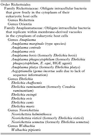
Table 1. Current classification of the order Rickettsiales
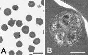
Fig. 1. Bovine erythrocytes infected with Anaplasma marginale. (A) Inclusion bodies are located at the periphery of the erythrocyte in a stained blood film. (B) An electron micrograph of an A. marginale inclusion that contains five organisms. A, bar=10 μm; B, bar=0·5 μm.
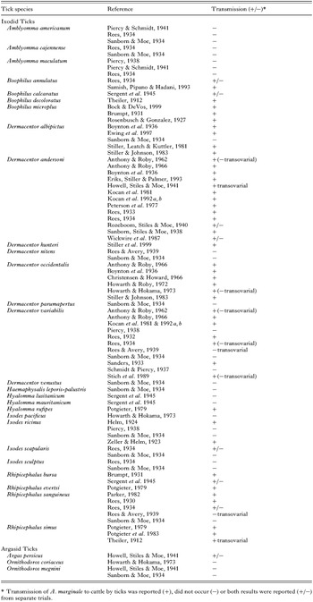
Table 2. Studies in which Ixodid and Argasid ticks were tested as vectors of anaplasmosis
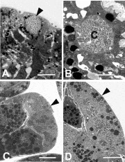
Fig. 2. Micrographs of colonies of Anaplasma marginale in tick gut and salivary gland cells. (A) A light micrograph of a large colony (arrow) in a tick gut cell. (B) An electron micrograph of a colony (C) in a tick gut cell. (C) A light micrograph of many colonies (arrow) in a salivary gland cell. (D) An electron micrograph of a tick salivary gland cell (arrow) that contains several A. marginale colonies. A, bar=5 μm; B, bar=5 μm; C, bar=10 μm; D, bar=5 μm.
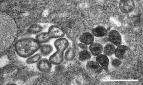
Fig. 3. Electron micrograph of the two developmental stages of Anaplasma marginale within colonies in tick cells. Reticulated forms within a colony on the left divide by binary fission and dense forms are within a second colony in a cultured tick cell. Bar=1 μm.
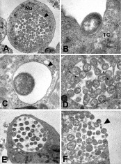
Fig. 4. Electron micrographs of A. marginale in cultured IDE8 tick cells. (A) A large colony of A. marginale adjacent to the nucleus (Nu) within a cultured tick cell. (B) An A. marginale adhered to the cell membrane of a cultured tick cell (TC). (C) A reticulated form of A. marginale within a membrane-bound colony. (D) A colony that contains reticulated forms of A. marginale that are dividing by binary fission (small arrows). (E) A colony that contains dense forms of A. marginale. (F) A. marginale being released (arrow) from a cultured tick cell. A, bar=5 μm; B, bar=0·5 μm; C, bar=0·5 μm; D, bar=5 μm; E, bar=5 μm; F, bar=1 μm.
 You have
Access
- 263
- Cited by