Introduction
Arrhythmogenic mitral valve prolapse syndrome is a recently recognised clinical condition that is associated with repolarisation disturbances and can lead to sudden cardiac death. Reference Kubala, Essayagh and Michelena1–Reference Chakrabarti, Bogun and Liang4 It is more common in females and is characterised by certain features including bileaflet mitral valve prolapse, regurgitation of the mitral valve, T-wave inversions in inferolateral leads on electrocardiogram, and complex ventricular ectopy. Reference Giudicessi, Rohatgi and Bos5,Reference Nagata, Bertrand and Levine6 In the past, some patients may have been misdiagnosed with atypical long QT syndrome before the identification of arrhythmogenic mitral valve prolapse syndrome. Reference Giudicessi, Rohatgi and Bos5 In this case, a 16-year-old adolescent girl experienced a sudden cardiac arrest during exercise. After further evaluation, she was diagnosed with arrhythmogenic mitral valve prolapse syndrome as the underlying cause of her cardiac event.
Case report
A healthy 16-year-old teenage girl was admitted to our hospital after experiencing an out-of-hospital cardiac arrest. The cardiac arrest occurred suddenly during a volleyball game. Prior to this event, she had no known health issues and no history of syncope or substance abuse.
She was transferred to the paediatric ICU where she received mechanical ventilation support. Therapeutic head hypothermia, oral allopurinol, and oral topiramate treatments were administered for neurologic protection. Laboratory results were normal except for elevated troponin and transaminase levels.
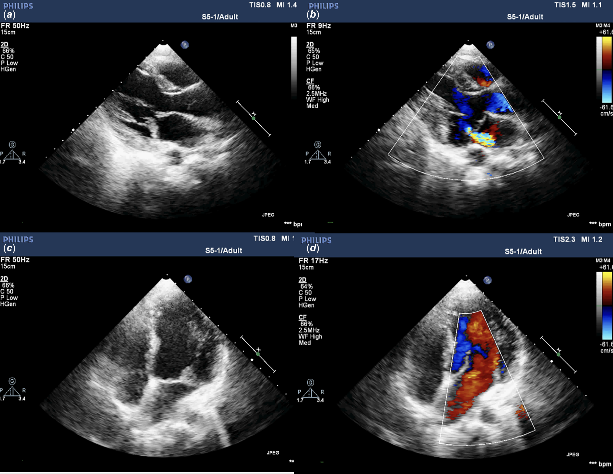
Two-dimensional echocardiography confirmed left ventricular enlargement, increased thickness, and prolapse of the mitral valve (Figure 1). A secondary degree of mitral valve insufficiency with a velocity of 4.2 m/s was detected. The ejection fraction was measured at 42%. Intravenous furosemide and enalapril treatments were administered due to the left ventricular enlargement.
The initial two-dimensional echocardiography findings. ( a ) Parasternal long axis view showing thickened and prolapsed leaflets of the mitral valve. ( b ) Colour Doppler imaging indicating 1st-2nd degree regurgitation flow in the mitral valve Mitral annular disjunction (MAD) is also observed. ( c ) Apical 4 chamber view showing thickened and prolapsed mitral valve leaflets. ( d ) Noticable widening of the left cavities.

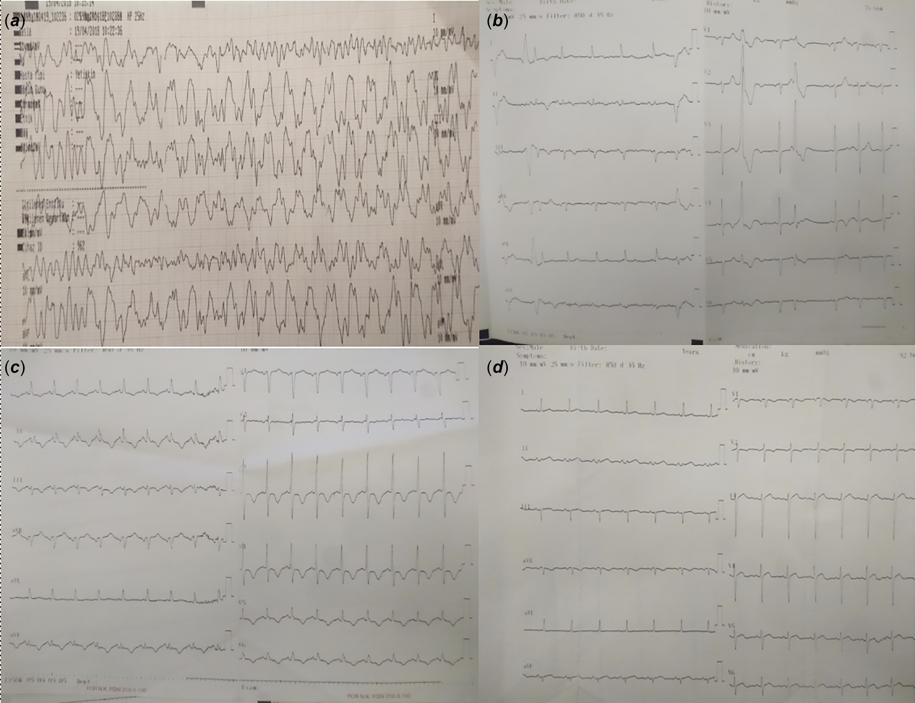
The QTc interval on the 12-lead electrocardiogram was measured at 500 milliseconds. T-wave inversions were observed in the inferolateral leads, and premature ventricular contractions in 3 different morphologies were present. To address the high QTc values, oral propranolol and magnesium were initiated for protective purposes. Due to the patient’s low left ventricular function, propranolol was started at a low dose of 1 mg/kg/day and gradually increased to 2.5 mg/kg/day. During the patient’s follow-up under beta blocker treatment, it was noted that T waves in the inferolateral leads intermittently became positive, and interestingly, the QTc values returned to normal during these times.
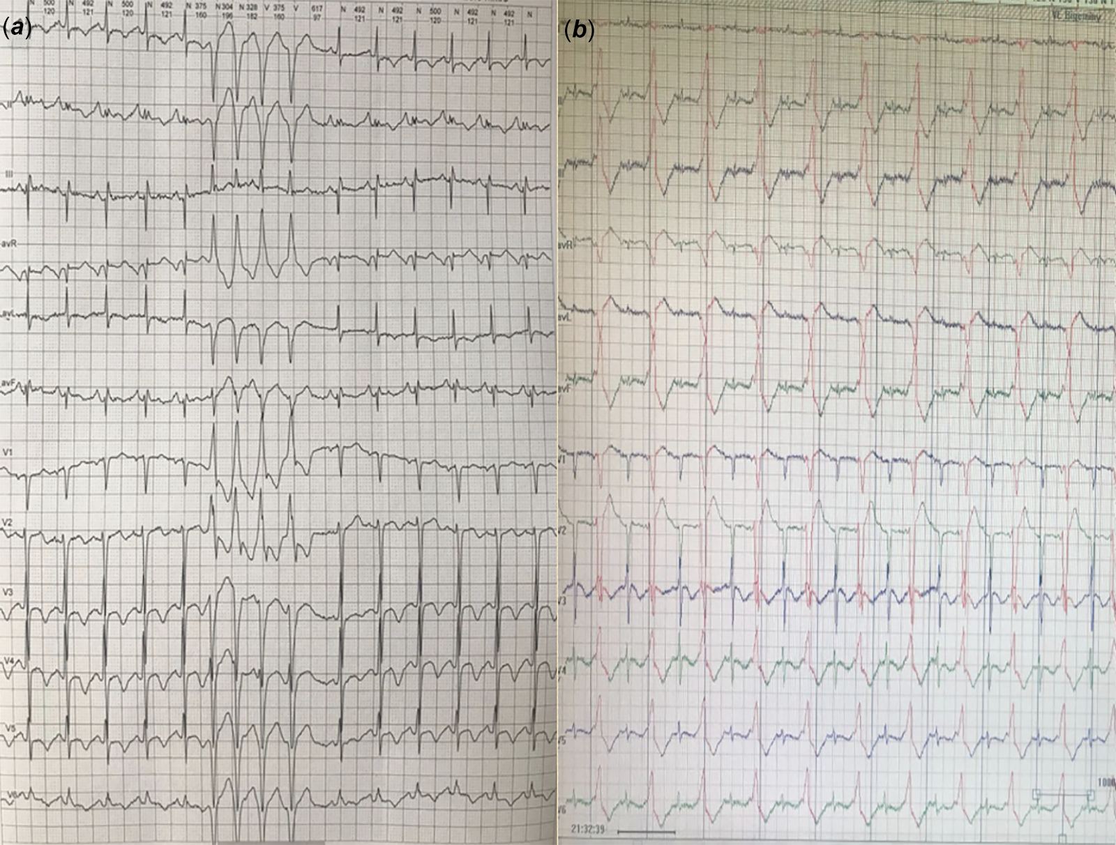
Premature ventricular contractions were observed in 3 different morphologies: RBBB superior axis with positive concordance, left axis RBBB northwest axis with relatively narrow QRS, and LBBB inferior axis with transition zone V3 (Figure 2). Non-sustained ventricular tachycardia occurred, with a maximum duration of 4 beats and a maximum rate of 190/min. During Holter monitoring, similar to the ECG findings, intermittent QTc values of 500 ms were observed, along with T wave negativity in D2, D3, aVF, V5, and V6 only during these time periods. There was also a noticeable decrease in QRS voltage at V6 (Figure 3).
The 12-lead ECGs after the resuscitation, first ECG. ( a ) The ECG of the patient taken while resuscitation was continuing in the ambulance. ( b ) In the first ECG taken in the hospital, the following findings were noted: polymorphic premature ventricular contractions, T wave inversions in leads II and III, flat or negative/positive T waves in lead aVF, and pure positive T waves in lead V1. Additionally, minimal changes in T wave morphology from beat to beat were observed. ( c ) After the patient was stabilised in terms of rhythm, the ECG performed while the patient was receiving only Propranolol showed prolonged QTc intervals and negative T waves in the inferior leads and positive T waves in V1. ( d ) However, interestingly, on another ECG taken on the same day, QTc values returned to normal when T wave polarities returned to normal.

Figure 2. Long description
Panel A: An ECG reading taken during resuscitation in an ambulance. The graph shows irregular and chaotic electrical activity, indicating severe arrhythmia. Panel B: The first ECG taken in the hospital. This graph displays polymorphic premature ventricular contractions, T wave inversions in leads II and III, flat or negative/positive T waves in lead aVF, and pure positive T waves in lead V1. Minimal changes in T wave morphology from beat to beat are observed. Panel C: An ECG performed after the patient was stabilized on Propranolol. This graph shows prolonged QTc intervals, negative T waves in the inferior leads, and positive T waves in V1. Panel D: Another ECG taken on the same day as Panel C. This graph shows QTc values returning to normal and T wave polarities returning to normal.
The 12‐lead Holter record findings ( a ) Non-sustained VT attack with northwest axis and RBBB pattern is observed ( b ) LBBB, inferior axis, most likely from outflow tract, bigemine PVCs seen.

Cardiac MRI revealed thickening, prolapse, and regurgitation of the mitral valve. The measurement of left ventricular width exceeded the standard limit, indicating enlargement, but overall cardiac function was normal. Although the T1 interval was prolonged regionally in the left ventricle, no significant pathological contrast enhancement was detected in late-stage contrast-enhanced images.
As part of the investigation following the aborted sudden death incident, Ajmaline and Adrenaline challenge tests were conducted. The Ajmaline provocation test yielded negative results. During the adrenaline provocation test, which was performed when T waves were in the normal polarity and QTc intervals were within normal range, a QTc prolongation of up to 25 msn was observed, but no ventricular arrhythmia was induced. Additionally, a coronary angiography was performed, which showed normal results. Interestingly, no abnormalities were found in the electrocardiograms or genetic scans of the family members.
A diagnostic electrophysiological study was conducted, which did not induce any ventricular arrhythmia. Due to the small percentage of premature ventricular contractions with three different morphologies and the absence of a dominant premature ventricular contraction, premature ventricular contraction ablation was not considered in the initial stage. Subsequently, a transvenous implantable cardiac defibrillator was implanted as a Class IA indication for the prevention of aborted sudden death. The patient was discharged with oral propranolol, mexiletine, furosemide, and enalapril treatments. However, due to availability issues, mexiletine was later replaced with flecainide.
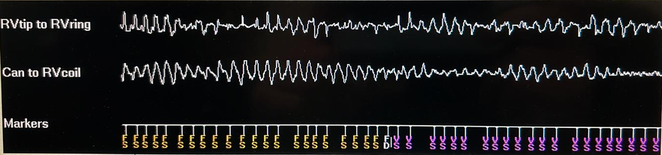
It was later discovered that the patient experienced a shock due to emotional stress during a period when she was irregularly taking her medication despite warnings. Upon reviewing the implantable cardiac defibrillator records, it was found that the patient had a genuine episode of Torsades de pointes and received an appropriate shock from the implantable cardiac defibrillator (Figure 4). Rare but polymorphic premature ventricular contractions that persisted despite dual antiarrhythmic treatment triggered these attacks. Bilateral sympathectomy was performed as the attacks were triggered by adrenergic stimuli such as exercise or emotional stress, both during the initial presentation and during follow-up. Although there was a noticeable decrease in appropriate shocks after sympathectomy, they were not completely eliminated.
The ICD recordings showed a Torsades de Pointes-like episode. This episode was terminated by an ICD shock.

The genetic analysis revealed heterozygote genomic changes in the KCNH2 gene and the 25th exon of the ANK2 gene. The patient was closely monitored for a period of 5 years following the initial presentation. Two years after undergoing sympathectomy, she experienced only one appropriate shock during a period when she was irregularly taking her medications. The level of mitral regurgitation, which was initially moderate, remained unchanged throughout the follow-up period.
Discussion
Arrhythmogenic mitral valve prolapse is a recently identified subgroup of mitral valve prolapse with a more severe clinical course. Reference Kubala, Essayagh and Michelena1–Reference Chakrabarti, Bogun and Liang4,Reference Korovesis, Koutrolou-Sotiropoulou and Katritsis7,Reference Zienciuk-Krajka, Daniçowicz-Szymanowicz and Dorniak8 It is characterised by specific findings, including central annular dysjunction, bileaflet prolapse of the mitral valve, QT prolongation on electrocardiogram, and ventricular ectopic beats, particularly originating from the left ventricle. Reference Sabbag, Essayagh and Barrera3,Reference Chakrabarti, Bogun and Liang4 It is worth noting that the response to established therapies for mitral valve prolapse and related conditions is not consistent in patients with arrhythmogenic mitral valve prolapse syndromev. Reference Thamman9,Reference Coutsoumbas and Pasquale10
From this case, several important findings were learned:
-
1. This case represents the first reported paediatric case of arrhythmogenic mitral valve prolapse in our country, indicating the importance of recognising and studying this condition in the paediatric population.
-
2. The presence of QT prolongation and T-wave inversion in inferolateral leads, which have been previously reported in association with arrhythmogenic mitral valve prolapse syndrome, was observed intermittently in this case. This highlights the variability and intermittent nature of these electrocardiographic findings in patients with arrhythmogenic mitral valve prolapse syndrome.
-
3. The intermittent nature of QT prolongation in these cases can complicate the selection of appropriate treatments and optimal dose adjustments. It suggests the need for close monitoring and individualised management strategies to address the intermittent QT prolongation and its impact on treatment decisions.
Mitral valve prolapse is the most common valvular anomaly observed in the Western population, and the role of mitral valve prolapse in sudden cardiac arrest remains controversial. Reference Coutsoumbas and Pasquale10,Reference Cipriani and Bauce11,Reference Marano, Lim and Sanchez12 Since 2017, when Giudicessi et al. described “arrhythmogenic mitral valve prolapse”, there has been a growing literature on the concept of arrhythmogenic mitral valve prolapse syndrome. Reference Miller, Dukkipati and Turagam2–Reference Chakrabarti, Bogun and Liang4,Reference Nagata, Bertrand and Levine6,Reference Korovesis, Koutrolou-Sotiropoulou and Katritsis7,Reference Basso, Iliceto, Thiene and Marra13
From an anatomical point of view, mitral regurgitation was initially recognised as an important component of the process because it disrupts the geometry and orientation of the left ventricle, leading to ventricular enlargement and sudden death. However, the coexistence of features such as T-wave inversion and ventricular arrhythmias in some patients with arrhythmogenic mitral valve prolapse without prominent mitral regurgitation later demonstrated that advanced mitral regurgitation is not an essential component of arrhythmogenic mitral valve prolapse. Reference Cipriani and Bauce11 In addition, fibrosis, particularly in the posterior papillary muscles and postero-basal region, previously attributed to continuous mitral regurgitation flux hitting the free wall during systole, was found to be an mitral regurgitation-independent process. Reference Miller, Dukkipati and Turagam2,Reference Cipriani and Bauce11 Another anatomical component that may be associated with arrhythmogenic mitral valve prolapse is mitral annular dysfunction. Mitral annular dysfunction refers to a degree of separation between the posterior mitral leaflet and the free wall. The degree of mitral annular separation can be measured quantitatively using MRI or even transthoracic ejection. Reference Kubala, Essayagh and Michelena1–Reference Chakrabarti, Bogun and Liang4,Reference Korovesis, Koutrolou-Sotiropoulou and Katritsis7,Reference Thamman9
Patients with arrhythmogenic mitral valve prolapse syndrome are believed to have areas of scar tissue intermixed with viable myocytes in the adjacent free wall of the left ventricle, papillary muscles, and infero-basal wall, independent of prolapse. Reference Chakrabarti, Bogun and Liang4 These areas of scar tissue have been demonstrated in histopathologic studies and through late gadolinium enhancement studies during MRI.
Borderline QT prolongation due to variable and prolonged repolarisation is not uncommon in patients with mitral valve prolapse. Reference Giudicessi, Rohatgi and Bos5,Reference Giudicessi, Rohatgi and Bos14 However, it is still unknown whether patients with QT prolongation have a worse prognosis compared to those without. It should be noted that QT prolongation can also be observed in patients with arrhythmogenic mitral valve prolapse syndrome. Reference Giudicessi, Rohatgi and Bos5,Reference Giudicessi, Rohatgi and Bos14 Interestingly, some case series in the literature have reported genetic mutations associated with long QT syndrome in patients diagnosed with arrhythmogenic mitral valve prolapse syndrome. However, these patients do not follow the expected course of classic Long QT syndrome and often show unusual responses to standard long QT syndrome treatments. As a result, these cases have been referred to as “atypical Long QT Syndrome” cases. Reference Giudicessi, Rohatgi and Bos5,Reference Giudicessi, Rohatgi and Bos14 On the other hand, approximately 2% of individuals with genetic positive long QT syndrome have coexisting mitral valve prolapse. Reference Giudicessi, Rohatgi and Bos5 While mitral valve prolapse alone is not considered a significant contributor to the long QT clinical phenotype, the combination of arrhythmogenic mitral valve prolapse syndrome and genetic positive long QT syndrome is a complex and potentially lethal association that poses challenges in terms of treatment. Reference Giudicessi, Rohatgi and Bos5 Some authors highlight arrhythmogenic mitral valve prolapse syndrome as the coexistence of three independent risk factors for sudden death: mitral valve prolapse, long QT syndrome, and papillary muscle ventricular tachycardia. Reference Kubala, Essayagh and Michelena1,Reference Chakrabarti, Bogun and Liang4,Reference Nagata, Bertrand and Levine6,Reference Thamman9 In a study by Ackerman et al., it was observed that patients with genetically positive long QT syndrome associated with arrhythmogenic mitral valve prolapse syndrome had more premature ventricular contractions with RBBB morphology during Holter monitoring, longer mean QTc intervals, a higher incidence of life-threatening events, and more positive shocks in implantable cardiac defibrillator recordings after sympathectomy compared to patients without arrhythmogenic mitral valve prolapse syndrome. Reference Giudicessi, Rohatgi and Bos5,Reference Giudicessi, Rohatgi and Bos14 The patient also has mutations in the KCNH2 and ANK2 genes whose clinical significance is unclear. The identified mutation in KCNH2, listed as VUS (variant of uncertain clinical significance) in the Franklin database, has the potential to cause abnormal splicing and cutting effects and is thought to be associated with the current clinical presentation.
T wave inversion in the inferolateral leads, reflecting changes in the direction and duration of repolarisation, is a more common finding than QT prolongation in arrhythmogenic mitral valve prolapse syndrome cases (2,3,10,13). In our case, we observed simultaneous QT prolongation and T wave negativity in the inferolateral leads, and interestingly, QT values returned to normal when the T wave became positive in those leads. We speculate that this may be due to myocytes utilising two different pathways for repolarisation. However, it remains uncertain whether this phenomenon increases the propensity for substrate instability or Torsades de Pointes. Nevertheless, the intermittent and dynamic changes in electrocardiogram findings highlight both electrical and anatomical alterations in arrhythmogenic mitral valve prolapse syndrome cases. These unique features, which fall within the spectrum of arrhythmogenic mitral valve prolapse syndrome but vary from case to case, also influence treatment planning. In our approach, we supplemented beta-blocker therapy with mexiletine to shorten the QTc values measured when T waves were negative by approximately 30 milliseconds. However, mexiletine significantly shortened the QTc values measured when T waves were positive, necessitating a reduction in dosage to avoid exceeding the therapeutic limit. This particular aspect of our case further adds to its complexity and underscores the need for individualised management.
In this patient, the abnormal mitral apparatus creates chronic mechanical traction, particularly on the posteromedial papillary muscle, which has a single blood supply, making it vulnerable to ischaemia and fibrotic remodelling. Mitral annular disjunction creates abnormal systolic twisting motion, generating repetitive mechanical triggers on the basal inferolateral wall. In mitral valve prolapse patients, accompanying autonomic dysregulation-particularly during physical exertion—may lead to excessive stimulation of sympathetic activation. Finally, the alternation of electrocardiogram findings suggests that it likely represents dynamic repolarisation heterogeneity driven by autonomic tone fluctuations on the diseased inferolateral substrate (posteromedial papillary muscle + basal inferior wall).
The role of sympathectomy in the treatment of patients with arrhythmogenic mitral valve prolapse syndrome remains uncertain. Reference Giudicessi, Rohatgi and Bos5,Reference Giudicessi, Rohatgi and Bos14,Reference Kelley, Chaudry and Syed15 While arrhythmogenic mitral valve prolapse syndrome may initially appear as a collection of anatomical defects, individuals with mitral valve prolapse syndrome are known to have increased sympathetic discharge compared to the general population. Reference Giudicessi, Rohatgi and Bos14,Reference Kelley, Chaudry and Syed15 Considering that life-threatening events in these cases result from the interaction between a pathological anatomical substrate and various triggers, sympathectomy may have potential benefits in selected cases (15). In our patient, given the clear adrenergic triggers such as exertion and emotional stress described before the event, we believed that she would benefit from sympathectomy. Although appropriate implantable cardiac defibrillator shocks were reduced following the procedure, they were not completely eliminated. It is worth noting that she experienced an appropriate implantable cardiac defibrillator shock during a period of irregular beta-blocker therapy.
Since the patient in our case had polymorphic premature ventricular contractions originating from multiple sites (posterior papillary muscle, posterior fascicle, and outflow), none of them being dominant and suitable for mapping, ablation was not considered as a primary treatment option. Instead, we focused on transvenous implantable cardiac defibrillator placement and sympathectomy procedures due to the complexity of the premature ventricular contraction patterns and the patient’s clinical presentation.
In conclusion, mitral valve prolapse is a common clinical finding in paediatric patients. However, it is crucial to identify which mitral valve prolapse patients are at a higher risk of sudden death. Recent advancements in data have allowed for the emergence and development of differential electrocardiographic and echocardiographic features that can help identify individuals at higher risk. Comprehensive assessments and algorithms that consider the patient’s personal and family history play a crucial role in accurately diagnosing and differentiating patients with arrhythmogenic mitral valve prolapse, thus avoiding both underdiagnosis and overdiagnosis.
Financial support
No financial or non-financial benefits have been received or will be received from any party related directly or indirectly to the subject of this article.
Competing interests
The authors have no conflicts of interest to declare.