Introduction
CHDs are among the most frequently encountered anomalies in clinical practice, occurring in roughly 1–6 out of every 1,000 live births. Reference Hoffman and Kaplan1 These defects cover a broad spectrum of malformations that vary significantly in clinical presentation and treatment strategies. Over the past decades, advances in surgical techniques and improvements in intensive care have led to marked increases in survival for children with CHD. As more children now live into adolescence and adulthood, the need for long-term follow-up and precise anatomical evaluation has grown considerably. Reference Chan, Aijaz and Zaidi2,Reference Das and Das3
Transthoracic echocardiography remains the cornerstone of CHD diagnosis because it is non-invasive, widely available, and free from radiation exposure. Reference Sachdeva and Gupta4 These qualities make it particularly useful in paediatric practice. However, echocardiography has limitations, especially in patients with complex anatomy, reconstructed cardiac structures after surgery, or when detailed assessment of coronary arteries and extracardiac anatomy is required. Reference Guzelbag, Bas, Ozcanoglu, Tanıdır, Hatemi and Ozturk5 In such cases, additional advanced imaging techniques are often needed to complement echocardiographic findings. Reference Sanphasitvong, Jim and Tantiwonglkosri6
The introduction of multi-detector CT has transformed imaging in CHD by allowing rapid, high-resolution scans with lower radiation doses and reduced contrast requirements. Reference Zucker7 These developments have positioned thoracic CT angiography as a valuable diagnostic tool, applicable in both pre-operative evaluation and post-operative surveillance. Reference Güzelbağ, Baş and Toprak8
In addition to excellent cardiac and vascular imaging, CT angiography provides high-quality visualisation of extracardiac structures, including the lungs, airways, pleura, chest wall, vertebrae, ribs, and upper abdominal organs. As a result, studies focusing on cardiovascular imaging often report incidental extracardiac findings. Reference Ramanathan, Ladumor, Francis, Allam and Alkuwari9,Reference Lazoura, Vassiou, Kanavou, Vlychou, Arvanitis and Fezoulidis10 While some of these are clinically insignificant, others may carry important consequences for patient care.
In adult coronary CT angiography studies, extracardiac findings have been reported in 7.1% to as many as 81% of patients, though their clinical relevance continues to be debated. Reference Lazoura, Vassiou, Kanavou, Vlychou, Arvanitis and Fezoulidis10–Reference Bhambhvani12 By contrast, evidence regarding extracardiac findings in paediatric patients undergoing CT angiography for CHD remains limited, with most available studies comprising relatively small cohorts and lacking systematic evaluation of their prevalence and clinical impact. Reference Malik, Hellinger, Servaes, Schwartz, Keller and Epelman13,Reference Nordjoe, Aubin Igombe and Chat14 Large-scale, single-centre data focusing on structured extracardiac assessment in paediatric CHD populations are therefore still lacking.
Therefore, the current study aims to determine the prevalence of non-cardiovascular findings in paediatric patients undergoing thoracic CT angiography for the evaluation of CHD. Additionally, we aim to evaluate the clinical significance of these findings and their impact on patient management strategies. Ultimately, we aim to raise awareness of these findings among clinical teams, particularly among cardiologists and radiologists.
Materials and methods
This research was designed as a single-centre, retrospective analysis. It included paediatric patients who underwent cardiac CT angiography in the paediatric cardiology department of our institution between January 1, 2022 and December 1, 2025. This retrospective study was conducted in accordance with the principles of the Declaration of Helsinki and was approved by the local institutional ethics committee (protocol number: 2025/151; approval date: June 14, 2025). Informed consent was waived due to the retrospective nature of the study.
All consecutive paediatric patients aged 0–17 years who underwent electrocardiography-gated contrast-enhanced cardiac CT angiography for the evaluation or follow-up of CHD during the study period were included. In this cohort, the maximum patient age was 13.9 years. All CT angiography examinations were independently reviewed by a radiologist with over 10 years of experience in congenital cardiovascular imaging. Exclusion criteria were incomplete imaging data, severe motion artefacts precluding adequate image interpretation, non-electrocardiography-gated CT examinations, non–contrast-enhanced CT studies, and CT examinations performed for non-cardiac indications. Demographic characteristics—including age, sex, height, weight, and body surface area, which was calculated using the Du Bois formula (body surface area = 0.007184 × height ^ 0.725 × weight ^ 0.425)—were systematically documented.
Cardiac CT angiography examinations in patients with CHD were evaluated using a structured and comprehensive approach. The CT angiography acquisition range extended from the thoracic inlet to the diaphragm, allowing for systematic assessment of extracardiac structures within the scan field. Extracardiac findings were evaluated using a systematic approach and categorised into three main groups: thoracic/respiratory system, upper abdominal system, and musculoskeletal system. Thoracic findings were further subcategorised into lung parenchyma, airways, pleura, and mediastinum, while upper abdominal findings included hepatic, splenic, and renal abnormalities when within the scan range. Musculoskeletal findings comprised vertebral, rib, and other skeletal anomalies.
The evaluation protocol consisted of a stepwise review of all extracardiac regions included in the scan range. This included (1) lung parenchyma (infection, oedema, and chronic changes), (2) airway anatomy (malacia, compression, and anatomical variations), (3) pleural space (effusion, thickening, and pneumothorax), (4) mediastinum (masses, thymic abnormalities, and vascular rings), (5) upper abdominal organs (hepatic, splenic, and renal abnormalities), and (6) musculoskeletal structures (vertebral and rib anomalies). Each category was assessed using predefined radiological criteria to ensure consistency and reproducibility.
For the purpose of subgroup analysis, CHDs were classified according to their embryological origin based on established developmental frameworks. The classification included the following groups: (1) laterality and looping disorders (e.g., heterotaxy syndromes and congenitally corrected transposition of the great arteries), reflecting abnormalities in left–right axis determination; (2) septation defects (e.g., atrial and ventricular septal defects), resulting from failure of intracardiac septation; (3) endocardial cushion defects (e.g., atrioventricular septal defects), associated with impaired atrioventricular canal development; (4) conotruncal defects (e.g., tetralogy of Fallot, transposition of the great arteries, double-outlet right ventricle, truncus arteriosus, and interrupted aortic arch), related to abnormalities in neural crest cell migration and outflow tract septation; (5) aortic arch anomalies (e.g., coarctation of the aorta and vascular ring anomalies), arising from abnormal remodelling of the pharyngeal arch arteries; (6) left-sided obstructive lesions (e.g., hypoplastic left heart syndrome, aortic and mitral valve anomalies); (7) right-sided obstructive lesions (e.g., pulmonary stenosis and pulmonary atresia); (8) anomalies of pulmonary venous return (e.g., total and partial anomalous pulmonary venous return); and (9) complex CHD (e.g., single-ventricle physiology, tricuspid atresia, and double-inlet ventricle). Each patient was assigned to a single primary embryological category based on the dominant underlying cardiac anatomy. This classification was used to evaluate the distribution of extracardiac findings across different embryological groups. Reference Li, Du and Deng15
CT acquisition protocol
All scans were obtained on a 640-slice CT scanner (Aquilion ONE GENESIS Edition, Canon Medical Systems, Otawara, Tochigi, Japan). Broad detector coverage (16 cm), iterative reconstruction algorithms, and Adaptive Iterative Dose Reduction (AIDR 3D Enhanced) technology were used to optimise imaging. Prospective electrocardiography-gated acquisition was performed within a single cardiac cycle. Images were acquired in Volume Axial mode with a rotation time of 0.35 s and a scan length of 80–160 mm. An exposure control system automatically adjusted the tube current to ensure optimal image quality. A tube voltage of 80 kV was used in all patients to optimise radiation dose in accordance with ALARA principles, without compromising image quality. Reference Baş, Alkara and Aliyev16 CT examinations were performed without general anaesthesia. Minimal sedation was used when necessary, and patients were carefully immobilised using age-appropriate positioning devices. Fast acquisition times, prospective electrocardiography gating, and wide-detector coverage enabled diagnostic image quality even in younger children.
Contrast protocol
Intravenous contrast medium (Kopaq 300 mgI/mL, Onko&Kocsel Pharmaceuticals, Turkey) was administered at a dose of 1.5 mL/kg, followed by a 10–20 mL saline flush using a dual-head injector (Bayer HealthCare, MEDRAD Europe, Netherlands). Injection rates varied between 0.7 and 3.5 mL/kg depending on catheter diameter and patient size. Contrast was administered undiluted. All examinations were supervised by an experienced radiologist specialising in imaging CHD. Data acquisition was timed to the first pass of contrast through the cardiovascular structures of interest. In patients with heart rates greater than 90 beats/min, the acquisition window was centred at 45% of the R-R interval, with phases of limited diagnostic value excluded. The optimal motion-free phase closest to the predefined window was selected for each patient.
Image reconstruction
Axial images were transferred to a post-processing workstation for further analysis. Reconstructions were performed with a slice thickness of 0.5 mm using standard kernels, followed by iterative reconstruction with AIDR 3D Enhanced. Advanced post-processing included multiplanar reconstructions, maximum intensity projections, and three-dimensional volume rendering.
Radiation dose evaluation
Radiation parameters—including scan range, dose-length product (DLP), and CT dose index volume (CTDI vol)—were documented. Values were calculated with reference to a 32 cm body phantom. For paediatric dose estimates, DLP values were multiplied by correction factors to account for differences in patient size and body composition. For neonates and infants, a coefficient of 0.039 mSv/(mGy·cm) was used. Reference Chu, Yu and Wang17,Reference Chu, Kofler and Mahendra18 The effective dose (ED) was determined using the formula: ED (mSv) = DLP (mGy·cm) × conversion factor, where chest-specific coefficients were defined by Deak et al. Reference Deak, Smal and Kalender19
Statistical analysis
Data analysis was performed using SPSS software. Descriptive statistics summarised demographic and clinical variables. Continuous data were expressed as mean, standard deviation, minimum, and maximum values, whereas categorical data were reported as counts and percentages.
Basic patient parameters—including age, sex, height, weight, body surface area, radiation dose, and contrast volume—were described with mean, standard deviation, minimum, maximum, and percentage distributions. The Shapiro–Wilk test was applied to assess normality. Additionally, histograms, skewness and kurtosis coefficients, and Q-Q plots were examined. For non-normally distributed data, median, interquartile range, and range values were provided. After confirming distribution assumptions, descriptive statistics were tabulated.
Results
This investigation retrospectively examined demographic and clinical characteristics of patients who underwent cardiac CT angiography within the Department of Pediatric Cardiology. A total of 1,336 patients who received CT angiography examination were included in the analysis. 56% of patients were male (n: 748) and 44% were female (n: 588). The mean patient age was 3.8 years, with a range of 0.1 to 13.9 years. Age distribution analysis revealed the following percentages: 0–2 years (57.4%, n: 766), 2–7 years (12.4%, n: 166), 7–12 years (10.8%, n: 144), and 12–17 years (19.4%, n: 260).
The mean patient weight was 15.8 kg (range: 3.1–49.7 kg), with a mean weight percentile of 35.9 (range: 23.9–44.1) and a weight standard deviation score of –0.36 (range: −0.52–0.87). Mean height measured 102.8 cm (range: 55.2–160.9 cm), with a mean height percentile of 42.7 (range: 26.87–41.7) and a height standard deviation score of −0.17 (range: −0.92–0.69). Mean body mass index was 14.9 kg/m2 (range: 9.8–21.9 kg/m2), with a body mass index percentile of 30.3 (range: 21.32–68.7) and a body mass index standard deviation score of −0.52 (range: −1.28–0.89). Mean radiation dose was 1.32 mSv (range: 0.8–2.3), and mean contrast volume utilised was 15.28 mL (range: 5.1–23.4 mL).
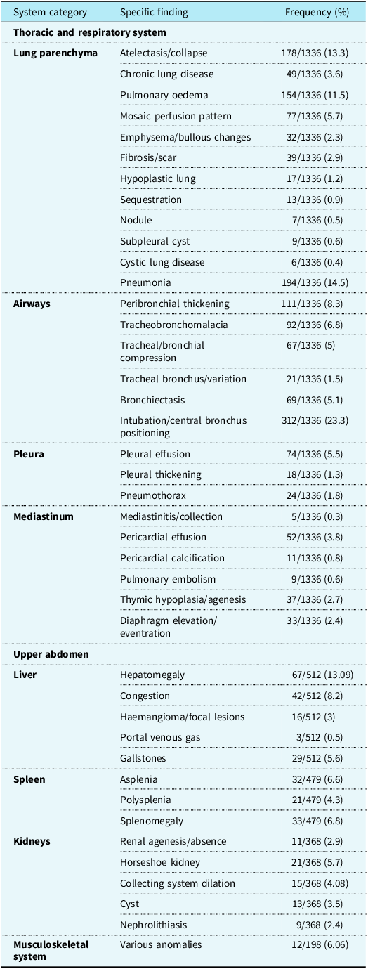
In these patients with CHD, beyond cardiovascular assessment, CT angiography allowed a structured review of extracardiac structures within the scan range. All patients underwent systematic evaluation of the thoracic and respiratory systems. In addition, hepatic anatomy was included in 512 scans, splenic anatomy in 479, and renal or collecting system anatomy in 368, allowing for the documentation of incidental or associated extracardiac findings.
Thoracic findings
Pulmonary parenchymal changes were the most frequent extracardiac abnormalities. Pneumonia was detected in 14.51% of patients and represented the leading diagnosis, followed by atelectasis or collapse (13.31%) and pulmonary oedema (11.52%). These findings are consistent with common complications and comorbidities in paediatric cardiac populations. Mosaic perfusion or attenuation (5.76%) and fibrotic or scar-related changes (2.92%) were also identified, suggesting chronic pulmonary alterations or sequelae of prior infections. Less common but clinically relevant abnormalities included hypoplastic lung (1.27%), sequestration (0.97%), subpleural cysts (0.67%), nodules (0.52%), and cystic lung disease (0.45%).
Airway abnormalities were also frequent. Intubation-related changes or main bronchus malposition were observed in 23.34% of cases, emphasising potential procedural complications or anatomical variants. Peribronchial thickening (8.3%), tracheobronchomalacia (6.88%), and airway compression (5%) were additional findings, reflecting structural modifications related to congenital anomalies or surgical interventions. Vascular ring anomalies causing airway compression were clearly demonstrated on CT angiography, including cases of double aortic arch with significant tracheal narrowing (Figure 1a and b), as well as pulmonary artery sling associated with airway compression (Figure 1c).
(a) Three-dimensional volume-rendered CT image demonstrates a double aortic arch forming a complete vascular ring with the first aortic arch indicated by the white arrow and the second arch indicated by the yellow arrow. (b) Three-dimensional volume-rendered CT image obtained from an alternative perspective further delineates the double aortic arch anatomy, with the white arrow indicating the first arch and the yellow arrow indicating the second arch. (c) Three-dimensional volume-rendered CT image demonstrating a pulmonary artery sling, in which the left pulmonary artery (yellow arrow) originates from the right pulmonary artery and courses between the trachea and oesophagus, potentially resulting in airway compression. These images illustrate the close anatomical and developmental relationship between congenital cardiovascular anomalies and their associated extracardiac airway findings.

Figure 1 Long description
The image shows a three-dimensional volume-rendered CT scan of a double aortic arch forming a complete vascular ring. The first aortic arch is indicated by a white arrow, while the second arch is marked by a yellow arrow. This anatomical configuration can lead to significant clinical implications due to the potential compression of surrounding structures. The detailed rendering provides a clear view of the complex vascular arrangement, highlighting the intricate relationship between the two arches. This type of imaging is crucial for precise anatomical evaluation and planning appropriate medical or surgical interventions.
In rare cases, CT angiography revealed complex thoracoabdominal arrangements, including a case of topsy-turvy syndrome associated with severe bronchial compression, illustrating the added value of comprehensive extracardiac assessment (Figure 2).
Imaging findings of a patient with topsy-turvy heart syndrome. (a) Three-dimensional volume-rendered CT image demonstrates the right ventricle located superiorly (yellow arrow) and the left ventricle located inferiorly (white arrow). (b) Three-dimensional volume-rendered CT image demonstrates an inferiorly positioned aortic arch (yellow arrow), with stars indicating the separate origins of the right subclavian artery, right common carotid artery, left common carotid artery, and left subclavian artery, all arising individually from the aortic arch. (c) Axial contrast-enhanced CT image shows severe tracheal luminal narrowing (red arrow) with associated post-obstructive changes in the right lower lobe (yellow arrow) and atelectasis in the left lower lobe (white arrow).

Figure 2 Long description
The image consists of three panels. Panel (a) displays a three-dimensional volume-rendered CT image of the heart, highlighting the right ventricle positioned superiorly with a yellow arrow and the left ventricle located inferiorly with a white arrow. Panel (b) shows another three-dimensional volume-rendered CT image, focusing on an inferiorly positioned aortic arch marked by a yellow arrow. Stars indicate the separate origins of the right subclavian artery, right common carotid artery, left common carotid artery, and left subclavian artery, all arising individually from the aortic arch. Panel (c) presents an axial contrast-enhanced CT image, revealing severe tracheal luminal narrowing indicated by a red arrow. Additionally, it shows post-obstructive changes in the right lower lobe marked by a yellow arrow and atelectasis in the left lower lobe highlighted by a white arrow.
Pleural abnormalities included pleural effusion (5.5%), pleural thickening (1.3%), and pneumothorax (1.8%), each with potential clinical implications for respiratory function and necessitating monitoring or intervention. Mediastinal findings comprised pericardial effusion (3.8%) and thymic hypoplasia/agenesis (2.7%), both of which may be associated with syndromic conditions. CT angiography also enabled the identification of anterior mediastinal masses adjacent to the cardiac chambers, including imaging features consistent with a cystic teratoma, findings that may have implications for surgical planning (Figure 3).
Contrast-enhanced axial CT image demonstrates an anterior mediastinal soft-tissue mass adjacent to the right atrium (yellow arrow) with internal calcification (yellow circle), imaging features suggestive of a cystic teratoma.

Figure 3 Long description
A contrast-enhanced axial CT image displays an anterior mediastinal soft-tissue mass located adjacent to the right atrium. The mass is indicated by a yellow arrow, and it contains internal calcification, which is highlighted by a yellow circle. These imaging features suggest the presence of a cystic teratoma. The right atrium is visible in the central region of the image, with the mass positioned to its right. The surrounding anatomical structures, including the lungs and other mediastinal components, are also discernible in the cross-sectional view.
Abdominal findings
Hepatomegaly was the most frequent liver abnormality, present in 13.09% of patients. Splenic abnormalities, such as splenomegaly (6.8%), asplenia (6.6%), and polysplenia (4.3%), were relevant due to their association with heterotaxy syndromes. Renal findings included a horseshoe kidney (5.7%), renal agenesis or absence (2.9%), dilatation or hydronephrosis of the collecting system (4.08%), renal cysts (3.5%), and nephrolithiasis (2.4%), all with potential implications for renal function and surgical planning.
Musculoskeletal findings
Musculoskeletal system anomalies were identified in 6.06% of cases, including scoliosis, vertebral malformations, costal deformities, osseous demineralisation, and complications related to implanted hardware. Although less frequent, these findings may influence patient mobility, posture, or thoracic development. Vertebral segmentation anomalies, including hemivertebra and butterfly vertebra, were among the musculoskeletal findings detected on CT angiography, as illustrated in a patient with tetralogy of Fallot (Figure 4).
Three-dimensional volume-rendered CT image demonstrating a hemivertebra deformity at the T7 level (yellow arrows) in a patient with tetralogy of Fallot, illustrating the coexistence of musculoskeletal and cardiac anomalies.

Figure 4 Long description
A three-dimensional volume-rendered CT image displays a hemivertebra deformity at the T7 level, highlighted by yellow arrows. The image illustrates the coexistence of musculoskeletal and cardiac anomalies in a patient with tetralogy of Fallot. The ribs and vertebrae are clearly visible, with the deformity located centrally in the thoracic spine. The yellow arrows point to the specific area of the hemivertebra, emphasizing the structural abnormality.
Additional musculoskeletal abnormalities included scoliosis and rib anomalies, as demonstrated in a patient with truncus arteriosus and VACTERL association (vertebral, anal, cardiac, tracheoesophageal, renal, and limb anomalies), further emphasising the coexistence of skeletal and cardiac anomalies (Figure 5).
(a) Three-dimensional volume-rendered CT image demonstrating a costal anomaly (yellow arrow) in a patient with truncus arteriosus and VACTERL association, highlighting the association between vertebral, costal, and cardiac anomalies. (b) Three-dimensional volume-rendered CT image showing scoliosis of the thoracic vertebral column in the same patient.

Figure 5 Long description
The image on the left is a three-dimensional volume-rendered CT scan highlighting a costal anomaly marked by a yellow arrow in a patient with truncus arteriosus and VACTERL association. This scan demonstrates the relationship between vertebral, costal, and cardiac anomalies. The image on the right shows scoliosis of the thoracic vertebral column in the same patient, captured through a three-dimensional volume-rendered CT image.
Extracardiac findings of the patients are presented in Table 1.
Comprehensive analysis of extracardiac findings in patient population

Table 1 Long description
The table presents extracardiac findings in patients, organized by system categories, specific findings, and their frequencies in percentage. The table has three columns: System category, Specific finding, and Frequency percentage. The Thoracic and respiratory system category includes findings such as Atelectasis/collapse at thirteen point three percentage, Chronic lung disease at three point six percentage, Pulmonary oedema at eleven point five percentage, and Pneumonia at fourteen point five percentage. The Airways category lists Peribronchial thickening at eight point three percentage, Tracheobronchomalacia at six point eight percentage, and Intubation/central bronchus positioning at twenty point three percentage. The Pleura category includes Pleural effusion at five point five percentage and Pneumothorax at one point eight percentage. The Mediastinum category lists Pericardial effusion at three point eight percentage and Thymic hypoplasia/agenesis at two point seven percentage. The Upper abdomen category includes Hepatomegaly at thirteen point zero nine percentage and Congestion at eight point two percentage. The Liver category lists Aspenia at six point six percentage and Splenomegaly at six point eight percentage. The Kidneys category includes Horseshoe kidney at five point seven percentage and Collecting system dilation at four point zero eight percentage. The Musculoskeletal system category lists Various anomalies at six point zero six percentage.
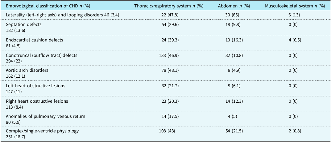
When extracardiac findings were analysed according to the embryological classification of CHDs, distinct distribution patterns were observed across different groups. Thoracic and respiratory system findings were most prevalent in conotruncal defects and aortic arch disorders, whereas abdominal findings were markedly higher in patients with laterality and looping abnormalities. Musculoskeletal findings were relatively infrequent across all groups. The distribution of extracardiac findings according to embryological classification is presented in Table 2. Percentages were calculated within each embryological CHD subgroup.
Distribution of extracardiac findings according to the embryological classification of CHD

Table 2 Long description
The table presents the distribution of extracardiac findings across different embryological classifications of congenital heart disease (CHD). It includes data for the thoracic/respiratory system, abdomen, and musculoskeletal system. The table has five columns: Embryological classification of CHD, Thoracic/respiratory system, Abdomen, and Musculoskeletal system. The rows list various CHD classifications and their corresponding percentages of extracardiac findings in each system. For instance, laterality and looping disorders show a high percentage of abdominal findings at sixty-five percentage, while conotruncal defects have a notable percentage of thoracic/respiratory findings at forty-six point nine percentage. Musculoskeletal findings are generally low across all classifications.
Values are presented as n (%).
Discussion
CT angiography has become an indispensable imaging modality for the comprehensive evaluation of CHD, offering high-resolution visualisation not only of cardiovascular structures but also of the extracardiac thoracic and upper abdominal regions. In our cohort, numerous abnormalities—most often involving the lungs and airways—were identified outside the primary cardiac structures. This situation underscores the importance of a systematic examination that goes beyond just the heart structures. The frequency and distribution of these findings in our cohort are broadly consistent with previously published paediatric CT angiography and MRI literature, yet our sample represents one of the largest single-centre paediatric CHD populations evaluated to date, thereby strengthening the generalisability of our observations.
Pulmonary and airway abnormalities constituted the majority of extracardiac findings in our population. Pneumonia, atelectasis, and pulmonary oedema were the most frequent parenchymal abnormalities. These findings align closely with the work of Malik et al., who reported pulmonary abnormalities in 58.7% of paediatric CHD patients, and Nordjoe et al., who found parenchymal and airway abnormalities in 41% of cases. Reference Malik, Hellinger, Servaes, Schwartz, Keller and Epelman13,Reference Nordjoe, Aubin Igombe and Chat14 The high prevalence of such findings across paediatric studies likely reflects shared pathophysiological mechanisms among CHD patients, including altered pulmonary blood flow, increased susceptibility to infections, prolonged mechanical ventilation, and post-operative respiratory complications. Our detection of mosaic perfusion patterns and chronic parenchymal changes also parallels prior observations suggesting long-standing haemodynamic disturbances or recurrent infections in this population.
Airway anomalies—such as bronchial malposition, peribronchial thickening, tracheobronchomalacia, and airway compression—were also commonly observed, with rates similar to those reported in previous paediatric CT angiography studies. Malik et al. described airway abnormalities in 34.3% of their patients, a figure comparable to the prominent airway involvement identified in our cohort. Reference Malik, Hellinger, Servaes, Schwartz, Keller and Epelman13 Such findings are clinically relevant as they may influence decisions regarding perioperative ventilation, bronchoscopy, or the timing of interventional procedures. Our findings reinforce the recommendation that airway evaluation should be an integral component of CT angiography interpretation in children with CHD.
Abdominal abnormalities were also documented at meaningful rates in our study, including hepatomegaly, splenic anomalies, and renal malformations. These results are consistent with observations by Mahani et al., who found extracardiac abnormalities in 16.5% of paediatric cardiac MRI studies, and by Malik et al., who reported hepatic abnormalities in 24% of patients. Reference Malik, Hellinger, Servaes, Schwartz, Keller and Epelman13,Reference Nordjoe, Aubin Igombe and Chat14,Reference Baş, Alkara and Aliyev16–Reference Mahani, Morani and Lu20 The presence of splenic abnormalities, such as asplenia or polysplenia, is particularly important as they are frequently associated with heterotaxy syndromes and may guide further diagnostic evaluation or immunisation strategies. Similarly, renal abnormalities—including horseshoe kidney and collecting system dilation—may have implications for surgical planning and long-term follow-up.
Although extracardiac findings have been more extensively studied in adult coronary CT angiography populations—with reported prevalence ranging from 7% to 81% depending on the study design and reporting criteria—paediatric populations tend to demonstrate a higher proportion of clinically relevant findings. Reference Lazoura, Vassiou, Kanavou, Vlychou, Arvanitis and Fezoulidis10–Reference Bhambhvani12,Reference Buckens, Verkooijen, Gondrie, Jairam, Mali and van der Graaf21–Reference Budoff and Gopal25 In adults, common extracardiac abnormalities include lung nodules, thyroid nodules, hepatic lesions, and adrenal abnormalities. However, the distribution of findings in children is characteristically different: pulmonary parenchymal and airway abnormalities dominate, likely due to the unique developmental, infectious, and haemodynamic context of paediatric CHD. The patterns observed in our population therefore reinforce the notion that extracardiac evaluation requires age-specific interpretation.
A notable proportion of abnormalities in our cohort were previously unsuspected, consistent with prior studies reporting that one-third to one-half of extracardiac findings in paediatric CT angiography may be newly diagnosed and clinically actionable. Reference Malik, Hellinger, Servaes, Schwartz, Keller and Epelman13 The identification of such unanticipated findings has the potential to modify treatment plans, influence the timing of surgery, or prompt additional subspecialty evaluation. Therefore, optimising radiological reporting standards to ensure systematic extracardiac assessment may meaningfully impact clinical outcomes.
The subgroup analysis based on embryological classification revealed distinct patterns in certain groups. Notably, laterality defects demonstrated a markedly higher prevalence of abdominal findings, consistent with the known association with splenic anomalies. Similarly, conotruncal and aortic arch defects showed a higher frequency of thoracic findings, particularly airway-related abnormalities. These associations can be partly explained by shared embryological pathways. Conotruncal and aortic arch anomalies are closely related to abnormalities in neural crest cell migration, which also contributes to the development of the aortic arch and great vessels, potentially affecting airway anatomy. Similarly, laterality defects arise from disturbances in left–right axis determination during early embryogenesis, which can result in abnormal arrangement of both thoracic and abdominal organs, including splenic anomalies. These overlapping developmental processes may underlie the coexistence of cardiac and extracardiac abnormalities observed in this cohort.
Despite these group-specific patterns, extracardiac findings were broadly distributed across all CHD subtypes. These findings are in line with previous studies reporting that extracardiac abnormalities in paediatric CHD are not confined to specific cardiac subtypes but may reflect complex developmental interactions between cardiovascular and extracardiac organogenesis. Nevertheless, certain extracardiac abnormalities appear to be more prevalent in specific CHD subgroups, as also observed in our cohort. Reference Lin, Ticho, Houde, Westgate and Holmes26,Reference Humphrey, Duncan and Fletcher27
Overall, our findings highlight the diagnostic value of incorporating a structured extracardiac review into routine paediatric CT angiography interpretation. By demonstrating a similar distribution of findings to previously published work but in a substantially larger cohort, this study contributes robust epidemiologic data to the limited paediatric literature. Future multicenter, prospective investigations are warranted to determine the long-term clinical implications of these extracardiac abnormalities and to evaluate whether standardised reporting protocols could improve outcomes in paediatric CHD care.
Conclusion
Cardiac CT angiography represents an essential methodology for diagnosing cardiac pathologies and identifying significant extracardiac anomalies in children with CHD. Due to the frequency and clinical significance of these findings, particularly in pulmonary and airway structures, radiologists and clinicians must systematically evaluate and report comprehensive imaging scan findings. Future prospective multicenter investigations are needed to provide additional data regarding long-term outcomes and management of these extracardiac findings. The present study highlights the crucial role of cardiac CT in detecting both cardiac and extracardiac abnormalities, underscoring the necessity of comprehensive and systematic approaches—especially in paediatric patients—since pulmonary and abdominal anomalies can directly affect prognosis and management strategies.
Limitations
This study has several limitations. First, due to its retrospective design, mild or subclinical extracardiac findings may have been overlooked. Second, the evaluation was conducted at a single centre, which may limit the generalisability of the results because imaging technology, scanning protocols, and patient characteristics may vary across institutions. Additionally, prospective follow-up data were not available to assess the long-term clinical impact of the extracardiac findings identified in this cohort. Nevertheless, the large sample size provides valuable insight into the prevalence of extracardiac abnormalities in the paediatric CHD population.
Data availability statement
The data that support the findings of this study are available from the corresponding author upon reasonable request. The data are not publicly available due to restrictions. Further inquiries can be directed to the corresponding author.
Author contributions
The authors conceived and designed the study. Data acquisition and analysis were performed by the authors. All authors critically reviewed the manuscript, approved the final version to be published, and agree to be accountable for all aspects of the work.
Financial support
This research received no external funding.
Competing interests
The authors declare that they have no conflicts of interest.
Institutional review board statement
This study was conducted in accordance with the Declaration of Helsinki and approved by the local ethics committee (protocol code: 2025/151; date: June 14, 2025).
Informed consent statement
Patient consent was waived due to the retrospective nature of this study.