Implications
Knowledge on the changes occurring in the gastrointestinal tract of mink kits during the weaning period is scarce. Such knowledge is important in order to understand the increased occurrence of gastrointestinal disorders around the time of weaning and the reduced growth rates often observed. This study shows that the enzymatic capacity is not reduced during the weaning period, but the reduced mucin-staining area and the lower vitamin E status suggests that the mink kits may be more susceptible to infections during the immediate post-weaning period.
Introduction
In the weaning period, the diet of young mammals changes from easily digestible milk to a more complex adult-type diet. Furthermore, the young mammals in livestock production are stressed by the absence of their mother. These factors have a profound influence on the physiology of the young mammals and especially on the gastrointestinal tract, which has to respond to a shift in diet composition.
Mink kits usually start to eat their mother’s diet around 4 weeks of age, and gradually decrease their intake of milk. Thus, when weaned at 6 to 7 weeks of age, they are adapted to the weaning diet (Jørgensen, Reference Jørgensen1985), in contrast to piglets and calves that are weaned abruptly, which is well known to cause major changes in the gastrointestinal tract (Le Huerou-Luron et al., Reference Le Huerou-Luron, Guilloteau, Wicker-Planquart, Chayvialle, Burton, Mouats, Toullec and Puigserver1992; Hedemann et al., Reference Hedemann, Højsgaard and Jensen2003; Hedemann and Jensen, Reference Hedemann and Jensen2004). The knowledge on the physiological changes occurring in the gastrointestinal tract of mink kits at weaning is limited.
Fat constitutes the major energy supply of mink kits. The fat source gradually changes from almost completely digestible milk fat to fat of mixed animal, marine and vegetable origin that with respect to chemical composition, physical structure, as well as digestibility is different. The digestion of fat relies on an interplay between the lipolytic enzymes secreted by the exocrine pancreas and the bile salts secreted from the gall bladder that serves to emulsify lipids and lipid solubles to form fat-containing micelles. Weaning has been shown to decrease the activity of lipase and carboxyl ester hydrolase (CEH) in pigs (Jensen et al., Reference Jensen, Jensen and Jakobsen1997b; Hedemann and Jensen, Reference Hedemann and Jensen2004), but whether this occurs in mink kits as well is unknown. Furthermore, the production and utilization of bile salts in animals is very limited at birth and during early developmental stages (Orban and Harmon, Reference Orban and Harmon2000) and it can be speculated that bile salt concentration would be limited after weaning and thus be a limiting factor for fat digestion.
Vitamin E is a lipid-soluble antioxidant important for the maturation and function of the immune system (Nakamura and Omaye, Reference Nakamura and Omaye2009). As with other lipid components, the vitamin E present in milk is well absorbed, whereas synthetic vitamin E added to the feed as all-rac-α-tocopheryl acetate has to be hydrolysed by CEH before it can be absorbed (Jensen et al., Reference Jensen, Engberg and Hedemann1999). The change in the concentration of all-rac-α-tocopherol in plasma at weaning is therefore a good marker of the change in the activity of CEH.
This experiment aimed to study the change in gut morphology, the activity of digestive enzymes, the concentration and composition of bile acids in the gall bladder, as well as the concentration and stereochemical composition of α-tocopherol in plasma and liver in mink kits during the weaning period.
Material and methods
Animals, feeding and housing
For the investigation we used male wild-type mink kits from eight litters with six to nine kits, born on 6 May. The animals were fed a standard Danish wet mink feed ration from the local feed kitchen (Holstebro Feed Kitchen). Diet composition (as fed): fish offal 18.25%, industrial fish 30%, boiled poultry offal 15%, swine lungs 15%, blood 7%, heat-treated barley 6%, ensiled hens 5.5%, haemoglobin meal 1.5%, arbocel 1.2%, dried pork scratchings 0.25%, vitamin and mineral mixture 0.2% and NaCl 0.1%. The animals were fed once a day, and given free access to tap water by means of an automatic system. The females were housed individually in wire netting rearing cages (L × W × H: 90 × 30 × 45 cm) with wooden nest boxes attached (L × W × H: 24 × 31 × 31 cm). The nests were furnished with straw. At weaning, at 42 days of age, the mother was removed from the litter. The littermates were kept together until the end of the investigation. All animal facilities were sheltered under permanent outdoor sheds.
Collection of samples
On days 34, 47 and 59 after birth one male kit from each litter was randomly selected. The body weight (BW) was recorded at the time of sampling. The mink kits were euthanized by an intra-peritoneal injection of an overdose of pentobarbital-Na 40% (1 ml/kg BW). A blood sample was taken by heart puncture using a syringe. The blood was transferred to a heparinized glass and subsequently centrifuged (1200 × g, 10 min). Plasma was stored at −20°C until analysis.
The abdominal cavity was opened and the liver was taken out and weighed and the content of the gall bladder was collected; both were put on ice and subsequently stored at −20°C until analysis. The entire gastrointestinal tract was removed. The pancreas was carefully dissected free and immediately frozen at −20°C. The small intestine was isolated, and the length determined. Tissue samples for microscopy (2 cm) and enzyme determination (7 cm) were taken at 25% and 75% of the small intestinal length (SI25 and SI75, respectively). The samples for microscopy were transferred to Lillie’s AAF (10% formalin, 5% glacial acetic acid and 85% absolute alcohol). The samples for enzyme determination were opened lengthwise, rinsed carefully with ice-cold 0.9% NaCl, blotted dry and stored at −20°C until enzyme analysis.
Samples for microscopy
After 24 h in Lillie’s AAF the tissue samples were carefully cleaned of remaining digesta using deionized water and then transferred to 99% ethanol. Subsequently, the samples were dehydrated and infiltrated with paraffin wax. A slide was prepared from each sample and each slide contained a minimum of four sections cut at 4 μm thickness, at least 50 μm apart.
The sections were deparaffinized and hydrated, and then were processed for carbohydrate histochemistry using the periodic acid-Schiff (PAS) reaction according to Kiernan (Reference Kiernan1990). The PAS reaction stains for neutral mucins (Kiernan, Reference Kiernan1990).
Carbohydrate histochemistry was evaluated as described previously (Brunsgaard, Reference Brunsgaard1997). In short, fifteen well-oriented villi and crypts were selected on each slide. For each villi and crypt, the area of mucin granules with a clear positive reaction for neutral mucins was determined using a computer-integrated microscope and an image analysis system (Leica QWin version 3.2.0, Leica Microsystems Imaging Solutions Ltd, Cambridge, UK) with a monitor. This area included the mucus material present in the crypt lumen. As the histochemical procedure used in our study stains the granules of all mucous cells (goblet cells and crypt secretory cells) as well as the apical secretion of these cells, these are all included in the measurements.
The slides were further used to determine the area, height and density of the crypts and villi. Moreover, the thickness of the Tunica muscularis was measured. The area of crypts or villi was determined as the area encircled by the basement membrane and the crypt mouth or the basement membrane and the end of the villi, respectively, on fifteen well-determined crypts or villi. The crypt depth was determined as the distance between the basement membrane and the crypt mouth, and the villi height was determined as the distance between the crypt mouth and the end of the villi. The density of crypt and villi was determined as the number of crypts or villi present over a defined distance across the luminal part of the mucosa.
Homogenization of tissue
The pancreas was thawed and weighed. It was homogenized in five volumes 0.9% NaCl using a homogenizer (Ultra Turrax T 25 equipped with a S25N-8G probe, IKA®, Staufen, Germany). The homogenate was centrifuged (13 800 × g for 20 min at 4°C) and aliquots of the supernatant were collected for enzyme activity determinations.
After thawing, mucosa of the intestinal segment was scraped off the underlying muscular layers. The mucosa was homogenized in aqueous Triton X-100 (1%, v/v; 6 ml/g mucosa) using a homogenizer. The homogenate was centrifuged (20 000 × g for 60 min at 4°C) and aliquots of the supernatant were collected for enzyme activity determinations.
The liver was homogenized in twice the amount of ethanol using an Ultra-Turrax homogenizer.
Enzyme assays
The activity of amylase, trypsin, lipase and CEH in pancreatic tissue homogenate was determined as described by Hedemann and Jensen (Reference Hedemann and Jensen2004). Amylase activity was measured using the Phadebas® amylase reagent (Pharmacia Diagnostics, Uppsala, Sweden). Tributyrin was used as a substrate for lipase in a titremetric method. Trypsinogen was activated with enterokinase (E 0632, Sigma, St. Louis, MO, USA; Jensen et al., Reference Jensen, Gabert, Jørgensen and Engberg1997a) and the trypsin activity was determined using Nα-benzoyl-d,l-arginine p-nitroanilide (B 4875, Sigma) as a substrate. The esterase activity of CEH was measured with p-nitrophenyl acetate (N 8130, Sigma) as a substrate.
The activity of aminopeptidase N (APN) was measured using l-alanine-4-nitroanilide (Merck 101014, Darmstadt, Germany) as a substrate (Hedemann et al., Reference Hedemann, Højsgaard and Jensen2003).
Lactase and sucrase activities were determined according to Dahlqvist (Reference Dahlqvist1968) using a glucose kit (166 391, Boehringer Mannheim, Mannheim, Germany) to determine the amount of liberated glucose.
The enzyme activities were expressed as units/mg mucosa. One unit was defined as the amount of enzyme that hydrolysed 1 μmol of substrate/min.
Tocopherol analyses
The concentrations of tocopherols were determined in feed, plasma and liver by HPLC after saponification and extraction into heptane. The stereochemical composition of α-tocopherol was determined after the methylation of α-tocopherol stereoisomers into their corresponding methyl ethers and subsequent separation by chiral HPLC (Jensen and Lauridsen, Reference Jensen and Lauridsen2007).
Determination of bile salts
Bile salts were quantified by reversed-phase HPLC according to Knarreborg et al. (Reference Knarreborg, Lauridsen, Engberg and Jensen2004).
Statistical analyses
Analysis of variance was carried out with the experimental day as a source of variation using the GLM procedure of SAS (SAS Institute Inc., Cary, NC, USA). The results are presented as means and s.e.m. Differences were considered significant at α = 0.05.
Results
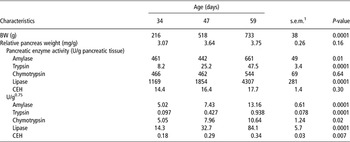
The mink kits used for the present experiment were all in good health conditions and showed no signs of illness. The mean BW of the mink kits increased from 216 ± 38 g at 34 days of age to 518 ± 38 g and 733 ± 38 g at 47 and 59 days of age, respectively (Table 1). The relative weight of the pancreas did not change (P > 0.05) during the experimental period.
The BW (g), relative pancreas weight (mg/g BW) and the activities of amylase, trypsin, chymotrypsin, lipase and CEH per gram pancreatic tissue (U/g) and total activities of pancreatic enzymes in relation to metabolic weight (U/g0.75) in mink kits at 34, 47 and 59 days of age (n = 8)

CEH = carboxyl ester hydrolase.
The mink kits were weaned at 42 days of age.
1Pooled s.e.m.
Pancreatic enzyme activity
The activity of amylase per gram pancreatic tissue (Table 1) did not change between 34 and 47 days of age, but the activity was increased (P = 0.01) at 59 days of age. The trypsin activity was very low at 34 days of age. It was tripled at 47 days of age and the activity at 59 days of age was twice that at 47 days of age (P < 0.0001). No changes in the chymotrypsin activity were observed between 34 and 59 days of age. Between 34 and 47 days of age no change in lipase activity was observed, whereas the activity doubled between 47 and 59 days of age (P < 0.0001). The activity of CEH did not change during the experimental period.
The activity of all the enzymes increased (P < 0.05) during the experimental period when the pancreatic enzyme activity was expressed as the total activity in the pancreatic tissue in relation to the metabolic BW (Table 1). The relative increase in activity differed between the enzymes. The activity of amylase, chymotrypsin and CEH was doubled, whereas the activity of lipase increased six times and the activity of trypsin increased nine times during the experimental period.
Mucosal enzyme activity
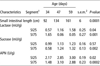
The length of the small intestine was 92, 134 and 161 ± 6 cm at days 34, 47 and 59, respectively (Table 2). Lactase activity in the proximal small intestine increased during the experimental period (P = 0.04). In contrast to this, lactase activity in the distal small intestine declined abruptly from 34 to 47 days of age (P = 0.001) and remained low from 47 to 59 days of age. In the proximal small intestine, sucrase activity did not change during the weaning period. However, in the distal small intestine an increase in sucrase activity was observed from 34 to 47 days of age (P = 0.002). The activity of APN increased in both the proximal and distal part of the small intestine from 34 to 47 days of age (P < 0.05), but the activity did not increase further in 59-day-old mink kits.
Small intestinal length (cm) and the activities of lactase, sucrase and APN/g intestinal mucosa in the proximal and distal small intestine of mink kits at 34, 47 and 59 days of age (n = 8)
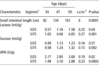
APN = aminopeptidase N.
The mink kits were weaned at 42 days of age.
1SI25 and SI75: samples taken at 25% and 75% of the small intestinal length.
2Pooled s.e.m.
Gut morphology
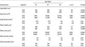
The crypt depth was strongly influenced by the age of the animal and it increased both in the proximal and distal small intestine during the experimental period (P = 0.0001; Table 3). Parallel to the increased crypt depth, the crypt area increased from 7831 μm2 in 34-day-old mink kits to 22 012 μm2 in 59-day-old mink kits in the proximal small intestine (P = 0.0001). In the distal small intestine, the crypt area increased from 7989 μm2 to 18 539 μm2 during the same period (P = 0.0002). The crypt density, the number of crypts per millimetre, was equal at all three sampling times in both the proximal and the distal small intestine.
Data describing the gut wall architecture at two positions in the small intestine of mink kits at 34, 47 and 59 days of age (n = 8)

The mink kits were weaned at 42 days of age.
1SI25 and SI75: samples taken at 25% and 75% of the small intestinal length.
2Pooled s.e.m.
The villous height increased (P < 0.0001) in the proximal small intestine of mink kits; it was 388, 525 and 628 μm at 34, 47 and 59 days of age, respectively. In contrast to this, the villous height was decreased in 47- and 59-day-old mink kits when compared to 34-day-old mink kits in the distal small intestine (P = 0.0006), whereas no difference was observed between the 47- and 59-day-old mink kits. The villous area followed the same trends as the villous height; increasing the area in the proximal small intestine and decreasing the area in the distal small intestine during the experimental period. The number of villi per millimetre was not affected by the age of the mink kits.
The thickness of the T. muscularis was 159 ± 29 μm in the proximal and 136 ± 19 μm in the distal small intestine of 34-day-old mink kits. At 59 days of age, it was 161 ± 29 μm and 154 ± 19 μm in the proximal and distal small intestine, respectively, and hence it did not change with the age of the animals.
Mucin staining characteristics
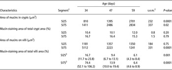
The area of neutral mucins in the crypts increased both in the proximal (P = 0.0001) and distal small intestine (P = 0.02) (Table 4). In the proximal small intestine, the increase was observed from 47 to 59 days of age and in the distal small intestine the area increased from 34 to 47 days of age but remained constant from 47 to 59 days of age (Table 4).
Mucin-staining area on villi and in crypts and Mucin-staining area of total villi and crypt area at two positions of the small intestine in mink kits at 34, 47 and 59 days of age (n = 8)

The mink kits were weaned at 42 days of age.
1SI25 and SI75: samples taken at 25% and 75% of the small intestinal length.
2Pooled s.e.m.
3Values are back-transformed least square means (lower and upper limits of the 95% CI).
When taking into account the differences in crypt size, the mucin-staining area of the total crypt area did not alter during the period that was examined. In the proximal small intestine, the mucin-staining area of the total crypt area was 10.1% to 12.0% and in the distal small intestine it was 15.3% to 16.7%.
The mucin-staining area on the villi in the proximal small intestine did not differ between mink kits at 34, 47 or 59 days of age. In the distal small intestine, the mucin-staining area was high, 4515 μm2, at 34 days of age after which it declined to 2223 μm2 and 1241 μm2 at 47 and 59 days of age, respectively (P = 0.0001).
The mucin-staining area of the total villi area decreased from 16.7% at 34 days of age to 6.1% at 59 days of age in the proximal small intestine (P = 0.001). In the distal small intestine, a very pronounced decrease in the mucin-staining area of total villi area was observed. Of the villi area 74.4% was covered with mucin at 34 days of age; this decreased to 13.9% and 6.4% at 47 and 59 days of age, respectively (P = 0.0001).
Total α-tocopherol and α-tocopherol stereoisomers in feed, plasma and liver
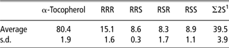
The content of α-tocopherol in the feed was 80.4 mg/kg feed (Table 5). The amount of the 2S-isomer (∼50%) indicates that the α-tocopherol in the diet is added, synthetic all-rac-α-tocopherol.
α-Tocopherol in feed, total and content of the individual stereoisomers in mg/kg feed

Average of two samples (9 June and 4 July).
1Sum of SSS-, SSR-, SRS- and SRR-α-tocopherol.
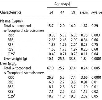
At 34 days of age, the total α-tocopherol content of the plasma of mink kits was 15 μg/ml and the natural isomer, RRR-α-tocopherol, made up 60% of the total α-tocopherol content (Table 6). The total content of α-tocopherol was unaltered at 47 and 59 days of age, but the content of RRR-α-tocopherol decreased (P = 0.003) from day 34 to day 47 and made up only 44% of the total content during this period. The proportion of the almost inactive synthetic isomers with 2S configuration was doubled from 0.40 μg/ml plasma to 0.78 μg/ml during the experimental period, whereas the proportion of the other synthetic isomers was unaltered from 34 to 59 days of age.
Content of α-tocopherol and absolute content of α-tocopherol stereoisomers in liver and plasma from mink kits at 34, 47 and 59 days of age (n = 8)

The mink kits were weaned at 42 days of age.
1Σ2S represents the sum of SSS-, SSR-, SRS- and SRR-α-tocopherol.
The total α-tocopherol content of the liver decreased significantly from 67 μg/g at day 34 to 25 μg/g at day 47 and an intermediate concentration of 37 μg/g at day 59. In particular, the concentration of RRR-α-tocopherol decreased from 26.3 to 5.5 μg/g. From day 47 to day 59, there was a significant increase in the 2S stereoisomers of α-tocopherol from 12 μg/g at day 47 to 19 μg/g at day 59, which corresponded to the concentration at day 34.
Concentration of bile salts in bile
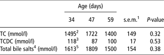
The bile salts in bile consisted exclusively of taurine-conjugated bile salts with taurocholate making up 94% of total bile acids and taurochenodeoxycholate making up the remaining 6% (Table 7). The concentration of bile salts did not change during the experimental period.
Biliary concentration of bile salts in mink kits at 34, 47 and 59 days of age (n = 8)

TC = taurocholate; TCDC = taurochenodeoxycholate.
The mink kits were weaned at 42 days of age.
1Pooled s.e.m.
2n = 5, s.e.m. = 188.
3n = 5, s.e.m. = 21.
4Sum of TC and TCDC.
5n = 5, s.e.m. = 194.
Discussion
Mink kits are weaned at 6 to 7 weeks of age. The kits are completely dependent on mother’s milk for nourishment during the first 24 to 25 days of life; from then the consumption of solid feed increased at the expense of milk. Hence, the transition from mother’s milk to solid feed is gradual and the mink kits are eating an appreciable amount of solid feed at weaning. This study showed that the relative weight of the pancreas was unaltered during the period around weaning. This is in agreement with previous results by Elnif et al. (Reference Elnif, Hansen, Mortensen and Sørensen1988) but in contrast to what has been observed in pigs in which the pancreas weight in relation to BW increases considerably at weaning (Cranwell, Reference Cranwell1995). A larger digestive system is needed to satisfactorily digest and absorb the less digestible post-weaning diet. The difference between the species reflects that in mink the dietary change is gradual in opposition to the abrupt change of diet in modern pig production.
The amylase activity in pancreatic tissue increased during the experimental period. This is in opposition to previous results that showed a decreasing amylase activity in mink kits during the same period (Elnif et al., Reference Elnif, Hansen, Mortensen and Sørensen1988), but in agreement with studies in kittens showing increasing amylase activity during the first month of life (Harper and Turner, Reference Harper and Turner2000). The production of pancreatic enzymes is considered to be induced by their substrates (Brannon, Reference Brannon1990) and hence increased activity of amylase in response to increased intake of starch is expected in mink kits during the transition from mother’s milk to solid feed. The amylase activity observed in 59-day-old mink kits is comparable to the activity observed in adult mink (Lærke et al., Reference Lærke, Hedemann and Hejlesen2004 and Reference Lærke, Hedemann and Hejlesen2006) and hence the capacity to digest starch seems to be fully developed at approximately 8 weeks of age.
In agreement with previous studies (Sangild and Elnif, Reference Sangild and Elnif1996), developmental changes in the activity of mucosal oligosaccharidases occurred during the period from 34 to 47 days of age, when the mink kits consumed decreasing amounts of milk and increasing amounts of solid food. The increasing lactase activity in the proximal small intestine is in opposition to what has been previously reported in rats (Motohashi et al., Reference Motohashi, Fukushima, Kondo and Sakuma1997) and pigs (Hampson, Reference Hampson1986). Sangild and Elnif (Reference Sangild and Elnif1996) reported that lactase activity in the small intestine of mink kits declined as observed for the distal small intestine in this study, however, their result, being an average for the whole small intestine, may cover up regional differences in lactase activity. The increasing activity of sucrase is in accordance with previous studies in mink kits and puppies (Buddington et al., Reference Buddington, Elnif, Malo and Donahoo2003) and corresponds to the increasing intake of starch and dietary disaccharides.
The proteolytic capacity in terms of trypsin, chymotrypsin and APN activity, increased in this study in the mink kits from 34 to 59 days of age. This is in line with the results obtained by Elnif et al. (Reference Elnif, Hansen, Mortensen and Sørensen1988). The digestibility of protein is low in mink kits (Skrede, Reference Skrede1978), but was found to stabilize around 19 weeks of age suggesting that the proteolytic capacity is limiting in mink kits. Elnif et al. (Reference Elnif, Hansen, Mortensen and Sørensen1988) found that maximum proteolytic capacity was obtained after an age of 3 months and we have observed in previous studies (Lærke et al., Reference Lærke, Hedemann and Hejlesen2004 and Reference Lærke, Hedemann and Hejlesen2006) that the trypsin and chymotrypsin activity in pancreatic tissue was 2 to 3 times higher in adult male mink than in 59-day-old mink kits.
There are, to our knowledge, no reports of pancreatic lipase and CEH development in mink. This study showed that lipase activity in relation to metabolic BW was increased six times from 34 to 59 days of age. However, lipase activity in 59-day-old mink kits is low compared to lipase activity in adult male mink, which was found to be 1800 to 2900 U/g0.75 (Lærke et al., Reference Lærke, Hedemann and Hejlesen2004 and Reference Lærke, Hedemann and Hejlesen2006). This corresponds to an increase of 20 to 35 times, but it is unknown at what time during the growth period the increase in lipase activity occurs. Studies in cats have shown that the digestibility of fat is low at weaning and that the digestibility of fat stabilizes at 24 weeks of age (Harper and Turner, Reference Harper and Turner2000). In puppies, lipase activity was not detected in pancreatic tissue until 9 weeks of age (Buddington et al., Reference Buddington, Elnif, Malo and Donahoo2003); at this time the puppies shifted from a dependency on milk lipase to pancreatic lipase.
The activity of CEH in pancreatic tissue of mink kits is comparable to that observed in adult male mink (Lærke et al., Reference Lærke, Hedemann and Hejlesen2004 and Reference Lærke, Hedemann and Hejlesen2006). CEH is identical to bile salt-stimulated lipase (Hernell and Bläckberg, Reference Hernell and Bläckberg1994), an enzyme that has been found in mother’s milk in several carnivorous species, for example, ferret, dog and cat. Whether bile salt-stimulated lipase is also present in mink milk is unknown, but the low activity of the enzyme in the exocrine pancreas of mink kits suggests that the enzyme is present in milk to compensate for low pancreatic lipase activity.
The decreasing concentration of RRR-α-tocopherol in plasma and liver is in agreement with previous studies in piglets (Lauridsen and Jensen, Reference Lauridsen and Jensen2005). RRR-α-tocopherol is the stereoisomer with the highest biological activity (Jensen and Lauridsen, Reference Jensen and Lauridsen2007) and a decrease in the concentration of this stereoisomer indicates that there is consumption of RRR-α-tocopherol. In mink (Jensen et al., Reference Jensen, Hansen and Clausen2004) as well as in piglets (Lauridsen and Jensen, Reference Lauridsen and Jensen2005), the intake of α-tocopherol is several folds higher with the weaner diet compared with the intake via milk, but despite this a significant drop in α-tocopherol status is seen in both species. In piglets, this phenomenon has been shown to correlate to a corresponding drop in CEH activity (Hedemann and Jensen, Reference Hedemann and Jensen1999), which together with bile acid is responsible for the intestinal hydrolysis of α-tocopheryl acetate before absorption. As previously stated, α-tocopheryl acetate provided with the weaner diet must be hydrolysed by CEH in order to be absorbable (Jensen et al., Reference Jensen, Engberg and Hedemann1999). The major part of α-tocopherol in milk from sows and mink is RRR-α-tocopherol and only very small percentage is 2S-α-tocopherol (Jensen and Lauridsen, Reference Jensen and Lauridsen2007). In this experiment, the major decrease in α-tocopherol concentration in the liver was observed from day 34 to 47, corresponding to the pre-weaning period, where the mink kits still suck their mother, but steadily increase the voluntary intake of the weaner diet. During this period all stereoisomers of 2R-α-tocopherol decreased significantly in the liver and RRR-α-tocopherol decreased in plasma (Table 6). However, the significant increase of 2S-α-tocopherol indicates that some α-tocopheryl acetate has been hydrolysed and made available for absorption. The increase in concentration of CEH during the weaning period to a concentration similar to adult mink (Lærke et al., Reference Lærke, Hedemann and Hejlesen2004) indicates that CEH is not the limiting factor for the absorption of α-tocopherol in weaning mink kits. This is further supported by the high and unaltered occurrence of conjugated bile acids, which has been shown to be the most efficient activator of CEH (Lauridsen et al., Reference Lauridsen, Hedemann and Jensen2001). In contrast, lipase activity in the weaning mink kits is 20 to 35 times lower than in adult mink (Lærke et al., Reference Lærke, Hedemann and Hejlesen2004). This low concentration of lipase may interfere with lipid absorption in general and thus indirectly with tocopherol absorption. However, in order to elucidate this connection, further studies of lipid absorption in weaning mink kits are necessary.
This study is, to our knowledge, the first to describe the morphological changes taking place in the small intestine of mink kits during the weaning period. The crypt depth is doubled in both the proximal and distal small intestine. This is probably an indication of high proliferative activity in the crypts reflecting the rapid growth of the small intestine during this period (Elnif and Sangild, Reference Elnif and Sangild1996). The development of the villous height displayed a dual pattern: the height increased in the proximal small intestine and decreased in the distal small intestine. At weaning in pigs, villous atrophy is generally observed and it has been associated with a usually low feed intake during the post-weaning period (Pluske et al., Reference Pluske, Hampson and Williams1997). In adult mink, it was observed that villous height was reduced both proximally and distally during 10 days of fasting (Bjørnvad et al., Reference Bjørnvad, Elnif and Sangild2004), suggesting that the changes observed in this study cannot be attributed to low feed intake during the weaning period. The changes are more likely reflecting the postnatal development of the gastrointestinal tract, resulting in longer villi in the proximal than the distal part of the small intestine, determined by genetic and endocrine factors (Sangild and Elnif, Reference Sangild and Elnif1996).
The knowledge on the postnatal development and the effect of weaning on the protective mucus gel covering the gastrointestinal tract is scarce. In this experiment, the proportion of the crypts covered with mucins remained constant during the experimental period, but on the villi a very pronounced decrease in the mucin-covered area was observed. Such changes may imply that the thickness of the mucus layer decreased (Hedemann et al., Reference Hedemann, Theil and Bach Knudsen2009). There are no previous reports on the development of the mucus layer around weaning in mink. In pigs, conflicting results have been reported. Bruininx et al. (Reference Bruininx, Schellingerhout, Lensen, Peet-Schwering, Schrama, Everts, den Hartog and Beynen2002) observed a decrease in the number of goblet cells, whereas Brown et al. (Reference Brown, Miller, Stokes, Blazquez and Bourne1988) reported that the number of goblet cells increased after weaning. Without a well-formed mucus gel layer, the underlying mucosa is more susceptible to attack by bacteria such as Escherichia coli (Specian and Oliver, Reference Specian and Oliver1991). The suckling mink rely on immunoglobulins from the mother’s milk and the innate defence of mucus as the acquired immune system is not fully functional in the intestine (Cebra, Reference Cebra1999). However, as the mucin-staining area in the gastrointestinal tract decreases, the mink kits may be more susceptible to infections during the period of weaning.
This study shows that considerable changes take place in the gastrointestinal tract of mink kits during the weaning period. The enzymatic capacity is not reduced during the weaning period, but the immune status of the mink kit may be impaired during the post-weaning period due to the lower concentration of RRR-α-tocopherol in plasma and liver; and as the mucin-staining area in the gastrointestinal tract decreases, the mink kits may be more susceptible to infections during this period.
Acknowledgements
This study was supported by the Danish Fur Breeders Research Center. The authors thank Kathrine Hansen Høirup, Elsebeth Lyng Pedersen, Mette Würtz Reh, Lisbeth Märcher and Marie Lilleris Nielsen for excellent technical assistance.