Introduction
Although typically rare, tellurium (Te) oxysalt minerals often have unique structures, many of which have neither been determined nor synthesised (Christy et al., Reference Christy, Mills and Kampf2016a). The rate of discovery of Te oxysalts has burgeoned in the last decade, aided greatly by the discovery of new Te mineral localities, including at Otto Mountain and the Blue Bell mine in the Mojave desert, near Baker, California (Marty et al., Reference Marty, Kampf, Housley, Mills and Weiß2010; Mills et al., Reference Mills, Kampf, Christy, Housley, Rossman, Reynolds and Marty2014; Christy et al., Reference Christy, Mills, Kampf, Housley, Thorne and Marty2016b) and the Masonic Mountain district mines in northern California (Kampf et al., Reference Kampf, Housley, Rossman, Marty and Chorazewicz2018). Tellurium oxysalt compounds are continuing to grow as an area of interest, in part due to the rich structural variation of Te bonding (e.g. Mills and Christy, Reference Mills and Christy2013). Tellurites (Te4+ oxysalts), especially synthetic analogues, have been investigated more thoroughly due to the asymmetrically bonded Te4+ cation (Christy and Mills, Reference Christy and Mills2013; Brugger et al., Reference Brugger, Liu, Etschmann, Mei, Sherman and Testemale2016); the inclusion of which allows for greater structural versatility than the more regularly coordinated Te6+, including the possibility of generating non-linear optical properties (Weil et al., Reference Weil, Shirkhanlou and Stürzer2018).
Mixed-valence Te oxysalts are uncommon as they require very specific conditions in which the tellurite and tellurate anions are stable enough to crystallise together. At surface conditions (25°C, ambient pressure), the border of stability between Te4+ and Te6+ occurs for log  $f_{O_2}$ values between –10 and 0 across a pH range of 0 to 14 (McPhail, Reference McPhail1995; Grundler et al., Reference Grundler, Brugger, Etschmann, Helm, Liu, Spry, Tian, Testemale and Pring2013). Tlapallite is a rare secondary Te mineral found at only a few North American localities and is one of many Te oxysalt minerals first recognised from the Moctezuma, Bambolla, Bambollita and Oriental mines of Sonora, Mexico (Williams and Duggan, Reference Williams and Duggan1978). Pb-rich tlapallite was discovered concurrently at Tombstone, Arizona. Since then, tlapallite has also been identified at other mines, including Otto Mountain, California and the North Star mine, Utah, and is thus more widespread than many other Te oxysalts. Tlapallite commonly occurs in association with carlfriesite (Williams and Gaines, Reference Williams and Gaines1975) – the only other naturally occurring mixed-valence Te oxysalt with a known crystal structure (Effenberger et al., Reference Effenberger, Zemann and Mayer1978). Tlapallite, occurring as powdery, paint-like coatings, was first described without a crystal structure (Williams and Duggan, Reference Williams and Duggan1978). The end-member formula was reported as H6Ca2Cu3(TeO3)4(TeO6)(SO4), or more generally H6(Ca,Pb)2(Cu,Zn)3(TeO3)4(TeO6)(SO4). The Te oxidation states and ratio of Te4+:Te6+ were determined by wet chemical methods (Williams and Duggan, Reference Williams and Duggan1978).
Carlfriesite was first identified from the Bambollita mine, Moctezuma (Williams and Gaines, Reference Williams and Gaines1975). First defined as containing only Te4+, the mineral was successfully synthesised and discovered to contain Te in both oxidation states (Effenberger et al., Reference Effenberger, Zemann and Mayer1978). It was amongst the first handful of compounds to have this feature, following the synthesis of Te4+Te6+O5 five years earlier (Lindqvist and Moret, Reference Lindqvist and Moret1973). Recently, ‘girdite’, which has since been discredited (Kampf et al., Reference Kampf, Mills and Rumsey2017) and eztlite, which was recently redefined by Missen et al. (Reference Missen, Mills, Spratt, Welch, Birch, Rumsey and Vylita2018), have been found not to be mixed-valence Te oxysalts, leaving (aside from tlapallite and carlfriesite) only three other minerals of this type. None of these three has a known crystal structure: oboyerite (Williams, Reference Williams1979), tlalocite (Williams, Reference Williams1975) and yecoraite (Williams and Cesbron, Reference Williams and Cesbron1985). Oboyerite typically forms ball-like growths, which seem to be composed of a mixture of phases based on a preliminary study incorporating some of the authors (OPM, ARK and SJM). Tlalocite and yecoraite are only known as powdery coatings, meaning that it will be difficult to determine the structures until more crystalline occurrences are found. Until these minerals have been investigated in greater detail, their status as mixed-valence Te oxysalts must be considered doubtful.
The present study is part of a broader one aimed at determining crystal structures for the ~20 secondary Te minerals without a known topology, based on Christy et al. (Reference Christy, Mills and Kampf2016a) and the official International Mineralogical Association (IMA) list of minerals (Pasero, Reference Pasero2018). There is a high likelihood that many of them will possess a unique structural arrangement, consistent with the majority of known secondary Te mineral structures having no known synthetic analogue at their time of discovery. For instance, the high charge of Te6+ coupled with the other cations it is likely to be associated with (e.g. Cu2+ and Pb2+) results in a diverse range of tellurates. The factors governing the variety of Te oxysalts found today are fully discussed in Christy et al. (Reference Christy, Mills and Kampf2016a).
Specimen and locality descriptions
Grains of cotype tlapallite from the Muséum Nationale d'Histoire Naturelle (MNHN), Paris collection (MNHN_MIN_180.92, henceforth referred to as 180.92) were first examined. This material originates from Bambollita Mine (Oriental Mine), Moctezuma, Sonora, Mexico; the sample was part of a suite of 35 samples originally described by Sidney Williams and bought by the MNHN after the IMA congress in Orléans (France) in 1980. Due to the powdery nature of cotype tlapallite, the crystal structure could not be determined. Powder X-ray diffraction (PXRD), Raman analysis and electron probe microanalysis (EPMA) were used to verify its identity. A high-quality Raman spectrum was recorded and was subsequently entered into the Caltech Raman database (see details below). This was then used for comparison with material from several Te-rich localities.
In the course of this study, tlapallite specimens have been identified from four localities (Bambollita mine, Moctezuma, Sonora, Mexico; Otto Mountain, California, USA; North Star mine, Utah, USA; and Wildcat prospect, Utah, USA), confirming the distinctive identity of what until now has been a somewhat doubtful mineral species. A tlapallite-like arsenate was also found from the Masonic district, California, USA, but it cannot be described at this time because the crystal blades are too small. This article focuses mainly on a cotype tlapallite specimen (180.92) and new material from the Wildcat prospect.
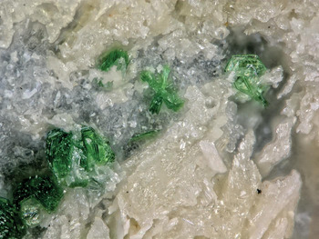
The specimen of tlapallite from which the single crystal was isolated for X-ray diffraction analysis was collected from the Wildcat prospect, in the Drum Mountains, Utah, USA, on 8 September 2018 by four of the authors (OPM, SJM, MFC and JM). The green plates were recognised in a vugh after a larger rock was cracked into two pieces (see Fig. 1). The piece of the specimen used for single-crystal X-ray diffraction is registered in the collections of Natural History Museum of Los Angeles County (catalogue number 67231), while the other half is registered in the collections of Museums Victoria (M54548). The crystals (if present) are often overgrown and intergrown, making single-crystal studies very challenging. The tlapallite crystals on these specimens are the best that have ever been reported for the species. Tlapallite occurs as several clusters of emerald green plates, nestled in cavities amongst quartz and carlfriesite. Other associated minerals include calcite, khinite, hedyphane and eckhardite (previously only identified at Otto Mountain, California; Kampf et al., Reference Kampf, Mills, Housley, Rossman, Marty and Thorne2013b). The plates of tlapallite reach 150 μm in diameter. A strong cleavage is observed parallel to the plates, indicative of the layered nature of the tlapallite crystal structure.
Pale emerald-green crystalline aggregates of tlapallite from Wildcat Prospect on specimen number 67231, closely associated with carlfriesite (yellow) and quartz. Field of view = 0.84 mm.

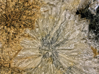
The carlfriesite crystal used in the new structure refinement was removed from an 8 cm × 7 cm × 3 cm fragment of dark grey jasperoid matrix. A radial spray of crystals on this carlfriesite specimen is shown in Fig. 2. The specimen was collected by one of the authors (MFC) at the Wildcat prospect in 2014 and it is now deposited in collections of the Natural History Museum of Los Angeles County (catalogue number 67227).
Pale yellow transparent crystals of carlfriesite from Wildcat Prospect on specimen number 67227. Field of view = 3.5 mm.

Chemistry
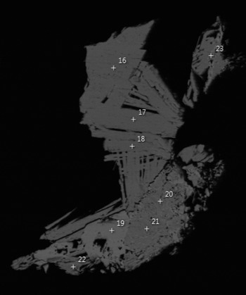
Quantitative chemical analyses of cotype tlapallite were performed on a Cameca SX100 Electron Microprobe (wavelength-dispersive mode, 12 kV, 10 nA, 5 μm beam diameter and PAP matrix correction) at the Imaging and Analysis Centre, Core Research Laboratories, Natural History Museum, London. Results are recorded to only one decimal place for Cu, Zn, Te and Pb as the low values of voltage and current used to reduce dehydration during analysis input less energy into the sample and, consequently, give lower counting statistics (and errors slightly greater than ± 0.1 wt.%) for these elements. Analyses on Wildcat tlapallite (67231) were performed at Caltech using a JEOL 8200 microprobe operating at 15 kV, 25 nA and using a 5 μm spot size. There are 4 grains consisting largely of tlapallite exposed in the polished surface of the section and probe analyses were obtained from 2 of them. A back-scattered electron image of one of these grains along with EDS analysis spots is shown in Fig. 3. The EDS showed the tlapallite blades to be homogeneous in composition.
Back-scattered electron image of one of the grains used in the Caltech microprobe analyses of tlapallite from Wildcat Prospect. Field of view is 100 μm. EDS analyses were recorded at the numbered spots. Spots 16, 17, 18, and 22 are tlapallite blades. Spot 19 is carlfriesite. Spots 20, 21, and 23 are fine-grained mixtures of the two.

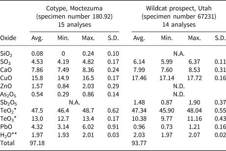
No beam damage was observed during the microprobe analyses. The standards used for cotype tlapallite were: synthetic fayalite (Si), baryte (S), wollastonite (Ca), Cu2O (Cu), sphalerite (Zn), gallium arsenide (As), TeO2 (Te) and vanadinite (Pb); while standards for specimen 67231 were: galena (Pb), anorthite (Ca), Cu metal (Cu), Te metal (Te), antimony metal (Sb), sodalite (Cl) and anhydrite (S). There was insufficient tlapallite for CHN analyses; therefore, H2O was calculated based on 6 H atoms per 41 O atoms in the formula unit, as determined by the crystal structure analysis (see below), with the rest of the cation content renormalised to obtain electroneutrality. The total average weight percent of tlapallite after including H2O remained slightly low, at 97.18 wt.% for the cotype and 93.77 wt.% for specimen 67231, most likely due to the thinness of the tlapallite plates. Analytical results are given in Table 1. These major-element analyses concur with the original data presented by Williams and Duggan (Reference Williams and Duggan1978).
The average composition of tlapallite determined by electron probe micro-analysis and structure determination.

* Te total split in a 4:1 ratio of TeO2:TeO3 for cotype tlapallite, calculated from the crystal structure, while the 4:1 split for Wildcat prospect takes Sb2O5 into account
** Calculated based on the crystal structure (using 41 total O atoms per formula unit as a reference)
N.A. – Not analysed; N.D. – Not determined; S.D. – standard deviation.
The analyses show that tlapallite contains essential Ca, Cu, S and Te (both Te4+ and Te6+, as determined by the crystal structure) and that the stoichiometric ratios of these major elements concur with the original data presented by Williams and Duggan (Reference Williams and Duggan1978). Pb substitutes for Ca and Zn (only present in cotype tlapallite) substitutes for Cu. Substitutions of Si and As for S were also detected for cotype tlapallite. A substitution of Sb, assumed to be Sb5+ substituting for Te6+ (as in joëlbruggerite, Mills et al., Reference Mills, Kolitsch, Miyawaki, Groat and Poirier2009b), was detected for specimen 67231. The empirical formula for cotype tlapallite (180.92) is Ca3.86Pb0.53Cu5.48Zn0.53Te4+8.19Te6+2.05S1.56As0.13Si0.04H6.00O41.00, which can be expressed as (Ca3.86Pb0.53)Σ4.39(Cu5.48Zn0.53)Σ6.01Te4+8.19Te6+2.05(S1.56As5+0.13Si0.04)Σ1.73H6.00O41.00, while for specimen 67231, the empirical formula is Ca3.82Pb0.12Cu6.56Te4+7.97Te6+1.59Sb5+0.25S2.06H6.00O41.00, which can be expressed as (Ca3.82Pb0.12)Σ3.94Cu6.56Te4+7.97(Te6+1.59Sb5+0.25)Σ1.84S2.06H6.00O41.00. The ideal end-member formula, based on the H2O content inferred from crystal structure determination is Ca4Cu6Te4+8Te6+2O30(SO4)2·3H2O. This requires CaO 8.82, CuO 18.76, TeO2 50.19, TeO3 13.81, SO3 6.29 and H2O 2.12, total 100 wt.%.
Raman spectroscopy
Raman spectra were recorded from a fine-grained crystalline aggregate of cotype tlapallite (180.92) using a Renishaw inVia spectrometer (Monash University) with an argon ion laser excitation operating at 514 nm. The spot was ~1 μm in diameter, with a 20 cm–1/s scan rate and ~1 mW at the sample when using 10% laser power. This high-quality spectrum was used for comparison purposes.
At Caltech, a Renishaw M-1000 spectrometer with a 514 nm solid-state laser was used in most of the Te-oxysalt scanning. Most scanning was done at 10% power and 20× magnification resulting in a 5 μm spot size. A polarisation scrambler was used to minimise polarisation effects and the energy scale was calibrated with silicon at 520.5 cm–1. Spectra from the microprobe polished section of tlapallite were obtained on a Renishaw inVia spectrometer also using a 514 nm solid-state laser and a polarisation scrambler. This spectrometer was also calibrated with a silicon standard and was equipped with a low-wavenumber filter that allowed spectra to be recorded down to 10 cm–1.
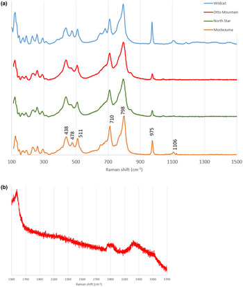
Spectra from the fine-grained Moctezuma cotype sample, the Otto Mountain fine-grained sample, and the North Star fine-grained sample are almost identical. We have recorded spectra from many different crystals in the Wildcat microprobe sample and, as might be expected when scanning different crystal orientations, they show variation in relative peak heights as well as lesser variations in the positions of some peaks; however, all are recognisable as tlapallite. The four spectra are shown between 100 and 1500 cm–1 in Fig. 4a. The lines at 710 and 798 cm–1 are in the ranges typically expected for tellurate and tellurite, while the lines at 975 cm–1 and 1106 cm–1 are in the range typically expected for sulfate. inVia data on the microprobe sample show that significant lines also occur at 58 and 91 cm–1 and that no other lines are present down to 10 cm–1. The broad, fairly weak feature at ~1600 cm–1 is at twice the shift of the strongest Raman line, suggesting that it could be due to a second-order Raman process. It is also in the range commonly observed for water bending but appears to be stronger than expected for that.
(a) Raman spectra of tlapallite from the four analysed localities, with key lines identified for the Moctezuma specimen. All units in cm–1. (b) Raman spectrum of Otto Mountain tlapallite between 1500 and 3700 cm–1.

There are also two features beyond 2800 cm–1. The very broad water-stretching feature between 3220 and 3540 cm–1 has the same shape in the randomly oriented Moctezuma and Otto Mountain (Fig. 4b) samples. This feature can be reasonably interpreted as moderately strongly hydrogen-bonded water. The feature between 2860 and 2950 cm–1 can be reasonably described as a strongly hydrogen-bonded water molecule; the value of the D…A distance calculated using the equation of Libowitzky (Reference Libowitzky1999) is 2.630 Å, within 2.5% of the D…A distance (2.689 Å) observed in the crystal structure (Table 5). This closeness also contributes to the overbonding of the OW site in the crystal structure (see below).
Crystallography
Powder diffraction
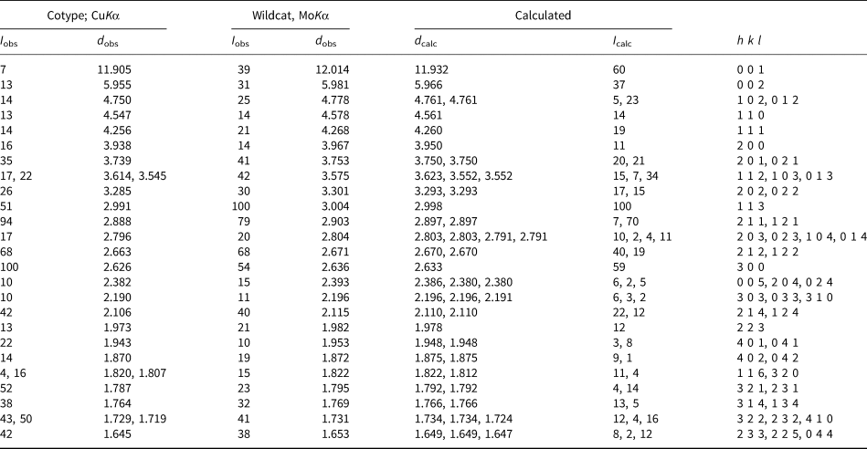
A powder aggregate of tlapallite, containing very minor quartz and calcite impurities, obtained from the cotype specimen (180.92) was mounted on a Rigaku R-Axis Rapid II curved imaging plate diffractometer (®Rigaku Oxford Diffraction) at the Natural History Museum, London and a dataset collected using CuKα radiation to 80°2θ. A Gandolfi-type randomised crystal movement was achieved by rotations on the φ and ω axes. Observed d hkl and reflection intensities of the cotype pattern were derived by profile-fitting using the Pawley method (Pawley, Reference Pawley1981) as implemented in the HighScore Plus software (Degen et al., Reference Degen, Sadki, Bron, König and Nénert2014). The PXRD for Wildcat tlapallite was recorded on a Rigaku R-Axis Rapid II diffractometer using MoKα radiation at the Natural History Museum of Los Angeles County, with conditions otherwise identical to the cotype data collection. Observed d-values and intensities were derived by profile fitting using JADE 2010 software (Materials Data, Inc, 2010). The unit-cell parameters refined for cotype tlapallite from the powder data are a = 9.0971(3) Å, c = 11.9135(8) Å and V = 853.83(7) Å3, and for Wildcat tlapallite the cell parameters are a = 9.12(2) Å, c = 11.960(1) Å and V = 861(2) Å3. Both patterns are in good agreement with the one calculated using PowderCell (Kraus and Nolze, Reference Kraus and Nolze1996) from the single-crystal structure (shown in Table 2). The powder data as indexed by Williams and Duggan (Reference Williams and Duggan1978) was incorrectly indexed as monoclinic with a = 11.97 Å, b = 9.11 Å, c = 15.66 Å, β = 90.60° and V = 1707 Å3. The unit-cell parameters of tlapallite indicate that Williams and Duggan (Reference Williams and Duggan1978) were working on the same mineral, but indexed the powder lines in a lower symmetry setting: a 1978 ≈ c 2019, b 1978 ≈ a 2019, c 1978 ≈ √3a 2019, β1978 ≈ 90° and V 1978 ≈ 2V 2019.
X-ray powder diffraction data for tlapallite to 60°2θ (Wildcat) and 80°2θ (cotype). Cutoff: averaged 10% intensities.

Key: I obs are observed intensities, d obs are observed d-values, d calc and I calc are calculated d-values and intensities.
Single crystal X-ray diffraction
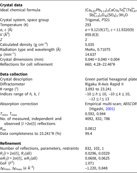
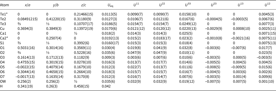
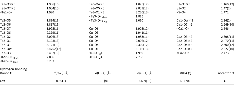
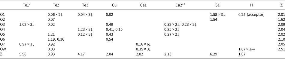
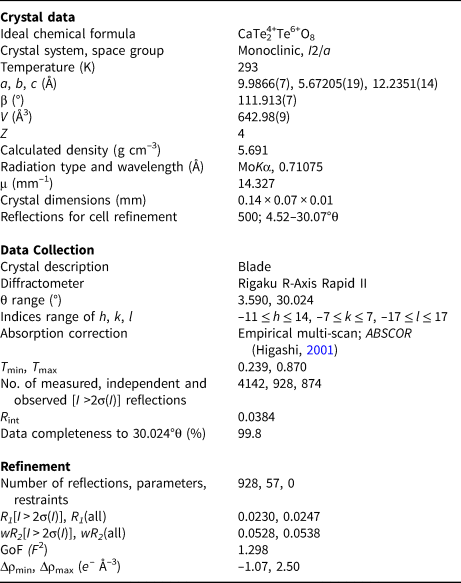
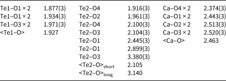
Single-crystal X-ray diffraction studies for both tlapallite and carlfriesite were carried out at the Natural History Museum of Los Angeles County using a Rigaku R-Axis Rapid II curved imaging plate microdiffractometer, with monochromatic MoKα radiation. The Rigaku CrystalClear software package was used for processing the structure data, including the application of empirical multi-scan absorption corrections using ABSCOR (Higashi, Reference Higashi2001). Initial structure models were obtained by the charge-flipping method using SHELXT (Sheldrick, Reference Sheldrick2015b). SHELXL-2016 (Sheldrick, Reference Sheldrick2015a) was used for the refinement of the structures. The Pb content of the Ca2 site was refined to 13%. Similar attempts to refine the Sb content of the Te1 site were unsuccessful, so the Sb:Te ratio was fixed based on EPMA. A difference-Fourier map revealed the location of one H site in the structure of tlapallite. The hydrogen atom position was refined with a soft restraint of 0.90(5) Å on the O–H distance with the U eq of the H set to 1.2 times that of the donor OW atom. The carlfriesite structure contains no H atoms. For tlapallite, data collection and refinement details are given in Table 3, atom coordinates and displacement parameters in Table 4, selected bond distances in Table 5 and a bond valence analysis in Table 6. For carlfriesite, data collection and refinement details are given in Table 7, atom coordinates and displacement parameters in Table 8, selected bond distances in Table 9 and a bond valence analysis in Table 10.
Summary of information relating to data collection and structure refinement of tlapallite.

Atom coordinates, site occupancies and atomic displacement parameters (Å2) for tlapallite. All full occupancies except for Te1 and Ca2.*

*Te1 is 0.8661 occupied by Te, 0.1339 occupied by Sb, fixed based on EPMA; **Ca2 is 0.871(5) occupied by Ca, 0.129(5) occupied by Pb.
Metal–oxygen bond lengths (Å) for tlapallite.

Bond valence sums (in valence units) for tlapallite.

*87% Te, 13% Sb (fixed in the crystal structure based on EPMA); **87% Ca, 13% Pb (calculated from the crystal structure).
Summary of information relating to data collection and structure refinement of carlfriesite.

Atom coordinates, site occupancies and atomic displacement parameters (Å2) for carlfriesite. All sites fully occupied.

Metal–oxygen bond lengths (Å) for carlfriesite.

Bond-valence sums (in valence units) for carlfriesite.

Crystal structure description
Tlapallite
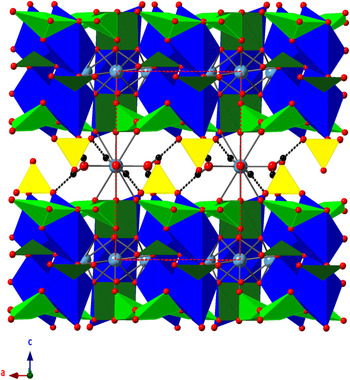
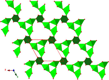
The crystal structure of tlapallite (Figs 5–8) has no known synthetic analogue, notably including the phyllotellurate anion [Te4+3Te6+O12]12– for the first time. Tlapallite forms a layered structure parallel to {001}. Te6+O6 octahedra share corners with three Te4+O4 disphenoids. The Te4+O4 disphenoid is a distorted trigonal bipyramid centred on Te4+, with one of the equatorial oxygen atoms replaced by the lone pair of Te4+. One primary Te4+–O bond in the disphenoid is considerably longer than the other three (2.379(11) compared to 1.884(11), 1.887(11) and 1.995(11) Å), a feature observed in some ‘[3 + 1]’ asymmetrical disphenoids (e.g. the Te4 site in β-Na2Te4+4O9; Lee and Ok, Reference Lee and Ok2014). Each disphenoid is corner-linked to two others in a ‘trimeric’ arrangement, building two infinite sheets in the ab plane. Between these two sheets are distorted Cu2+O6 octahedra and Te4+O3 trigonal pyramids. The Cu2+O6 square pyramids are connected to each other via edge-sharing to form Cu2+2O10 dimers. Each Cu2+O6 octahedron connects to a Te6+O6 octahedron by corner-sharing an O3 atom and connects to two Te4+O4 disphenoids via a corner-sharing O6 with one and an edge-sharing of O5 and O6 with another. The Te4+O3 trigonal pyramids link three Cu2+2O10 dimers via corner-sharing with the bridging O4 atoms of the dimers. CaO8 polyhedra (comprising three-quarters of the Ca atom sites) are also found within the layers, which they reinforce by sharing oxygen ligands with Cu and all three Te sites.
The structure of tlapallite, showing stacking of two layers in the c direction. Cu2+O6 octahedra are in dark blue, Te4+ in dark green (surrounding oxygen atoms trigonal pyramidal) or in light green (surrounding oxygen atoms disphenoidal), SO4 tetrahedra in yellow, Ca2+ cations in light blue, O atoms in red, and H atoms in black. H bonds are shown as dotted lines.

The new sheet-forming phyllotellurate anion, [Te4+3Te6+O12]12–. A portion of the infinite sheet formed by these anions is shown.

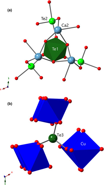
Three-connecting sites in tlapallite. (a) Each Te6+O6 octahedron is linked to three Te4+O4 disphenoids and three Ca2 cations. (b) The trigonal pyramidal Te4+O3 bridges three Cu2+2O10 dimers.

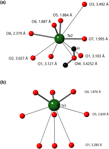
Secondary bonding environments in tlapallite. (a) For Te4+O4 with four primary and five secondary bonds (noting that one secondary bond is on the same side as the primary), and (b) for Te4+O3 with three primary and six secondary bonds. Primary bonds are shown as thicker bonds with secondary as thin bonds.

The Cu site in tlapallite displays a rather extreme example of Jahn-Teller type distortion, in a [4 + 1+1] arrangement. The four square-planar Cu–O bonds have an average length of 1.959 Å, in agreement with short Cu–O bonds having an average length ≈1.97 Å (Eby and Hawthorne, Reference Eby and Hawthorne1993). The Cu–O bond to the near apex (O4) of the Cu2+O6 octahedron within the main layer is relatively short at 2.360(12) Å, and it is this edge along which the Cu2+O6 octahedra share faces. Cu also displays an extremely weak Cu–O bond to the other octahedral apex at 3.116(13) Å, which extends into the interlayer, as this O1 site is one of the four oxygen atoms strongly bonded to S in the SO4 group. In fact, this O1 site is further from Cu than the nearest Te atom, the Te3 site of the Te4+O3 trigonal pyramids, at a distance of 3.000(3) Å. The average long Cu–O bond length is 2.738 Å, longer than the expected ~2.44 Å of Eby and Hawthorne (Reference Eby and Hawthorne1993), but within the upper bounds of Jahn-Teller distortion, in which the longest apical bonds may reach ~3.15 Å (Eby and Hawthorne, Reference Eby and Hawthorne1993).
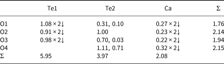
The coordination environments around Te4+ cations in tlapallite display two different one-sided geometries. The coordination of the Te3 site has typical one-sided trigonal pyramidal geometry, with three short, strong Te4+–O4 primary bonds at 1.876(12) Å, in good agreement with the 1.911 ± 0.077 Å expected for the three short Te4+–O trigonal pyramidal bonds (Mills and Christy, Reference Mills and Christy2013). Te3 forms six longer secondary bonds to more distant oxygen atoms, on the same side of the cation as the lone pair – which for this Te site does not seem to be highly stereoactive. Three of these are 2.839(12) Å in length and connect with the O5 site, which is involved in the edge-linking of Cu2+O6 octahedra and Te4+O4 disphenoids, while the other three are longer at 3.280(13) Å, extending into the interlayer to the O1 sites of the SO4 tetrahedra. The other half of the Te4+ cations (the Te2 site) are in disphenoidal fourfold coordination as described above. The Te2 site also possesses five longer secondary bonds, ranging in length from 3.027(13) to 3.492(10) Å, linking the O3 site (shared by the trigonal pyramidal Te3 site and octahedral Cu2+), the OW site and some of the SO4 oxygen atoms. The lone pair of the Te2 site is more stereoactive than that of Te3 and is oriented into the interlayer channels. There is an average of 5.5 secondary bonds for the two Te4+ cations, in the expected range for Te4+ secondary bonds (Christy and Mills, Reference Christy and Mills2013). The Te6+ site (Te1) displays typical octahedral coordination, with three Te6+–O bonds at 1.906(10) Å and another three at 1.934(10) Å, with an average length of 1.920 Å, in excellent agreement with the 1.923 ± 0.041 Å expected for Te6+–O bonds (Mills and Christy, Reference Mills and Christy2013).
Ca(H2O)3O6 polyhedra (comprising the remaining quarter of the Ca sites), SO4 tetrahedra and H2O groups are found between the layers, which also incorporate several long Te4+–O secondary bonds from both Te4+ sites. Oxygen atoms in SO4 tetrahedra also form the long vertex of the Cu2+O6 octahedra and secondary bonds to both of the Te4+ centres within the main framework. Thus, the polytellurate sheets and other strongly-bound polyhedra CuO6, SO4 and TeO3 form a double-layered structural unit {Cu6[Te4+3Te6+O12]2(Te4+O3)2(SO4)2}8–. The long Ca1–O bonds, in particular, are involved in linking the layers together along [001], although this interlayer contains weaker connecting bonds than the rest of the tlapallite structure, resulting in a strong {001} cleavage. Only one of the two H atom positions of the interlayer H2O groups is involved in a hydrogen bond. This H-bond from the OW site via the H atom is accepted by one of the O1 sites of the SO4 tetrahedra, providing further stability within the interlayer, in addition to the Te4+–O secondary bonds and linking Ca–O bonds.
The oxidation states assigned to the elements are confirmed by bond-valence analysis (Table 6), using the parameters of Gagné and Hawthorne (Reference Gagné and Hawthorne2015) for all elements except Te (Mills and Christy, Reference Mills and Christy2013), Sb (Mills et al., Reference Mills, Christy, Chen and Raudsepp2009a), Pb (Krivovichev and Brown, Reference Krivovichev and Brown2001) and H (Brown, Reference Brown2002). The OW site is notably overbonded due in part to the short O–H distance (see above).
Carlfriesite
The nanoporous [Te4+2Te6+O8]2– framework structure of carlfriesite presented here (Figs 9–10) is identical in topology to that presented by Effenberger et al. (Reference Effenberger, Zemann and Mayer1978), in which a more detailed description of the structure and synthesis of carlfriesite are presented. Eight-coordinate Ca2+ cations are found within the channels. The stoichiometric Sr-analogue of carlfriesite, SrTe4+2Te6+O8, has since been synthesised (Weil and Stöger, Reference Weil and Stöger2007), although it contains a significantly different topology in its [Te4+2Te6+O8]2– framework, leading to narrower nanoporous channels than in carlfriesite, ~4 Å in diameter compared to ~6 Å (Christy et al., Reference Christy, Mills and Kampf2016a). Both of these compounds are also enumerated and briefly (re-)described in the compendium of Te oxysalt structures compiled by Christy et al. (Reference Christy, Mills and Kampf2016a).
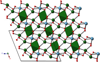
The crystal structure of carlfriesite. Te6+O6 octahedra are shown in dark green, Te4+ cations in dark green, Ca2+ cations in light blue, and O atoms in red. Note that the Ca2+ cations fill pore space in the three-dimensional framework.

Secondary bonding environments in carlfriesite. Primary bonds are shown as thicker bonds, secondary as thin bonds.

Te4+ cations in carlfriesite form an unusual Te4+O5 network of primary bonds, with four bonds having lengths between 1.916(3) Å and 2.104(3) Å, and one longer primary bond to O1 at 2.445(3) Å. Te4+ in carlfriesite also possesses two secondary bonds at 2.899(3) and 3.380(3) Å, providing extra crosslinks within the nanoporous framework. The lone pairs of the Te4+ cations occupy the 5.091 Å space in the a direction between each Te4+⋯Te4+ pair. Bond valence analysis (Table 10) for Ca (Gagné and Hawthorne, Reference Gagné and Hawthorne2015) and Te4+ and Te6+ (Mills and Christy, Reference Mills and Christy2013) concurs with the expected oxidation states for Ca, Te4+ and Te6+ in carlfriesite.
Relationships to other Te oxysalt structures
For either tlapallite or carlfriesite, there is no exact analogue in the catalogue of 703 Te oxycompounds with crystal structures compiled by Christy et al. (Reference Christy, Mills and Kampf2016a), in which 26 mixed-valence Te oxysalts are reported. One of these 26 is the original carlfriesite refinement. No previously reported mixed-valence Te oxysalt contains Te4+ that is both trigonal prismatic and disphenoidal in its primary bonding. In addition, tlapallite also contains the first reported occurrence of the phyllotellurate anion, [Te4+3Te6+O12]12–. As mentioned above, the Te environments in carlfriesite are different from those of tlapallite. Both carlfriesite and tlapallite display Te4+ dominance over Te6+, with Te4+:Te6+ ratios of 2:1 and 4:1, respectively. Both minerals also contain calcium cations, although no synthetic mixed-valence Te oxysalt contains Ca. Tlapallite is the first mixed-valence Te oxysalt to also contain SO4 (previous additional anions are limited to chloride and hydroxide in synthetic mixed-valence Te oxysalts). Tlapallite is similar to several other SO4-containing Te oxysalt minerals in that the SO4 is found between layers composed of TeOn polyhedra and MO6 octahedra, where M is an octahedrally coordinated transition metal cation. A large-radius cation, such as Ca2+ or Pb2+, also may be incorporated in the interlayer space with sulfate. Both tellurite- and tellurate-sulfate minerals are known with SO4 in interlayer channels, including poughite (Gaines, Reference Gaines1968; Kampf and Mills, Reference Kampf and Mills2011) and bairdite (Kampf et al., Reference Kampf, Mills, Housley, Rossman, Marty and Thorne2013a).
Supplementary material
To view supplementary material for this article, please visit https://doi.org/10.1180/mgm.2019.9
Acknowledgements
Equipment used in this study at Museums Victoria has been partly funded by The Ian Potter Foundation grant ‘tracking tellurium’ to SJM and a Museums Victoria 1854 Student Scholarship awarded to OPM, which we gratefully acknowledge. Equipment at the Natural History Museum of Los Angeles County was funded by the John Jago Trelawney Endowment. Support funding has also been provided to OPM by an Australian Government Research Training Program (RTP) Scholarship and a Monash Graduate Excellence Scholarship (MGES). Microprobe work on the cotype specimen was funded through Natural Environment Research Council grant NE/M010848/1 ‘Tellurium and Selenium Cycling and Supply’ to Chris J. Stanley (Natural History Museum, London).
We thank Brendan Abrahams (University of Melbourne) and Joël Brugger (Monash University) for their insightful comments. We also thank Bill Birch (Museums Victoria) for his assistance in making the mount for EPMA analysis of the cotype, Finlay Shanks (Monash University) for his assistance in acquiring the Raman data on cotype tlapallite and Jens Najorka (Natural History Museum, London) for help in acquiring the powder pattern on cotype tlapallite. The Raman spectroscopy at Caltech was done in the laboratory of George Rossman, and it and microprobe analyses at Caltech were supported by a grant from the Northern California Mineralogical Association. Three anonymous reviewers and Structures Editor Peter Leverett are thanked for their helpful comments, which improved the manuscript.