Implications
This paper is a review on the mechanisms of milk secretion. The understanding of these mechanisms is fundamental to the research on milk. Research on milk and its safety as a source of substances that are beneficial to growth and health in children and adults is a major concern in developed countries. The production of dairy products as basic nutrients is a priority in the fight against under-nourishment. Research on milk quality and milk composition is important for a sustainable dairy production.
Introduction
The mammary epithelium finely orchestrates the uptake of blood-borne molecules at its basal side (endocytosis), intracellular trafficking across cells and the release of products into milk at its apical side (exocytosis). In this way, the mammary gland has a remarkable capacity to control the concentrations of milk constituents (nutrients, growth factors, hormones, immunoglobulins, minerals and ions, etc.) as well as total secretion. To achieve their ends, cells must carry components to the correct place using specific receptors and intracellular compartmentalization. As described in a pioneering work, ‘The cell membranes are making products and undergoing transformations simultaneously. More specifically, milk components packaged in membranes are moving through and out of the cell. Thus, there is product flow and membrane flow’ (Patton, Reference Patton1976a). An important goal for the physiologists is therefore to understand the cellular mechanisms leading to optimum milk secretion.
The mammary epithelial cell (MEC) is atypical among the exocrine cells because of the huge quantity of plasma membrane, enrobing milk fat droplets, which is pinched off and expelled from the cell into milk. Thus, the mechanisms required to maintain membrane homeostasis in mammary cells differ from those governing membrane recycling in other exocrine glands (exocrine pancreas, lacrimal and parotid glands) (Meldolesi, Reference Meldolesi1974; Herzog and Farquhar, Reference Herzog and Farquhar1977), and from coupling of the fusion and recycling of vesicles in the presynaptic nerve terminal (Gundelfinger et al., Reference Gundelfinger, Kessels and Qualmann2003; Ryan, Reference Ryan2006). The highly polarized MEC represents a cell type in which the transfer of material between intracellular compartments occurs by means of membrane-bound vesicles without compromising the identity of the compartments. Numerous questions, however, remain concerning the process of membrane reconstitution in the mammary cell. Does a process of internalization of a portion of the basolateral plasma membrane correspond to an equivalent incorporation of membranes of the secretory vesicles at the apical membrane? How is the apical membrane reconstituted after release of the portion enveloping the lipid droplet?
Another unique feature of MEC functioning is the hormonal control of milk secretion. The lactogenic hormones released at each suckling or milking not only control the gene expression of milk proteins but also maintain cellular architecture and stimulate the mechanisms of MEC intracellular transport by secretagogue effects (Ollivier-Bousquet and Devinoy, Reference Ollivier-Bousquet and Devinoy2005 and references therein; Lollivier et al., Reference Lollivier, Marnet, Delpal, Rainteau, Achard, Rabot and Ollivier-Bousquet2006). In addition, some blood-borne hormones are carried to the milk across MECs (Ollivier-Bousquet, Reference Ollivier-Bousquet1993). Moreover, in the case of prolactin (PRL), its intracellular transport is required for its secretagogue effect.
How are events occurring at the basolateral side and apical side related? Are endocytosis and intracellular transport required for the triggering of different intracellular signals? Is endocytosis itself regulated by hormonal effects?
We attempt here to review some of the old and new data available on the relationship between endocytosis and exocytosis in MECs. Because these events have often been studied in mammary gland from lagomorphs and rodents, we mainly refer to these species. However, an important aim is to use the data obtained in these species to understand the functioning of the ruminant mammary gland. We will first describe some of the routes used by different blood constituents to enter cells. Some aspects of the exocytosis of milk constituents and its regulation will also be presented. Finally, the possible crosstalks between the two mechanisms will be discussed.
Endocytosis
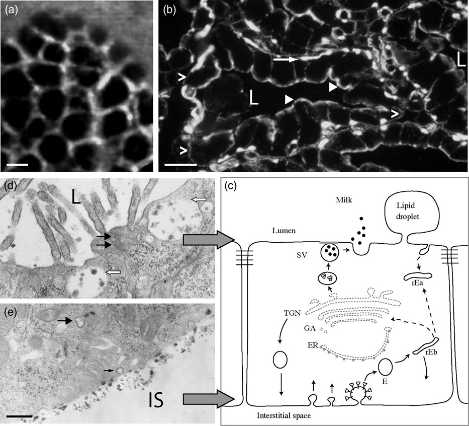
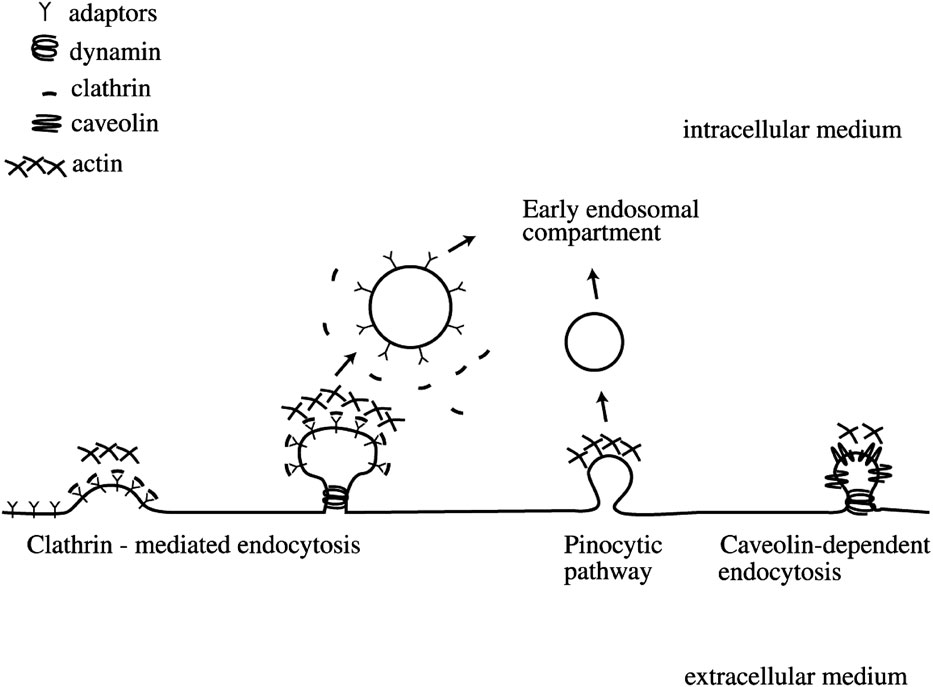
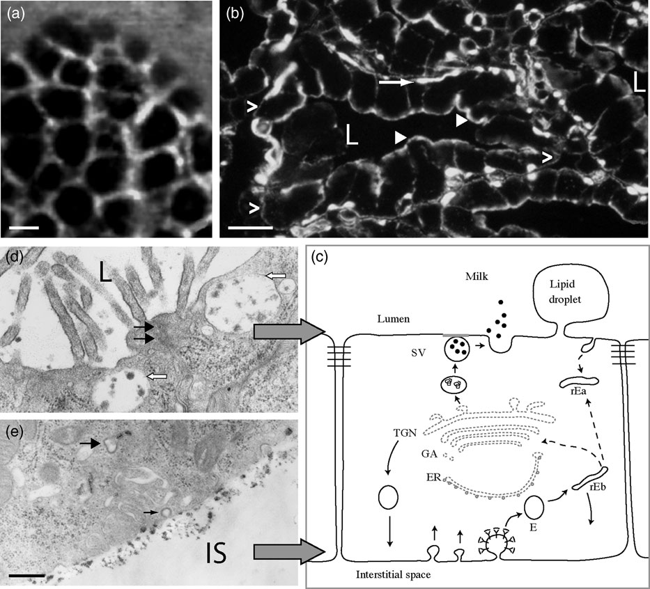
The microvasculature constituted by a basket-like capillary network surrounding the mammary acini ensures the supply of nutrients to the mammary epithelium (Djonov et al., Reference Djonov, Andres and Ziemiecki2001) (Figure 1a). After crossing the capillary endothelium, blood nutrients and other components are sequestered or diffused in the extracellular space by unknown mechanisms. They have access to the basolateral membrane of the MEC up to the tight junctions that separate the basal and apical sides of the epithelium (Pitelka and Hamamoto, Reference Pitelka and Hamamoto1983). The general molecular mechanisms underlying endocytosis are well described in different cell types (Gundelfinger et al., Reference Gundelfinger, Kessels and Qualmann2003 and references therein) (Figure 2). The basolateral membrane of MECs from lactating animals offers a large surface for the endocytosis of blood supplies. Besides convulated interdigitations of epithelial and myoepithelial surfaces, the basal membrane contains clathrin-coated pits and vesicles as well as non-coated vesicles. Caveolae are abundant in myoepithelial cells and the capillary endothelium but very rare at the basolateral membranes of MECs (Pitelka and Hamamoto, Reference Pitelka and Hamamoto1983; Hue-Beauvais et al., Reference Hue-Beauvais, Péchoux, Bouguyon, Chat, Truchet, Pauloin, Le Gouar and Ollivier-Bousquet2007; Figure 1). To understand how the mammary epithelium regulates the internalization of milk-constituent precursors, it is necessary to characterize the routes used by different blood supplies to enter the MECs.
Organization of exocytosis and endocytosis sites in the mammary epithelium. (a) The microvasculature constitutes a basket-like capillary network around the mammary acini. Capillaries were visualized with fluorescein isothiocyanate dextran injected in the vein of the tail of lactating rat and detected by Fibered Confocal Fluorescence Microscopy (image kindly provided by R. Boisgard, CEA-Saclay, France). Bar 50 μm. (b) Section of lactating rat mammary tissue. Actin was visualized with tetramethyl rhodamine isothiocyanate-phalloidin. Actin is abundant in myoepithelial cells (arrow) and appears concentrated in the vicinity of both the basolateral (empty arrowheads) and the apical plasma membrane (arrowheads) in the MECs. L = lumen. Bar 20 μm. (c) Membrane trafficking in the MECs. Endocytosis mainly occurs at the basolateral side of the MEC and materials are then transported to the recycling endosomes. At this level, internalized components can be addressed to different cellular compartments. Proteins that are synthesized by the MEC follow the classical secretory pathway and are targeted to the basolateral or the apical side of the MEC. E = endosome; rEb = recycling basal endosome; rEa = recycling apical endosome; ER = endoplasmic reticulum; GA = Golgi apparatus; TGN = trans-Golgi network; SV = secretory vesicle. (d) Electron micrograph of the apical region of a MEC from a lactating rabbit. Secretory vesicles containing casein micelles close to the apical membrane (empty arrows). Tight junction (black arrows). L = lumen. (e) Electron micrograph of the basal region of a MEC from a lactating rabbit incubated in the presence of cationized ferritin for 30 min at 4°C. Cationized ferritin accumulated close to the basal membrane and was internalized in tubulo-vesicular endosomes (arrow). A coated vesicle does not contain the marker (small arrow). IS = interstitial space. Bar 0.5 μm.
Different endocytic internalization pathways. Clathrin-mediated endocytosis is involved in receptor-mediated internalization of different hormones and biologically active molecules (transferrin, Immunoglobulin A, etc.). Pinocytic endocytosis relies on membranes microdomains and mediates the cellular entry of dextran and GPI-AP (glycosylphospatidylinositol-anchored protein). Caveolin-dependent endocytosis takes place at some membrane microdomains (caveolae) containing caveolin and promotes the internalization of the cholera toxin B subunit.

Ions
The movement of sodium (Na+), potassium (K+) and chloride (Cl−) across the basal membrane contributes to the gradients between interstitial and intracellular fluids (Linzell and Peaker, Reference Linzell and Peaker1971; Shennan and Peaker, Reference Shennan and Peaker2000). Based on histochemical localization, the presence of Na+-K+ ATPases on the basolateral membrane supports the scheme of a functional Na+-K+ pump on the blood-facing side of the mammary epithelium. A Na+-K+-2Cl− co-transport system has also been described. In mammary explants and cell cultures, a long-term effect of PRL up-regulates K+ uptake (Shennan and Peaker, Reference Shennan and Peaker2000 and references therein). Moreover, in HC11 cells, PRL exerts an early effect (after 5 and 10 min of stimulation) on Cl− transport (Selvaraj et al., Reference Selvaraj, Omi, Gibori and Rao2000) via phosphorylation of the Na+-K+-2Cl− co-transporter. Another example of a transport system localized on the basolateral membrane of mammary cells is given by the NA+/I− symporter (Cho et al., Reference Cho, Léveillé, Kao, Rousset, Parlow, Burak, Mazzaferri and Jhiang2000), the expression of which is dependent on PRL and oxytocin.
During lactation, the mammary gland extracts large quantities of calcium (Ca2+) from plasma (Neville and Peaker, Reference Neville and Peaker1979). The total calcium content of blood is about 3 mM. Milk calcium is very highly concentrated (from approximately 8 mM in humans to about 100 mM in rabbits) and is found associated with casein micelles (about 20%), free ionized or non-ionized (about 32%) or complexed to inorganic anions such as phosphate and citrate (about 46%). The mechanism for calcium entry into mammary cells may be related to the presence of calcium channels in the basolateral membrane. The intracellular compartmentalization of calcium is dependent on cytoplasmic binding proteins (VanHouten and Wysolmerski, Reference VanHouten and Wysolmerski2007).
Trace elements
Trace elements such as iron (Fe), copper (Cu) and zinc (Zn) are essential nutrients in all mammalian tissues. Fe is essential for the oxygen metabolism, mitochondrial function, normal growth and maturation of tissues. Cu is necessary for cell metabolism as a cofactor of metabolic enzymes, and Zn for both DNA and protein synthesis and cell division (Kelleher and Lönnerdal, Reference Kelleher and Lönnerdal2005; Lutsenko et al., Reference Lutsenko, LeShane and Shinde2007). Although high fetal stores of trace elements are mobilized during the early neonatal period, adequate supply of trace elements from milk is crucial to ensure neonate survival. At a given stage of lactation, trace element concentrations remain remarkably stable, independently of the mother’s diet (Lönnerdal, Reference Lönnerdal2007 and references therein). These stable levels are achieved by the mammary epithelium that tightly regulates both the uptake of elements from the blood and their release into milk.
Fe concentrations in mouse milk are approximately three times those found in serum (Zhang et al., Reference Zhang, Sawicki, Lewis, Hanson, Monks and Neville2000). Fe uptake by the mammary cell mainly occurred by the transferrin–transferrin receptor (Tf–TfR) pathway, as described in other cell types. Plasmatic diferric Tf binds to the TfR at the basal membrane of MEC and is internalized in clathrin-coated vesicles (Ollivier-Bousquet, Reference Ollivier-Bousquet1998 and references therein; Zhang et al., Reference Zhang, Sawicki, Lewis, Hanson, Monks and Neville2000). Fe is released from Tf due to the acidic environment of endosomes, Tf and TfR being recycled to the plasma membrane. Fe is transferred to the cytoplasm by an unknown transport mechanism where it is either bound to a divalent ion transporter-1 (DMT1) or to a ferroportin, or sequestered by ferritin for storage. The internalization of Fe depends on the Tf–TfR complex, whereas, the quantity of Fe internalized and secreted in milk is not correlated with TfR expression, suggesting that receptor-dependent uptake is not the only step regulating its concentration in milk (Lönnerdal, Reference Lönnerdal2007).
Cu intake is essential for the neonate and, despite high Cu stores at birth, milk constitutes an important dietary supply. Specialized transport mechanisms tightly regulate cellular Cu levels in MECs (Lönnerdal, Reference Lönnerdal2007 and references therein). A Cu transporter-1 (Ctr1) is believed to form a membrane channel by multimerization of several Ctr1 proteins. Such channels are located in both plasma and intracellular membranes and are likely involved in Cu import into the cell. The intracellular trafficking of Cu is mediated by the metallochaperone Atox1 which interacts with the two Cu-ATPases: ATP7A and ATP7B. ATP7B is localized in a perinuclear region associated with the trans-Golgi network (TGN). ATP7B promotes the transport of cytoplasmic Cu into this compartment, where it could bind to ceruloplasmin, the major copper-carrying protein. ATP7A is a cytosolic protein, which relocalized to the plasma membrane during lactation. On the other hand, PRL stimulates the transport of Ctr1 and ATP7A to the plasma membrane in HC11 cells and may also stimulate copper transport (Kelleher and Lönnerdal, Reference Kelleher and Lönnerdal2006). Moreover, human ATP7A accumulates at the basolateral side of MEC when over-expressed in the mammary gland of transgenic mice. However, the concentrations of Cu in mammary cells and milk of these animals are decreased, suggesting that the role of this transporter may not be to carry Cu to the milk but rather to remove excess Cu from mammary cells (Llanos et al., Reference Llanos, Michalczyk, Freestone, Currie, Linder, Ackland and Mercer2008).
Another example of the regulation of trace element transport from the maternal circulation to milk is that of Zn transport. In humans, 0.5–1 mg of Zn2+/day is secreted in milk. A Zn-specific transport protein (Zip3) has been characterized in the mammary gland (Kelleher and Lönnerdal, Reference Kelleher and Lönnerdal2003). Based on the localization of Zip3 in HC11 cells after in vitro differentiation and 2 h of PRL exposure, it has been suggested that PRL may regulate the relocalization of Zip3 from the intracellular perinuclear region to the basal membrane, thus facilitating Zn import into mammary cells.
Much is still unknown regarding the precise nature of internalization compartments. However, taken together, the results referred to above reveal that specific carriers for trace elements, located in the basolateral region are controlled by the hormonal status of MECs.
Glucose
Glucose is the obligate precursor for lactose synthesis in MEC. In some species it is also used to synthesize lipids and amino acids. Therefore the uptake of glucose and its transport across the basolateral membrane are important processes during milk synthesis. The glucose transporter (GLUT1) is present at the basolateral membrane of rodent MEC (Madon et al., Reference Madon, Martin, Davies, Fawcett, Flint and Baldwin1990; Nemeth et al., Reference Nemeth, Tsang, Geske and Haney2000) and is the predominant glucose transporter in the lactating bovine mammary gland (Zhao and Keating, Reference Zhao and Keating2007). The expression of genes encoding the glucose transporters is under the control of lactogenic hormones in the mammary gland during pregnancy and lactation (Shennan and Peaker, Reference Shennan and Peaker2000 and references therein; Zhao and Keating, Reference Zhao and Keating2007). In addition, the compartmentalization of GLUT1 between the cell surface and the intracellular site of lactose synthesis has been described in CIT3, a mouse mammary cell line. Following a short exposure to PRL (a few minutes to a few hours), GLUT1 was targeted to a brefeldin-A sensitive intracellular compartment via the endosomal pathway (Haney, Reference Haney2001; Riskin et al., Reference Riskin, Nannegari and Mond2008).
Thus, in addition to a long-term effect of lactogenic hormones on the expression of various transporters during mammary gland differentiation, mention should also be made of the early effects of PRL in highly differentiated mammary cells. This hormone may control the localization of numerous transporters between intracellular sites of synthesis and the basolateral membrane via vesicular transport.
Hormones and growth factors
The basolateral membrane is the binding site of plasma-borne polypeptidic hormones and growth factors to their receptors. For most polypeptidic hormones, internalization of the hormone–receptor complex leads to sorting towards the lysosomes, with recycling or degradation of the receptor and ligand. In contrast, numerous hormones and growth factors are carried from the plasma to the milk in MEC during lactation. For example, after binding to its receptor, PRL is internalized and transported in an intact and biologically active form to the milk (Ollivier-Bousquet, Reference Ollivier-Bousquet1993). The mechanisms governing the initial steps of internalization of PRL in MECs have only been partly described. The involvement of clathrin-coated vesicles in PRL–PRL receptor (PRLR) endocytosis has been shown in different cell lines transfected with PRLR. However, PRL mainly binds to non-coated domains of the basal membrane in lactating MECs. PRL is then targeted to endosomes, multivesicular bodies and to a specific pathway that meets the exocytotic pathway (Ollivier-Bousquet, Reference Ollivier-Bousquet1998 and references therein; Castino et al., Reference Castino, Delpal, Bouguyon, Demoz, Isidoro and Ollivier-Bousquet2008). The fate of PRLR is not known in MEC secreting milk. In different cell types transfected with PRLR, the ubiquitination of PRLR may lead to its degradation in the lysosomes (Varghese et al., Reference Varghese, Barrière, Carbone, Banerjee, Swaminathan, Plotnikov, Xu, Peng, Goffin, Lukacs and Fuchs2008). In MECs from lactating animals incubated with various antibodies directed against PRLR, the route followed by the complex PRLR-antibody depends on the antibody used (Ollivier-Bousquet, Reference Ollivier-Bousquet1998 and references therein).
Internalization by fluid-phase endocytosis or by association with membranes
The albumin present in milk originates from plasma and is carried to the milk by transcytosis. Whether albumin transport across the mouse mammary epithelium is receptor-mediated or a function of fluid-phase endocytosis remains unclear. In an interstitial space preparation of mouse mammary gland, albumin and IgA (Immunoglobulin A) were internalized via a clathrin-mediated endocytosis. In endosomes, albumin colocalized with IgA, whereas Tf followed a different pathway (Monks and Neville, Reference Monks and Neville2004).
The highly active membrane traffic of labeled basolateral membrane is illustrated by the localization of some membrane markers. Concanavalin A, a classical mannose-binding glycane, interacts with sugars on the cell membrane and is subsequently internalized. Cationized ferritin binds to negative charges on the cell surface in mammary fragments incubated in vitro. This marker is present on the basolateral membrane and in tubulovesicular structures in the basal region of cells as soon as 1 min (Figure 1), and in different vesicles of the Golgi region and secretory pathway after 5 to 15 min. It should also be noted that an early effect of PRL (1 min at 37°C) is an accumulation of cationized ferritin in the Golgi region (Ollivier-Bousquet, Reference Ollivier-Bousquet1998; Castino et al., Reference Castino, Delpal, Bouguyon, Demoz, Isidoro and Ollivier-Bousquet2008).
Taken together, these examples show that in MEC from lactating animals, endocytosis at the basolateral side is a very strictly controlled process, which implies the sorting of the different constituents and the hormonal regulation of internalization.
Exocytosis
General features of exocytosis in mammary cells
Extensive overviews have summarized the major routes of secretion across the mammary epithelium (Linzell and Peaker, Reference Linzell and Peaker1971; Shennan and Peaker, Reference Shennan and Peaker2000; Boisgard et al., Reference Boisgard, Chanat, Lavialle, Pauloin and Ollivier-Bousquet2001). Transport of newly synthesized proteins proceeds in MECs according to the general pathway described in exocrine glands. Entry in the endoplasmic reticulum (ER) requires a signal peptide. Once translocated into the lumen of the ER, the polypeptide chains undergo various co-translational modifications (formation of S–S bonds, N-glycosylation or oligomerization). The proteins that are completely folded are transported into and across the Golgi complex by a process associating a vesicle shuttle mechanism and the maturation of Golgi saccules. Then, proteins are carried to the TGN. Enzymes, which reside in the Golgi saccules and in the TGN, carry out functions including glycosylation, phosphorylation, sulfation and proteolytic processings. The exact site where casein micelle formation begins is not clear. Closely packed casein micelles are clearly seen in the secretory vesicles (Vilotte et al., Reference Vilotte, Whitelaw, Ollivier-Bousquet and Shennan2003). The mechanism of lipid droplet growth and translocation within the cell remain hypothetical. Droplets are believed to form in the bilayer of the ER membrane, then bud from the ER into the cytoplasm as droplets surrounded by a phospholipids monolayer derived from the ER. Different cytosolic proteins (adipophilin, TIP 47, etc.), known to be related to membrane trafficking, have been detected in lipid droplets and in the milk fat globule membrane. These proteins may be involved in the growth and the translocation of lipid droplets within the cell (Ollivier-Bousquet, Reference Ollivier-Bousquet2002 and references therein). However, numerous questions remain regarding the last step of secretion at the apical membrane.
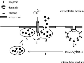
The release of the milk constituents occurs at the apical pole of MECs from lactating females, through two main processes: (i) the envelopment of lipid droplets in the plasma membrane followed by budding of the lipid globules secreted in milk (Keenan, Reference Keenan2001); (ii) the exocytosis of secretory vesicles content by fusion with the apical membrane (Figure 1). General features of exocytosis are well described for the release of neurotransmitters and may be used as a model for the mammary cell (Figure 3). The secretory vesicles of the MEC contain casein micelles, soluble proteins, lactose, ions, minerals and numerous biologically active molecules (Vilotte et al., Reference Vilotte, Whitelaw, Ollivier-Bousquet and Shennan2003 and references therein). The morphological features of the association between secretory vesicles and the plasma membrane of the MEC have been extensively described (Franke et al., Reference Franke, Lüder, Kartenbeck, Zerban and Keenan1976). However, it is still not known whether the secretory vesicles localized in the vicinity of the apical membrane are docked and then readily releasable under stimulation.
Coupling of exocytosis and endocytosis in the context of neurotransmitters release. Synaptic vesicles (SV) containing neurotransmitters are first transported to the presynaptic active zone by the cytoskeleton elements (a). These vesicles then dock to the plasma membrane and are primed in an ATP-dependent manner and become competent for fusion (b). Calcium (Ca2+) increase (c) in response to a membrane depolarization induces the rapid fusion of the vesicle with the plasma membrane that leads to the opening of the fusion pore and the release of the vesicle content (d). After exocytosis, membrane retrieval occurs through clathrin-mediated endocytosis (e) that allows the recycling and the refilling of synaptic vesicles (f) but also eliminates the excess of membrane created by the bulk of neurotransmitter secretion.

Directed movements in eukaryotic cells involve the cytoskeleton. The disruption of microtubules, by colchicine in vivo and in vitro, provokes an accumulation of secretory vesicles in MECs and inhibits milk secretion (Ollivier-Bousquet and Denamur, Reference Ollivier-Bousquet and Denamur1973; Patton, Reference Patton1976b). Most microtubules are found in the apical and medial cytoplasm (Nickerson and Keenan, Reference Nickerson and Keenan1979), suggesting that they are involved in the final steps of secretion. Moreover, a dense web containing actin (Figure 1b) lines the apical side of the mammary epithelium. Disruption of actin cytoskeleton by cytochalasin B results in an increase of casein release (Ollivier-Bousquet, Reference Ollivier-Bousquet1979), thus demonstrating the barrier role of this network.
Transcellular calcium transport in MEC is an important process regarding both the transfer of calcium from mother to neonate and its role as an intracellular second messenger in many cellular processes and the exocrine function. Calcium entry by channels, its storage in the endoplasmic reticulum and its intracellular transport by specific calcium transporters, have partly been described (VanHouten and Wysolmerski, Reference VanHouten and Wysolmerski2007). As for the role of calcium as a signaling molecule, previous and recent studies demonstrated that PRL does not significantly modify the influx and efflux of calcium in MECs (Ollivier-Bousquet, Reference Ollivier-Bousquet1983; Anantamongkol et al., Reference Anantamongkol, Takemura, Suthiphongchai, Krishnamra and Horio2007). Activation of the calcium-sensing receptor (CaR), a G-protein cell surface receptor located at the basal side, increases Ca-ATPase activity in cultured MECs. This activation implies the expression of PMCA2, an ATPase located at the apical membrane (VanHouten et al., Reference VanHouten, Neville and Wysolmerski2007). Its activation also regulates the secretion of parathyroid hormone-related protein (PTHrP) by normal mammary cells (Mamillapalli et al., Reference Mamillapalli, VanHouten, Zawalich and Wysolmerski2008 and references therein). Thus the availability of calcium linked to activation of the CaR may be coupled with a control of apical secretion (also see section ‘Molecular mechanisms and regulation of exocytosis in MECs’). Taken together, these results suggest that calcium concentration affects different types of signaling through micro-localized modifications.
Hormonal control of exocytosis in MECs
Milk protein synthesis and secretion are dependent on the action of lactogenic hormones (Ollivier-Bousquet, Reference Ollivier-Bousquet1993). A question arises as to whether lactogenic hormones play a role in the specific mechanism of exocytosis. This seems to be the case for oxytocin that, through its oxytocin receptors, acts directly on MECs (Lollivier et al., Reference Lollivier, Marnet, Delpal, Rainteau, Achard, Rabot and Ollivier-Bousquet2006). Within one minute, oxytocin was shown to induce an increase in the intracellular transport of caseins to the lumen of acini. This effect was accompanied by an increase in the number of secretory vesicles containing casein micelles in the apical region and in close contact with the apical membrane. We could then ask whether the oxytocin play a role in the vesicular transport and/or membrane fusion involved in exocytosis.
PRL exerts a secretagogue effect on MECs. The disappearance of secretory vesicles after bromocriptine treatment that markedly decreases the levels of plasma-borne PRL, suggests a role of PRL in maintaining the complex network of intracellular membranes (Ollivier-Bousquet, Reference Ollivier-Bousquet1978; Akers et al., Reference Akers, Bauman, Goodman, Capuco and Tucker1981). Interestingly, a very early effect of PRL added in vitro to MECs of lactating rats and rabbits, is to induce within a few minutes the release of caseins which had been newly synthesized 45 min prior to addition of the hormone (Lkhider et al., Reference Lkhider, Petridou, Aubourg and Ollivier-Bousquet2001). These results demonstrated the rapid control, by PRL, of the last steps of apical transport and possibly the exocytosis of caseins. One important actor in signaling of the secretagogue response to the action of PRL is arachidonic acid, a polyunsaturated fatty acid (PUFA) which is a component of membrane phospholipids, which is transiently released a few seconds after PRL binds to its receptor. Arachidonic acid mimics the secretagogue effect of PRL, either directly or via its metabolites (Ollivier-Bousquet, Reference Ollivier-Bousquet1998).
It should be noted that heregulin β1, a combinatory ligand for human epidermal growth factor receptors 3 and 4, has been proposed as a regulator of vesicles trafficking in MEC (Vadlamudi et al., Reference Vadlamudi, Wang, Talukder, Adam, Johnson and Kumar2000). The physiological significance of these results obtained in cell culture remains to be confirmed in vivo.
Molecular mechanisms and regulation of exocytosis in MECs
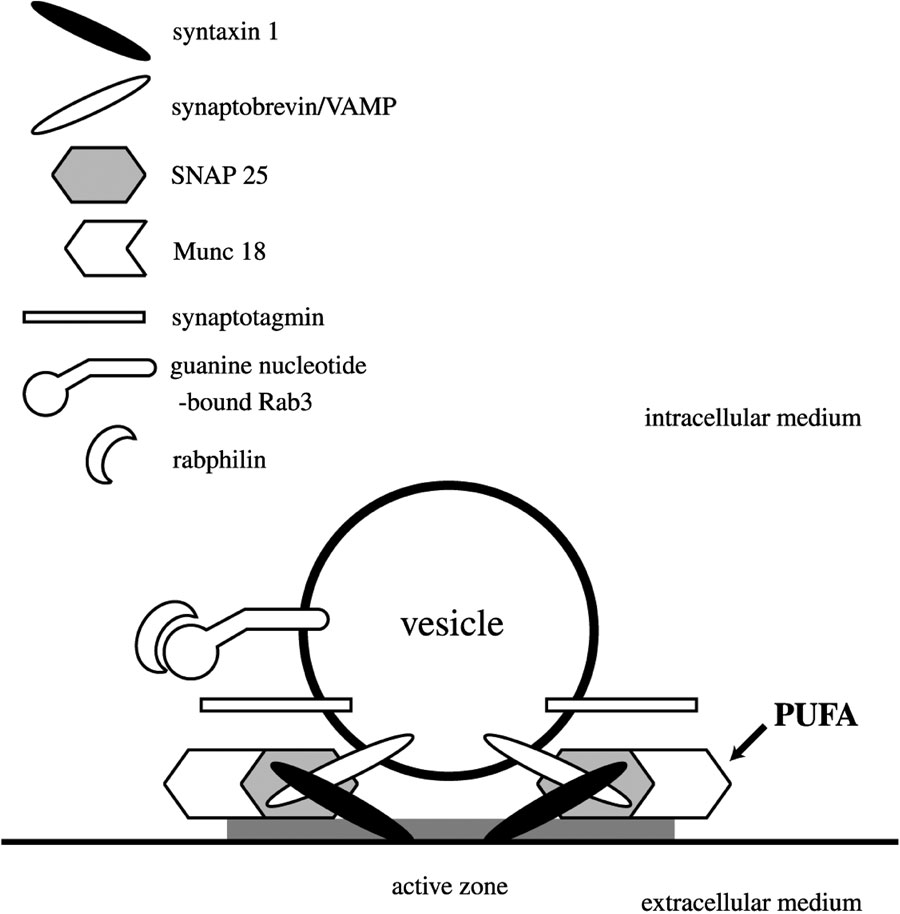
The molecular machinery responsible for membrane fusion has been particularly well described in the case of evoked neurotransmitters release (Geppert and Südhof, Reference Geppert and Südhof1998) (Figure 4). Few data, however, are available concerning the molecular control of exocytosis in MECs. Regulated exocytosis essentially relies on the SNARE (Soluble N-Ethylmaleimide-Sensitive Factor Attachment Protein Receptor) proteins that mediate specific fusion of transport vesicles with target cellular membranes. In response to intracellular calcium increase, the vesicular SNARE (v-SNARE) links cognate SNAREs located on the target membrane (t-SNAREs). Thus, these proteins form a SNARE complex that promotes the docking and the tethering of the vesicle to the membrane. The whole process of exocytosis is highly regulated by numerous proteins working in close association with the SNARE complex. In MECs, to date, only a v-SNARE (VAMP8/endobrevin), also involved in the regulated exocytosis in pancreatic, parotid and lacrimal acinar cells, has been described (Wang et al., Reference Wang, Shi, Guo, Ng, Li, Gan, Liew, Leinomen, Rajaniemi, Zhou, Zeng and Hong2007). On the other hand, several studies have shown that SNARE proteins are the targets of arachidonic acid in different neuroendocrine cell types (Darios and Davletov, Reference Darios and Davletov2006; Latham et al., Reference Latham, Osborne, Cryle and Meunier2007). An intriguing possibility is that the SNARE proteins may be the targets of arachidonic acid produced in response to PRL thus providing a link between signal transduction, secretagogue effect and exocytosis in MECs (also see section ‘Coupling between endocytosis and exocytosis in the MECs’). The expression of other proteins involved in the docking and fusion of vesicles has also been described in normal and cancerous mammary cells. For example, the expression of the small GTPase Rab3A is increased during pregnancy and post-weaning period. Heregulin β1 promotes the accumulation of Rab3A-associated vesicles in the mammary cell lines MCF-7 and HC11, suggesting its role in vesicle trafficking. These cells also express a number of proteins involved in exocytosis (rabphilin 3A, Sec8, syntaxin 4, SNAP 25, rabaptin and Doc2) (Vadlamudi et al., Reference Vadlamudi, Wang, Talukder, Adam, Johnson and Kumar2000). A possible role for Annexin II in apical exocytosis has also been suggested in MECs (Handel et al., Reference Handel, Rennison, Wilde and Burgoyne1991). Future work, however, is needed to elucidate the molecular mechanisms and regulations involved in caseins secretion during lactation. Differences may also exist in the regulation of secretion between species such as mouse, rabbit and cow, due to differences in frequency of suckling. Regulations leading to differences in the quality and quantity of milk, in relation to the stage of lactation in dairy cows, may also be relevant to these mechanisms.
Schematic representation of proteins involved in the release of neurotransmitters by synaptic vesicles. Guanine nucleotide-bound Rab3A binds to vesicles that recruit rabphilin3A before docking on the presynaptic plasma membrane (active zone) through mechanisms involving GTP hydrolysis in a calcium-dependent manner. Local increase of calcium concentration is sensed by synaptotagmin associated with the vesicular membrane. Once docked to the target membrane, the vesicle is primed and the SNARE complex is formed. In the case of neurotransmitters release, the SNARE complex results of the interaction of one v-SNARE (synaptobrevin or VAMP1/2) with t-SNAREs (syntaxin 1) and SNAP-25. Formation of the SNARE ternary complex is regulated by cytosolic proteins such as Munc18. Polyunsaturated fatty acids, such as arachidonic acid, may also exert immediate effects on the fusion machinery by direct interaction with SNARE proteins and with regulatory partners of the complex.

Coupling between endocytosis and exocytosis in the MECs. Questions and perspectives
In neurons and neuroendocrine cells, a compensatory process has been described, corresponding to the tightly balanced delivery of membranes by exocytosis and re-internalization by endocytosis (Gundelfinger et al., Reference Gundelfinger, Kessels and Qualmann2003 and references therein). In β-cells secreting insulin, a direct coupling between insulin granule exocytosis and subsequent endocytosis occurs locally at the site of the fusion pore (Min et al., Reference Min, Leung, Tomas, Watson, Gaisano, Halban, Pessin and Hou2007).
As reported above, the processes of endocytosis and exocytosis in MECs are complex and a coupling of the two, through a compensatory process, remains elusive.
Two examples of temporally coordinated events in MECs raise more precise questions concerning a cross-connection between endocytosis and exocytosis. The first example is the regulation of these two events occurring on the same side of the mammary epithelium. A release of different milk constituents occurs at the basolateral side of the mammary epithelium (Devinoy et al., Reference Devinoy, Stinnakre, Lavialle, Thépot and Ollivier-Bousquet1995). Interestingly, endosome-lysosome like vesicles localized at the basal side of MECs release cathepsin D (CD) into the extracellular medium. After binding to its receptor on the same side, PRL increases the release of CD by these vesicles (Castino et al., Reference Castino, Delpal, Bouguyon, Demoz, Isidoro and Ollivier-Bousquet2008). Questions then arise as to whether endocytosis of the hormone may be compensated by a supply of membrane due to the exocytosis of CD containing vesicles at the same side of the mammary epithelium.
The second example is the different intracellular movements described for molecules associated with the secretagogue effect of PRL. After binding to its receptor, PRL is internalized but there are no data available regarding the first steps of endocytosis process. By contrast, the regulation of PRL transport in endosomes is well described and has been shown to be impaired when the composition of membrane phospholipids is modified. A marked decrease in n-6 family PUFAs induces an accumulation of PRL in early endosomes, whereas the addition of arachidonic acid restores PRL transport to the secretory pathway. Furthermore, the inhibition of the intracellular transport of PRL also leads to the disappearance of its secretagogue effect. In parallel, it has been described that various phospholipases A2 (PLA2) are involved in mediating the secretagogue effect of PRL (Ollivier-Bousquet, Reference Ollivier-Bousquet1998 and references therein; Lkhider et al., Reference Lkhider, Petridou, Aubourg and Ollivier-Bousquet2001). It should be noted that the intracellular site where PLA2 may be active is determinant for arachidonic acid release and the subsequent PRL secretagogue effect (Ollivier-Bousquet et al., Reference Ollivier-Bousquet, Radwanyi and Bon1991). Moreover, an early effect of PRL added to mammospheres from a lactating rabbit, is associated with a change, in a few seconds, in the localization of PLA2 (Ollivier-Bousquet, unpublished results). Overall, these data strongly suggest that the early effects of PRL linked to its internalization have repercussions on its biological response.
These data raise questions regarding the precise links between these effects and the structural components of MECs. New perspectives have arisen with recent data on the role of PUFAs in regulating the SNARE machinery in neuronal plasma membranes (also see section ‘Molecular mechanisms and regulation in MECs’). Arachidonic acid is involved in the assembly of the SNARE complex in the presence of the cytoplasmic protein Munc18, intervening in functioning of the complex (Rickman and Davietov, Reference Rickman and Davietov2005; Latham et al., Reference Latham, Osborne, Cryle and Meunier2007). Moreover, arachidonic acid has also been shown to be able to directly interact with some SNARE proteins, thus promoting the formation of SNARE complex (Latham et al., Reference Latham, Osborne, Cryle and Meunier2007). The new concept of fatty acids ‘adding grease to exocytosis’ (Rohrbough and Broadie, Reference Rohrbough and Broadie2005; Verhage, Reference Verhage2005) will now make it possible to envisage a role for arachidonic acid in the regulation of the exocytotic mechanism in MECs. Of interest is the fact that MECs offer a model for the hormonal regulation of PLA2-mediated phospholipid hydrolysis, leading to the release of micromolar concentrations of arachidonic acid at local sites, where the different elements of the SNARE complex reside. A future challenge will be to understand the physiological relevance of these molecular interactions within the secretory machinery.
Conclusion
One key question that remains to be answered concerns the extent to which endocytic proteins play roles that are interconnected with the cellular processes leading to secretion in MECs. At present, the experimental data reported in this review underline the relationships between these two processes, either in the field of receptor-mediated signaling, or in that of membrane trafficking. Recent data have shown that the strength of the signaling response induced by transcription factors carried by the endocytic pathway is related to endocytic membrane trafficking. Thus interesting questions regarding endosomal proteins as trafficking regulators are now clearly posed (McShane and Zerial, Reference McShane and Zerial2008 and references therein).
Why are these questions now fundamental not only for biologists but also in terms of dairy production? Research on milk and its safety as a source of substances that are beneficial to growth and health in children and adults is a major concern in developed countries. On the other hand, the production of dairy products as basic nutrients is a priority in the fight against under-nourishment. In both cases, research on milk and its safety may be useful in optimization of sustainable dairy production. The control of milk production requires an understanding of the molecules involved in the cellular machinery (sorting of different constituents in endosomes leading to different types of intracellular signaling?), compartmentalization (regulation of pathways leading to the synthesis of milk components?) and membranes trafficking. It will be useful to explore these points using model systems. Moreover, it is crucial to validate the conclusions under physiological conditions, as a function of environmental parameters.
Acknowledgements
We are grateful to Dr R. Boisgard and S. Delpal for the micrographs, to M.-E. Marmillod for secretariat assistance and to B. Nicolas and M. Weber for their contribution to prepare the digital files for the figures.