




No CrossRef data available.
Defining the neurocircuitry of borderline personality disorder: Functional neuroimaging approaches
Published online by Cambridge University Press: 12 December 2005
Abstract
Functional neuroimaging recently has been used to localize brain dysfunction in borderline personality disorder (BPD). Initial studies have examined baseline activity or emotional reactivity, and our group has investigated what we consider to be a crucial interaction between negative emotion and behavioral (dys)control. This research is beginning to identify abnormal frontolimbic circuitry likely underlying core clinical features of this condition. We review the evidence for dysfunction in specific frontolimbic regions, leading to a mechanistic model of symptom formation in BPD. In addition, we offer an integration of these neuroimaging findings with developmental perspectives on the emergence of borderline psychopathology, focusing on the ways in which early psychosocial experience may interact with developing brain systems. We also consider possible mechanisms of psychotherapeutic change at the neural systems level in BPD. Finally, we propose that future neuroimaging studies of BPD should integrate multiple levels of observation (structural, functional, neurochemical, genetic, and clinical) in a model-driven fashion to further understand the dynamic relationship between biological and psychological factors in the development and treatment of this difficult condition.
Information
- Type
- Research Article
- Information
- Copyright
- © 2005 Cambridge University Press
Using a variety of imaging technologies and methodologies, neuroimaging investigations are shedding light on brain structure and function in both normal and pathological states including psychotic, mood, anxiety, and developmental disorders (Dougherty & Rauch, 2001; Stern & Silbersweig, 2001). These varied approaches include using structural neuroimaging, positron emission tomography (PET), and functional magnetic resonance imaging (fMRI) in the setting of symptom provocation studies, cognitive activation paradigms, and resting state studies. By defining the dysfunctional neural circuitry in individuals with these conditions, neuroimaging studies provide the possibility of enhanced diagnostic specificity, novel targets for therapeutic interventions, and more reliable predictors of treatment response.
Initially focused on the investigation of Axis I disorders, psychiatric neuroimaging researchers are beginning to study the functional neuroanatomy of Axis II disorders, including borderline personality disorder (BPD). BPD is a complex psychiatric disorder characterized by affect dysregulation and behavioral impulsivity. Individuals with this condition experience rapid alterations of mood and intense emotional responses, as well as impulsive, potentially self-destructive behaviors (substance abuse, self-mutilations, suicidal behaviors, binge eating, sexual promiscuity). Focusing on these domains of emotion and behavior, neuroimaging studies have begun to elucidate the neural correlates of this disorder.
Functional neuroimaging studies using cognitive activation paradigms in which subjects are presented with emotional stimuli have begun to examine neural dysfunctions associated with emotional reactivity in individuals with BPD. An fMRI study using negative versus neutral valenced images showed activations in bilateral amygdala, bilateral fusiform gyri, left medial prefrontal (Brodmann area [BA] 10), and right ventrolateral prefrontal (BA 47) cortices in BPD subjects compared with normal controls (Herpertz et al., 2001). In another fMRI study, investigators found greater activation in the left amygdala in response to facial expressions of emotion (happy, sad, fearful) in BPD patients versus normal control subjects (Donegan et al., 2003). A PET study by Schmahl, Elzinga, et al. (2003), in which subjects listened to personalized scripts of abandonment experiences, found increased activity in bilateral dorsolateral prefrontal cortex (DLPFC; BAs 8, 9, 10) and decreased activity in right dorsal anterior cingulate (BAs 24, 32), left superior/middle temporal gyri, left fusiform gyrus (BA 37), left visual association cortex (BA 19), and right amygdala/hippocampus in BPD subjects compared with non-BPD subjects who had comparable sexual abuse histories. Although the direction of change in neural activation between BPD subjects and controls varies in these studies, probably related to the specific methodologies used, these results nonetheless support the broad notion of dysfunction in limbic and prefrontal regions in this disorder. In addition, frontolimbic (amygdala/hippocampus, orbitofrontal cortex [OFC]) volume loss (Rusch et al., 2003; Schmahl, Vermetten, Elzinga, & Bremner, 2003; Tebartz van Elst et al., 2003) provides further evidence of dysfunction in these regions, and suggests an underlying neuropathological correlate in BPD in regions that can be associated with the symptomatology of this disorder.
[18F]Fluorodeoxyglucose PET (FDG-PET) imaging studies of BPD subjects consistently have demonstrated evidence of frontal dysfunction, most prominent in the prefrontal cortical areas (De la Fuente et al., 1997; Juengling et al., 2003; Soloff, Meltzer, Greer, Constantine, & Kelly, 2000). Metabolic challenge FDG-PET neuroimaging studies using fenfluramine, a serotonergic agonist, have found blunted neural responses in a variety of regions including medial, orbital, dorsolateral, and ventromedial prefrontal cortices, as well as in anterior cingulate cortex in BPD subjects (Siever et al., 1999; Soloff et al., 2000). Overall, these studies support the hypothesis that diminished serotonergic function in PFC, a region with neurotransmitter and neuroanatomical systems known to be involved in emotional regulation and inhibitory behavioral control, may constitute a biologic diathesis for disinhibition, impulsivity, and affect dysregulation in individuals with BPD.
Using PET with α-[11C]methyl-l-tryptophan (α-MTrp, an l-tryptophan analog) to assess brain serotonin (5-HT) synthesis capacity, Leyton et al. (2001) found significantly lower α-MTrp trapping in male BPD subjects compared with healthy controls in bilateral medial frontal cortices extending into the OFC, bilateral anterior cingulate cortex, left superior temporal gyrus (BA 22), bilateral fusiform gyri (BAs 19, 37), left globus pallidum, and the right hippocampal formation (BA 35). Correlational analysis between α-MTrp trapping and commission error rates on a Go/No-Go Task revealed significant negative correlations in the medial frontal gyrus, anterior cingulate cortex, and striatum in both male and female BPD subjects. Because these areas are involved in mediating the planning, initiation, and inhibition of behavior, low 5-HT synthesis capacity in these regions in individuals with BPD may contribute to the behavioral disinhibition characteristic of this patient population. Likewise, a preliminary fMRI study of response inhibition in patients with BPD and antisocial personality disorder has found that such individuals activate different neural networks than normal controls to inhibit prepotent responses (Vollm et al., 2004), perhaps a compensatory system resulting from deficits in the normal prefrontal inhibitory circuits.
FDG-PET resting state studies comparing BPD subjects to normal controls have demonstrated significantly reduced activity in a number of cortical and subcortical regions including the bilateral medial OFC (Soloff et al., 2003), temporoparietal cortices (Lange, Kracht, Herholz, Sachsse, & Irle, 2005), as well as the right premotor (BA 6), bilateral PFC (BA 9), mesial and anterior PFC (BAs 10, 32, 46), bilateral anterior cingulate cortex (BA 25), right ventral striatum, bilateral thalamus, and bilateral caudate (De la Fuente et al., 1997). These findings suggest dysfunction in a cortical–striatal–thalamic–frontal network that normally subserves important behavioral control functions.
Frontolimbic Circuitry in BPD
Prefrontal and limbic neural systems mediate both the processing of and responses to emotional stimuli (Phillips, Drevets, Rauch, & Lane, 2003), and therefore dysfunction in these circuits could mediate a number of core symptoms of BPD (Figure 1). Orbitofrontal dysfunction may underlie the impulsivity, aggression, and mood instability seen in BPD because lesions of this region have been linked with such behaviors in both animals and humans (Mega & Cummings, 1994). Neurobehavioral studies in animals suggest that an interplay between PFC, particularly the OFC, and the amygdala is important for affective style in humans (Davidson, 2002). In fact, extensive reciprocal connections between the OFC and the amygdala are well known, and these regions function as part of an integrated neural system guiding socioemotional decision making and adaptive response selection (Baxter, Parker, Lindner, Izquierdo, & Murray, 2000).
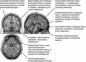
Frontotemporo–limbic and subcortical brain regions relevant to neural models of BPD. Selected functional affiliations of these regions (see text) are briefly noted.
Amygdala dysfunction has been implicated in playing an important role in the development of BPD because this limbic structure is known to be involved in the processing of negative valence stimuli, the establishment of fear conditioning, the production of strong negative emotional states, and vigilance for environmental stimuli with important survival value (Davis & Whalen, 2001; Isenberg et al., 1999; LeDoux, 1996). Findings of amygdala dysfunction in the above-mentioned fMRI studies likely reflect abnormal processing of aversive paradigm stimuli and/or the activation of emotional memories induced by such stimuli (Breiter, Rauch, et al., 1996). Furthermore, increased amygdala activation in BPD subjects may reflect an oversensitization to aversive emotional stimuli or a relative failure of normal habituation. A strong habituation of the BOLD fMRI signal in response to emotional stimuli has been observed in normal controls (Breiter, Etcoff, et al., 1996; LaBar, Gatenby, Gore, LeDoux, & Phelps, 1998), and may be dysfunctional in BPD individuals with long-lasting, intense emotional reactions.
Hippocampal dysfunction is not surprising as well, given the susceptibility of this neural structure to stress in the setting of increased cortisol and excitatory amino acids (McEwen & Magariños, 2001) and the prevalence of early traumatization experienced by those with BPD (Zanarini, 2000). Hippocampal volumes in individuals with BPD have been negatively correlated with the extent and duration of self-reported traumatic experiences, suggesting the presence of stress-induced structural changes in this disorder (Driessen et al., 2000). Because the hippocampus mediates fear responses to contextual features of the environment (Phillips & LeDoux, 1992), dysfunction of this structure may result in a maladaptive overgeneralization of fear responses (e.g., fear of abandonment) to a broad range of interpersonal situations as observed in those with BPD.
Activation of fusiform gyrus (BA 37), a region implicated in complex visual feature detection and recognition of facial expression (George et al., 1993), could be due to backprojections from the amygdala for the purpose of strengthening visual attention to emotionally relevant stimuli, a finding consistent with the hypervigilance for emotional aspects of the environment seen in individuals with BPD. Additional evidence for an emotion-based attentional bias in BPD is provided by Arntz, Appels, and Sieswerda's (2000) emotional Stroop study in which BPD subjects had longer response times than normal controls to negative versus neutral valence words. Interestingly, this finding was not specific to words reflecting BPD themes, but rather seemed to reflect a more general hypervigilance for all negative valence stimuli.
Dysfunction in medial prefrontal areas, in particular anterior cingulate cortex, a region involved in emotional regulation and with important inhibitory connections to the amygdala that mediate the extinction of fear responses (Carmichael & Price, 1995; Devinsky, Morrell, & Vogt, 1995), might underlie the difficulty modulating anxiety and other intense affects seen in individuals with BPD. Medial PFC lesions in humans result in emotional dysfunction and deficits in interpersonal functioning that requires the correct interpretation of the emotional expressions of others (Damasio, Grabowski, Frank, Galaburda, & Damasio, 1994). Although normal individuals tend to have increased activity in the anterior cingulate during the induction of angry affect states (Dougherty et al., 1999; Kimbrell et al., 1999), perhaps reflecting adaptive modulating functions, individuals with BPD have been shown to have decreased function of (De la Fuente et al., 1997; Goyer et al., 1994) and reduced gray matter volume in (Hazlett et al., in press) this area. In addition, anterior cingulate cortex recently has been implicated in the processing of the sensory and affective dimensions of pain (Hofbauer, Rainville, Duncan, & Bushnell, 2001; Olausson et al., 2001), and diminished activity in this region may account for the significantly attenuated pain perception among individuals with BPD and self-injurious behaviors (Bohus et al., 2000).
Abnormal activity of ventrolateral PFC (Herpertz et al., 2001), with its direct connections with the basal nucleus of the amygdala, could result in compromised inhibition of amygdala-driven emotional responses thereby undermining top-down control of this limbic region in BPD (Drevets, 1999). Activation of DLPFC (Schmahl, Elzinga, et al., 2003) has been observed in prior imaging studies using personalized scripts in women with abuse-related posttraumatic stress disorder (PTSD; Bremner et al., 1999; Shin et al., 1999), possibly reflecting the role of this prefrontal region in the encoding and retrieval of verbal memories (Tulving, Kapur, Craik, Moscovitch, & Houle, 1994) and/or its emerging role in aspects of emotional processing (Davidson, Jackson, & Kalin, 2000) and emotional–cognitive interactions (Liotti & Mayberg, 2001; Sergerie, Lepage, & Armony, 2005). Additional evidence of DLPFC dysfunction comes from Tebartz van Elst et al.'s (2001) proton magnetic resonance spectroscopy study demonstrating subtle prefrontal neuropathology as indicated by reduced N-acetylaspartate concentrations in this prefrontal region in individuals with BPD.
Interaction of Emotion and Behavior in BPD
As summarized above, functional neuroimaging studies have investigated the neural systems underlying emotional dysregulation in BPD using a variety of neuropsychological probes. Numerous other imaging studies with normal subjects and various patient populations have examined the neural basis of emotion and behavior, but rarely the interaction between these two domains. Our lab has developed an emotional Go/No-Go fMRI paradigm to specifically investigate the functional neuroanatomy underlying the interaction of emotion and inhibitory behavioral control in both normals and individuals with BPD. We believe that this important interaction has relevance to a wide range of human experience and behavior in health and disease. The BPD work is being performed in collaboration with Dr. Otto Kernberg's research group at the Personality Disorders Institute of the Weill Medical College of Cornell University. This approach can shed light on the neural circuits underlying the clinically observed phenomenon of behavioral impulsivity in the context of intense negative affect states so commonly seen in this patient population. The methodological approach of using neuropsychological probes that conjoin emotional stimuli with nonemotional behavioral task demands is an emerging line of investigation within the cognitive neurosciences to further investigate important interactions between emotion, cognition, and behavior (Dolan, 2002; Elliott, Rubinsztein, Sahakian, & Dolan, 2002).
Our fMRI paradigm is a variation on the classic Go/No-Go Task (Drewe, 1975) used extensively in investigational neuropsychology to probe response inhibition, and involves the execution or inhibition of a prepotent motor response triggered by a go or no-go stimulus, respectively. Our subjects are instructed to execute or inhibit a response to visually presented emotional stimuli. By experimentally manipulating the emotional valence of these stimuli that reflect themes salient for individuals with BPD, we are able to investigate the modulation of behavioral task demand (go or no-go) by emotional valence, thereby probing the neural systems subserving the interaction of emotion and behavioral control in this population.
We have hypothesized and detected dysfunction in medial OFC in individuals with BPD while performing this emotional Go/No-Go Task, a finding consistent with this region's presumed role in regulating behavior under changing contingencies and its role in guiding adaptive behavioral responses within socioemotional contexts (Damasio et al., 1994; Elliott et al., 2002; Rolls, 2000). Specifically, we have hypothesized differential neural activity in this region in BPD subjects compared with normals, reflecting the former's greater difficulty modulating behavioral responses under conditions of negative emotion.
Medial OFC is believed to process viscerosomatic afferent information as cues reflecting the personal relevance of environmental contexts, and to integrate such emotional information into decision making and response selection (Bechara, Damasio, & Damasio, 2000; Dolan, 1999). According to Damasio's somatic marker hypothesis (1994), dysfunction of medial OFC results in an impaired capacity to adaptively integrate emotional information into behavioral response selection. This may have particular relevance for individuals with BPD who, while experiencing intense negative affect states, have greater difficulty regulating behavioral responses. In addition, medial OFC appears to be involved in the suppression of context-activated emotional memories irrelevant to current cognitive/behavioral demands (Treyer, Buck, & Schnider, 2003), thereby allowing for more adaptive responses to the immediate environment. Individuals with BPD, however, seem to respond in characteristically inflexible and maladaptive ways based not upon current social contexts, but rather according to implicit emotional memories of past interpersonal experiences, possibly reflecting OFC dysfunction.
Integration With Developmental Perspectives
Evidence of dysfunction in limbic and prefrontal neural systems subserving affect regulation and behavioral control is consistent with developmental theories of BPD. Clarkin, Yeomans, and Kernberg's (1999) psychoanalytic developmental theory of BPD posits both constitutional (i.e., neurobiologic) and early environmental factors giving rise to a psychic structure populated by distorted, unidimensional mental representations of self and others, which leads to maladaptive perceptions of and responses to current interpersonal reality. According to this model, the normal infant's early experiences with caregivers lead to a multitude of distinct and contradictory internalized relationship patterns; for example, the satisfied, calm self linked with an affect of love to the nurturing other, and the needy, frustrated self linked with an affect of hate to the depriving other. Ideally, over the course of development these split-off mental representations are integrated, resulting in a more complex and realistic image of the other who can both satisfy and frustrate, allowing for more adaptive interpersonal functioning. Adaptive integration of these early, distorted representations of self and others, and the intense affects associated with them, likely requires sufficient affect regulation. Individuals who develop BPD appear not to reach this developmental stage, perhaps due to a primary (genetic) neurobiological deficit in those neural systems subserving affect regulation, effects of early trauma on these developing systems, and/or deficits in parental modulation of this emerging self-regulating capacity.
Deficits in affect regulation and impulse control during early development may lead to particularly conflictual experiences with caregivers, experiences that become internalized as prototypes for future relationships, eventually reflected in adult psychopathology. In addition, it is possible that these conflict-ridden interpersonal experiences and the resultant internalized mental representations act back upon the neurobiological substrate to produce further dysfunction in neural circuits required for affect/behavioral regulation. In animal models, chronic psychosocial and behavioral stress have been shown to produce neuronal reorganization and atrophy in medial PFC and hippocampus (Magariños, McEwen, Flugge, & Fuchs, 1996; Radley et al., 2004), as well as neuronal growth in the amygdala (McEwen, 2004), in part mediated via glucocorticoid action on these brain regions. Findings of reduced prefrontal and hippocampal volumes in individuals with BPD from human neuroimaging studies (Brambilla et al., 2004; Driessen et al., 2000; Irle, Lange, & Sachsse, 2005; Tebartz van Elst et al., 2003) are consistent with these animal findings, and may reflect early psychosocial stress-induced changes in these areas. For example, Posner et al. (2003) have suggested that the anterior cingulate and the self-regulating functions it subserves may be susceptible to the effects of trauma during early development, possibly accounting for the deficits in effortful control observed in adults with BPD.
Epidemiological data strongly suggest that numerous types of early psychosocial stress (sexual/physical abuse, neglect) are involved in the development of BPD (Zanarini, 2000), perhaps accounting for the high comorbidity of PTSD (50%) seen in BPD patients (McGlashan et al., 2000). The effects of psychosocial stress on the developing brain may be mediated in part by genetic factors. Just as a polymorphism of the serotonin transporter (5-HTT) gene has been shown to moderate the influence of stressful life events on the emergence of depression (Caspi et al., 2003), certain genes may render some individuals more susceptible to developing BPD in response to environmental stressors via functional and structural changes in the relevant neural circuits discussed above. Although the functional consequences of these morphological changes in prefrontal and limbic areas remain unclear, they could result in ongoing difficulties in affect regulation and behavioral control leading to continued psychosocial stress, resulting in a pathological reciprocal interaction between neurobiology and the environment in those with BPD.
An alternative to this psychosocial stress-induced model of BPD comes from Fonagy, Gergely, Jurist, and Target (2002), who propose that the development of effective affect regulation takes place within the context of the moment to moment interactions between infant and caregiver. They propose a social biofeedback model of parental affect mirroring according to which the parent's attuned mirroring of the infant's emotional expressions is vital for the child's development of higher order mental representations of his internal feeling states, eventually providing the basis for the infant's emerging capacity to reason about and to regulate his own affects. According to this theory, insufficient affect mirroring leads to a developmental deficit in the ability to mentalize about emotional states and may result in the relative failure of effective cognitive control over one's affect states observed in individuals with BPD. The emerging neuroimaging evidence of prefrontal dysfunction in BPD, a region involved in the top-down cognitive control of limbic areas, may in some cases reflect this early failure of affect mirroring and provides another developmental model for the effects of early social experience on the development of neural systems required for adaptive affect regulation.
Potential Neural Mechanisms of Psychotherapeutic Change in BPD
Based upon recent neuroscientific findings of dysfunctional neurocircuitry in BPD and these developmental theories of borderline psychopathology, we can begin to conceptualize putative mechanisms of psychotherapeutic action at the neural systems level in the treatment of this disorder. The psychotherapeutic use of language to describe and talk about emotional experience provides and fosters a representational/symbolic means to become aware of, to reason about, and ultimately to modulate affect states. This may be similar to the developmental emergence of mentalization (Fonagy et al., 2002), and may reflect the therapeutic enhancement of dysfunctional prefrontal regions underlying the capacity to mentalize. In addition, the activation of unconscious emotional memories of early interpersonal experiences with significant others within the therapeutic relationship may present the opportunity for enhanced declarative remembering of the past and more adaptive reconsolidation of such pathological memories (Ross, 2003). In fact, evidence from animal models demonstrates the possibility of altering emotional memories via disruption of reconsolidation at the level of the amygdala (Debiec & LeDoux, 2004). Finally, psychodynamically oriented psychotherapies that focus on the patient's transference to the therapist (the patterned and inflexible repertoire of emotional/behavioral responses based on internalized representations of past relationships; Clarkin et al., 1999) may lead to a greater flexibility of responses via alterations of frontolimbic circuitry. For example, changes in the OFC and hippocampus that mediate emotional/behavioral responses to contextual features of the social environment could allow for a greater capacity to process and respond to current interpersonal reality in novel, more adaptive ways rather than in repetitive, inflexible ways based upon past experience. In addition, transference-focused psychotherapies might promote higher order prefrontal processes that mediate the capacity for more complex mental representations of self and others, thereby changing the very quality of the subjective interpersonal world of those with BPD.
Conclusions and Future Directions
Neuroimaging studies of BPD using a variety of methodological approaches have identified structural and functional abnormalities in specific frontolimbic regions. Findings of dysfunction in these areas that normally subserve affect regulating and behavioral control functions are consistent with the phenomenology of this disorder and may provide a neuropathological correlate, as well as a dynamic, mechanistic understanding, of BPD. However, numerous methodological limitations must be considered when interpreting the results of individual studies. These may include small sample sizes, histories of comorbid Axis I and II disorders, and gender differences between patients and controls. Past histories of major depressive disorder and PTSD, highly prevalent comorbidities in the BPD population, are important issues (although not necessarily confounds) because these other disorders also appear to involve dysfunction in similar prefrontal and limbic regions (Drevets, 2001; Elliott et al., 2002; Shin et al., 2004). In fact, BPD, depressive disorders, and PTSD may be conceptualized as a spectrum of chronic stress-related disorders involving stress-induced changes in these neural circuits (Schmahl, Elzinga, et al., 2003).
Future neuroimaging investigations should begin to parse out the specific similaritites and differences in the functional neuroanatomy underlying these disorders by comparing across diagnostic groups with the aim of providing greater specificity to these initial findings in BPD. Using emotional stimuli with particular salience for individuals with BPD in the context of relevant neurobehavioral interactions, as our lab has done, may enable neuroimaging investigators to more specifically and mechanistically probe neural systems of interest in this patient population and to distinguish among diagnostic groups. In addition, correlational analyses of neuroimaging data with a variety of clinical and behavioral measures (e.g., measures of impulsivity, mood instability, suicidality, quality of interpersonal relationships) may elucidate the relationships between functional imaging brain abnormalities and the different symptom clusters of BPD. Multimodal imaging studies combining structural, functional, and neurochemical information will be highly informative as well. Moreover, studies utilizing neuroimaging to identify an endo-/intermediate phenotype, bridging clinical phenotype and genotype (e.g., measures of allelic variation), will be important. Understanding the neural basis of BPD symptomatology ultimately can lead to greater diagnostic specificity, new targets for therapeutic interventions, and more effective ways to predict treatment response in this debilitating condition.
References
REFERENCES
Arntz, A.,
Appels, C., &
Sieswerda, S.
(2000).
Hypervigilance in borderline personality disorder: A test with the
emotional Stroop paradigm.
Journal of Personality Disorders
14,
366–373.CrossRefGoogle Scholar
Baxter, M. G.,
Parker, A.,
Lindner, C. C.,
Izquierdo, A. D., &
Murray, E. A.
(2000).
Control of response selection by reinforcer value requires
interaction of amygdala and orbital prefrontal cortex.
Journal of Neuroscience
20,
4311–4319.Google Scholar
Bechara, A.,
Damasio, H., &
Damasio, A. R.
(2000).
Emotion, decision making and the orbitofrontal cortex.
Cerebral Cortex
10,
295–307.CrossRefGoogle Scholar
Bohus, M.,
Limberger, M.,
Ebner, U.,
Glocker, F. X.,
Schwarz, B.,
Wernz, M.,
et al.
(2000).
Pain perception during self-reported distress and calmness in
patients with borderline personality disorder and self-mutilating
behavior.
Psychiatry Research
95,
251–260.Google Scholar
Brambilla, P.,
Soloff, P. H.,
Sala, M.,
Nicoletti, M. A.,
Keshavan, M. S., &
Soares, J. C.
(2004).
Anatomical MRI study of borderline personality disorder
patients.
Psychiatry Research: Neuroimaging
131,
125–133.CrossRefGoogle Scholar
Breiter, H. C.,
Etcoff, N. L.,
Whalen, P. J.,
Kennedy, W. A.,
Rauch, S. L.,
Buckner, R. L.,
et al.
(1996).
Response and habituation of the human amygdale during visual
processing of facial expression.
Neuron
17,
875–887.CrossRefGoogle Scholar
Breiter, H. C.,
Rauch, S. L.,
Kwong, K. K.,
Baker, J. R.,
Weisskoff, R. M.,
Kennedy, D. N.,
et al.
(1996).
Functional magnetic resonance imaging of symptom provocation in
obsessive–compulsive disorder.
Archives of General Psychiatry
53,
595–606.CrossRefGoogle Scholar
Bremner, J. D.,
Narayan, M.,
Staib, L. H.,
Southwick, S. M.,
McGlashan, T., &
Charney, D. S.
(1999).
Neural correlates of childhood sexual abuse in women with and
without posttraumatic stress disorder.
American Journal of Psychiatry
156,
1787–1795.Google Scholar
Carmichael, S. T., &
Price, J. L.
(1995).
Limbic connections of the orbital and medial prefrontal cortex in
macaque monkeys.
Journal of Comparative Neurology
363,
615–641.CrossRefGoogle Scholar
Caspi, A.,
Sugden, K.,
Moffitt, T. E.,
Taylor, A.,
Craig, I. W.,
Harrington, H.,
et al.
(2003).
Influence of life stress on depression: Moderation by a polymorphism
in the 5-HTT gene.
Science
301,
386–389.CrossRefGoogle Scholar
Clarkin, J. F.,
Yeomans, F. E., &
Kernberg, D. F.
(1999).
Psychotherapy for borderline personality.
New York:
Wiley.
Damasio, H.,
Grabowski, T.,
Frank, R.,
Galaburda, A. M., &
Damasio, A. R.
(1994).
The return of Phineas Gage: Clues about the brain from the skull of
a famous patient.
Science,
264,
1102–1105.CrossRefGoogle Scholar
Davidson, R. J.
(2002).
Anxiety and affective style: Role of prefrontal cortex and
amygdala.
Biological Psychiatry
51,
68–80.CrossRefGoogle Scholar
Davidson, R. J.,
Jackson, D. C., &
Kalin, N. H.
(2000).
Emotion, plasticity, context, and regulation: Perspectives from
affective neuroscience.
Psychological Bulletin
126,
890–909.CrossRefGoogle Scholar
Davis, M., &
Whalen, P. J.
(2001).
The amygdala: Vigilance and emotion.
Molecular Psychiatry
6,
13–34.CrossRefGoogle Scholar
De la Fuente, J. M.,
Goldman, S.,
Stanus, E.,
Vizuete, C.,
Morlán, I.,
Bobes, J.,
et al.
(1997).
Brain glucose metabolism in borderline personality disorder.
Journal of Psychiatric Research
31,
531–541.CrossRefGoogle Scholar
Debiec, J., &
LeDoux, J. E.
(2004).
Disruption of reconsolidation but not consolidation of auditory fear
conditioning by noradrenergic blockade in the amygdala.
Neuroscience
129,
267–272.CrossRefGoogle Scholar
Devinsky, O.,
Morrell, M. J., &
Vogt, B. A..
(1995).
Contributions of anterior cingulate to behavior.
Brain
118,
279–306.CrossRefGoogle Scholar
Dolan, R. J.
(1999).
On the neurology of morals.
Nature Neuroscience
2,
927–929.CrossRefGoogle Scholar
Dolan, R. J.
(2002).
Emotion, cognition, and behavior.
Science
298,
1191–1194.CrossRefGoogle Scholar
Donegan, N. H.,
Sanislow, C. A.,
Blumberg, H. P.,
Fulbright, R. K.,
Lacadie, C.,
Skudlarski, P.,
et al.
(2003).
Amygdala hyperreactivity in borderline personality disorder:
Implications for emotional dysregulation.
Biological Psychiatry
54,
1284–1293.CrossRefGoogle Scholar
Dougherty, D. D., &
Rauch, S. L.
(2001).
Psychiatric neuroimaging research: Contemporary strategies.
Washington, DC:
American Psychiatric Press
Dougherty, D. D.,
Shin, L. M.,
Alpert, N. M.,
Pitman, R. K.,
Orr, S. P.,
Lasko, M.,
et al.
(1999).
Anger in healthy men: A PET study using script-driven
imagery.
Biological Psychiatry
46,
466–472.CrossRefGoogle Scholar
Drevets, W. C.
(1999).
Prefrontal cortical–amygdalar metabolism in major
depression.
Annals of the New York Academy of Sciences
877,
614–637.CrossRefGoogle Scholar
Drevets, W. C.
(2001).
Neuroimaging and neuropathological studies of depression:
Implications for the cognitive–emotional features of mood
disorders.
Current Opinion in Neurobiology
11,
240–249.CrossRefGoogle Scholar
Drewe, E. A.
(1975).
Go–no go learning after frontal lobe lesions in humans.
Cortex
11,
8–16.CrossRefGoogle Scholar
Driessen, M.,
Herrmann, J.,
Stahl, K.,
Zwaan, M.,
Meier, S.,
Hill, A.,
et al.
(2000).
Magnetic resonance imaging volumes of the hippocampus and the
amygdala in women with borderline personality disorder and early
traumatization.
Archives of General Psychiatry
57,
1115–1122.CrossRefGoogle Scholar
Elliott, R.,
Rubinsztein, J. S.,
Sahakian, B. J., &
Dolan, R. J.
(2002).
The neural basis of mood-congruent processing biases in
depression.
Archives of General Psychiatry
59,
597–604.CrossRefGoogle Scholar
Fonagy, P.,
Gergely, G.,
Jurist, E. L., &
Target, M.
(2002).
Affect regulation, mentalization, and the development of the
self.
New York:
Other Press.
George, M. S.,
Ketter, T. A.,
Gill, D. S.,
Haxby, J. V.,
Ungerleider, L. G.,
Herscovitch, P.,
et al.
(1993).
Brain regions involved in recognizing facial emotion or identity: An
oxygen-15 PET study.
Journal of Neuropsychology
5,
384–394.Google Scholar
Goyer, P. F.,
Andreason, P. J.,
Semple, W. E.,
Clayton, A. H.,
King, A. C.,
Compton–Toth, B. A.,
et al.
(1994).
Positron-emission tomography and personality disorders.
Neuropsychopharmacology
10,
21–28.CrossRefGoogle Scholar
Hazlett, E. A.,
New, A. S.,
Newmark, R.,
Haznedar, M. M.,
Lo, J. N.,
Speiser, L. J.,
et al.
(in press).
Reduced anterior and posterior cingulate gray matter in borderline
personality disorder.
Biological Psychiatry.
Herpertz, S. C.,
Dietrich, T. M.,
Wenning, B.,
Krings, T.,
Erberich, S. G.,
Wilmes, K.,
et al.
(2001).
Evidence of abnormal amygdale functioning in borderline personality
disorder: A functional MRI study.
Biological Psychiatry
50,
292–298.CrossRefGoogle Scholar
Hofbauer, R. K.,
Rainville, P.,
Duncan, G. H., &
Bushnell, M. C.
(2001).
Cortical representation of the sensory dimension of pain.
Journal of Neurophysiology
86,
402–411.Google Scholar
Irle, E.,
Lange, C., &
Sachsse, U.
(2005).
Reduced size and abnormal asymmetry of parietal cortex in women with
borderline personality disorder.
Biological Psychiatry
57,
173–182.CrossRefGoogle Scholar
Isenberg, N.,
Silbersweig, D.,
Engelien, A.,
Emmerich, S.,
Malavade, K.,
Beattie, B.,
et al.
(1999).
Linguistic threat activates the human amygdala.
Proceedings of the National Academy of Sciences of the United States
of America
96,
10456–10459.CrossRefGoogle Scholar
Juengling, F. D.,
Schmahl, C.,
Hesslinger, B.,
Ebert, D.,
Bremner, J. D.,
Gostomzyk, J.,
et al.
(2003).
Positron emission tomography in female patients with borderline
personality disorder.
Journal of Psychiatric Research
37,
109–115.CrossRefGoogle Scholar
Kimbrell, T. A.,
George, M. S.,
Parekh, P. I.,
Ketter, T. A.,
Podell, D. M.,
Danielson, A. L.,
et al.
(1999).
Regional brain activity during transient self-induced anxiety and
anger in healthy adults.
Biological Psychiatry
46,
454–465.CrossRefGoogle Scholar
LaBar, K. S.,
Gatenby, J. C.,
Gore, J. C.,
LeDoux, J. E., &
Phelps, E. A.
(1998).
Human amygdala activation during conditioned fear acquisition and
extinction: A mixed-trial fMRI study.
Neuron
20,
937–945.CrossRefGoogle Scholar
Lange, C.,
Kracht, L.,
Herholz, K.,
Sachsse, U., &
Irle, E.
(2005).
Reduced glucose metabolism in temporo-parietal cortices of women
with borderline personality disorder.
Psychiatry Research
139,
115–126.Google Scholar
LeDoux, J.
(1996).
The emotional brain: The mysterious underpinnings of emotional
life.
New York:
Touchstone.
Leyton, M.,
Okazawa, H.,
Diksic, M.,
Paris, J.,
Rosa, P.,
Mzengeza, S.,
et al.
(2001).
Brain regional
alpha-[11C]methyl-l-tryptophan trapping in
impulsive subjects with borderline personality disorder.
American Journal of Psychiatry
158,
775–782.CrossRefGoogle Scholar
Liotti, M., &
Mayberg, H. S.
(2001).
The role of functional neuroimaging in the neuropsychology of
depression.
Journal of Clinical and Experimental Neuropsychology
23,
121–136.CrossRefGoogle Scholar
Magariños, A. M.,
McEwen, B. S.,
Flugge, G., &
Fuchs, E.
(1996).
Chronic psychosocial stress causes apical dendritic atrophy of
hippocampal CA3 pyramidal neurons in subordinate tree shrews.
Journal of Neuroscience
16,
3534–3540.Google Scholar
McEwen, B. S.
(2004).
Protection and damage from acute and chronic stress: Allostasis and
allostatic overload and relevance to the pathophysiology of psychiatric
disorders.
Annals of the New York Academy of Sciences
1032,
1–7.CrossRefGoogle Scholar
McEwen, B. S., &
Magariños, A. M.
(2001).
Stress and hippocampal plasticity: implications for the
pathophysiology of affective disorders.
Human Psychopharmacology: Clinical and Experimental
16,
S7–S19.Google Scholar
McGlashan, T. H.,
Grilo, C. M.,
Skodol, A. E.,
Gunderson, J. G.,
Shea, M. T.,
Morey, L. C.,
et al.
(2000).
The collaborative longitudinal personality disorders study: Baseline
Axis I/II and II/II diagnostic co-occurrence.
Acta Psychiatrica Scandinavica
102,
256–264.CrossRefGoogle Scholar
Mega, M. S., &
Cummings, J. L.
(1994).
Frontal–subcortical circuits and neuropsychiatric
disorders.
Journal of Neuropsychiatry and Clinical Neuroscience
6,
358–370.Google Scholar
Olausson, H.,
Ha, B.,
Duncan, G. H.,
Morin, C.,
Ptito, A..,
Ptito, M.,
et al.
(2001).
Cortical activation by tactile and painful stimuli in
hemispherectomized patients.
Brain
124,
916–927.CrossRefGoogle Scholar
Phillips, M. L.,
Drevets, W. C.,
Rauch, S. L., &
Lane, R.
(2003).
Neurobiology of emotion perception I: The neural basis of normal
emotion perception.
Biological Psychiatry
54,
504–514.CrossRefGoogle Scholar
Phillips, R. G., &
LeDoux, J. E.
(1992).
Differential contribution of amygdala and hippocampus to cued and
contextual fear conditioning.
Behavioral Neuroscience
106,
274–285.CrossRefGoogle Scholar
Posner, M. I.,
Rothbart, M. K.,
Vizueta, N.,
Thomas, K. M.,
Levy, K. N.,
Fossella, J.,
et al.
(2003).
An approach to the psychobiology of personality disorders.
Development and Psychopathology,
15,
1093–1106.CrossRefGoogle Scholar
Radley, J. J.,
Sisti, H. M.,
Hao, J.,
Rocher, A. B.,
McCall, T.,
Hof, P. R.,
et al.
(2004).
Chronic behavioral stress induces apical dendritic reorganization in
pyramidal neurons of the medial prefrontal cortex.
Neuroscience
125,
1–6.Google Scholar
Rolls, E. T.
(2000).
The orbitofrontal cortex and reward.
Cerebral Cortex
10,
284–294.CrossRefGoogle Scholar
Ross, J. M.
(2003).
Preconscious defence analysis, memory and structural change.
International Journal of Psychoanalysis
84,
59–76.CrossRefGoogle Scholar
Rusch, N.,
van Elst, L. T.,
Ludaescher, P.,
Wilke, M.,
Huppertz, H. J.,
Thiel, T.,
et al.
(2003).
A voxel-based morphometric MRI study in female patients with
borderline personality disorder.
Neuroimage
20,
385–392.CrossRefGoogle Scholar
Schmahl, C. G.,
Elzinga, B. M.,
Vermetten, E.,
Sanislow, C.,
McGlashan, T. H., &
Bremner, J. D.
(2003).
Neural correlates of memories of abandonment in women with and
without borderline personality disorder.
Biological Psychiatry
54,
142–151.CrossRefGoogle Scholar
Schmahl, C. G.,
Vermetten, E.,
Elzinga, B. M., &
Bremner, J. D.
(2003).
Magnetic resonance imaging of hippocampal and amygdala volume in
women with childhood abuse and borderline personality disorder.
Psychiatry Research: Neuroimaging
122,
193–198.CrossRefGoogle Scholar
Sergerie, K.,
Lepage, M., &
Armony, J. L.
(2005).
A face to remember: Emotional expression modulates prefrontal
activity during memory formation.
Neuroimage
24,
580–585.CrossRefGoogle Scholar
Shin, L. M.,
McNally, R. J.,
Kosslyn, S. M.,
Thompson, W. L.,
Rauch, S. L.,
Alpert, N. M.,
et al.
(1999).
Regional cerebral blood flow during script-driven imagery in
childhood sexual abuse-related PTSD: A PET investigation.
American Journal of Psychiatry
156,
575–584.Google Scholar
Shin, L. M.,
Orr, S. P.,
Carson, M. A.,
Rauch, S. L.,
Macklin, M. L.,
Lasko, N. B.,
et al.
(2004).
Regional cerebral blood flow in the amygdala and medial prefrontal
cortex during traumatic imagery in male and female Vietnam veterans with
PTSD.
Archives of General Psychiatry
61,
168–176.CrossRefGoogle Scholar
Siever, L. J.,
Buchsbaum, M. S.,
New, A. S.,
Spiegel–Cohen, J.,
Tsechung, W.,
Hazlett, E. A.,
et al.
(1999).
d,l-Fenfluramine response in impulsive personality
disorder assessed with [18F]fluorodeoxyglucose
positron emission tomography.
Neuropsychopharmacology
20,
413–423.CrossRefGoogle Scholar
Soloff, P. H.,
Meltzer, C. C.,
Becker, C.,
Greer, P. J.,
Kelly, T. M., &
Constantine, D.
(2003).
Impulsivity and prefrontal hypometabolism in borderline personality
disorder.
Psychiatry Research: Neuroimaging
123,
153–163.CrossRefGoogle Scholar
Soloff, P. H.,
Meltzer, C. C.,
Greer, P. J.,
Constantine, D., &
Kelly, T. M.
(2000).
A fenfluramine-activated FDG-PET study of borderline personality
disorder.
Biological Psychiatry
47,
540–547.CrossRefGoogle Scholar
Stern, E., &
Silbersweig, D. A.
(2001).
Advances in functional neuroimaging methodology for the study of
brain systems underlying human neuropsychological function and
dysfunction.
Journal of Clinical and Experimental Neuropsychology
23,
3–18.CrossRefGoogle Scholar
Tebartz van Elst, L.,
Hesslinger, B.,
Thiel, T.,
Geiger, E.,
Haegele, K.,
Lemieux, L.,
et al.
(2003).
Frontolimbic brain abnormalities in patients with borderline
personality disorder: A volumetric magnetic resonance imaging
study.
Biological Psychiatry
54,
163–171.CrossRefGoogle Scholar
Tebartz van Elst, L.,
Thiel, T.,
Hesslinger, B.,
Lieb, K.,
Bohus, M.,
Hennig, J.,
et al.
(2001).
Subtle prefrontal neuropathology in a pilot magnetic resonance
spectroscopy study in patients with borderline personality
disorder.
Journal of Neuropsychiatry and Clinical Neuroscience
13,
511–514.CrossRefGoogle Scholar
Treyer, V.,
Buck, A., &
Schnider, A.
(2003).
Subcortical loop activation during selection of currently relevant
memories.
Journal of Cognitive Neuroscience
15,
610–618.CrossRefGoogle Scholar
Tulving, E.,
Kapur, S.,
Craik, F. I. M.,
Moscovitch, M., &
Houle, S.
(1994).
Hemispheric encoding/retrieval asymmetry in episodic memory:
Positron emission tomography findings.
Proceedings of the National Academy of Sciences of the United States
of America
91,
2016–2020.CrossRefGoogle Scholar
Vollm, B.,
Richardson, P.,
Stirling, J.,
Elliott, R.,
Dolan, M.,
Chaudhry, I.,
et al.
(2004).
Neurobiological substrates of antisocial and borderline personality
disorder: Preliminary results of a functional fMRI study.
Criminal Behavior and Mental Health
14,
39–54.CrossRefGoogle Scholar
Zanarini, M. C.
(2000).
Childhood experiences associated with the development of borderline
personality disorder.
Psychiatric Clinics of North America
23,
89–101.CrossRefGoogle Scholar

Frontotemporo–limbic and subcortical brain regions relevant to neural models of BPD. Selected functional affiliations of these regions (see text) are briefly noted.
 You have
Access
- 57
- Cited by