Introduction
The South American sea lion, Otaria flavescens, and the South American fur seal, Arctocephalus australis, are common pinnipeds along the eastern and western coasts of South America. The South American sea lion and the South American fur seal are distributed from Peru to southern Brazil (Vaz-Ferreira, Reference Vaz-Ferreira1982; Crespo, Reference Crespo1988; Sanfelice et al., Reference Sanfelice, De Castro Vasques and Crespo1999; Túnez et al., Reference Túnez, Cappozzo and Cassini2008). Along only the Argentine coast, over 100 colonies of sea lion and 17 of fur seal have been reported to date (Crespo et al., Reference Crespo, Schiavini, Pérez and Cappozzo1999; Dans et al., Reference Dans, Crespo, Pedraza and Alonso2004). Not surprisingly, there is a great deal of information about the feeding ecology, life history parameters and population dynamics for both species of otariid (Crespo et al., Reference Crespo, Pedraza, Dans, Koen Alonzo, Reyes, García and Coscarella1997, Reference Crespo, Schiavini, Pérez and Cappozzo1999; Lima & Páez, Reference Lima and Páez1997; Koen Alonso et al., Reference Koen Alonso, Crespo, Pedraza, García and Coscarella2000; Naya et al., Reference Naya, Arim and Vargas2002; Suarez et al., Reference Suarez, Sanfelice, Cassini and Cappozzo2005; Cappozzo & Perrin, Reference Cappozzo, Perrin, Perrin, Würsig and Thewissen2009, and references therein).
Several studies have also addressed diverse aspects about the helminth fauna of South American sea lions and fur seals, including single-species records (Southwell & Walker, Reference Southwell and Walker1936; Dailey, Reference Dailey1975; Lauckner, Reference Lauckner and Kinne1985; Zdzitowieki, 1991; Aznar et al., Reference Aznar, Hernández-Orts, Suárez, García-Varela, Raga and Cappozzo2012, and references therein), taxonomic issues (Zdzitowiecki, Reference Zdzitowiecki1986; George-Nascimento & Urrutia, Reference George-Nascimento and Urrutia2000; Timi et al., Reference Timi, Sardella and Mattiucci2003; Sardella et al., Reference Sardella, Mattiucci, Timi, Bastida, Rodríguez and Nascetti2005) and population studies of some parasite species (George-Nascimento & Marin, Reference George-Nascimento and Marin1992; George-Nascimento et al., Reference George-Nascimento, Lima and Ortiz1992; Aznar et al., Reference Aznar, Cappozzo, Taddeo, Montero and Raga2004; Berón-Vera et al., Reference Berón-Vera, Crespo, Raga and Pedraza2004). What is lacking, however, is a community perspective in the study of the helminth fauna from both species of pinnipeds. This integral, quantitative approach is fundamental to address key questions about the factors that determine the diversity and composition of parasite faunas in carnivores (Lindenfors et al., 2007) or vertebrates in general (Poulin, Reference Poulin1995). To our knowledge, only the study by George-Nascimento & Carvajal (Reference George-Nascimento and Carvajal1981) has provided a partial quantitative account of the gastrointestinal helminth fauna of the South American sea lion based on a sample of seven male individuals collected on the coast of Chile.
In this study we report, for the first time, on the intestinal helminth communities of the South American sea lion and the South American fur seal from northern Patagonia, Argentina. In particular, we assess the importance of specificity in shaping community diversity. Recently, some authors (Mateu et al., Reference Mateu, Raga and Aznar2011; Aznar et al., Reference Aznar, Hernández-Orts, Suárez, García-Varela, Raga and Cappozzo2012) have pointed out that many putative hosts for trophically transmitted parasites from marine mammals could actually be non-hosts in which the parasite passes through the gut without further development (i.e. the parasite never establishes). This phenomenon may have important implications: (1) to assess the role of putative non-hosts in the population dynamics of helminths; and (2) to define the ‘true’ community of helminths of a given host species. This discussion provides the context to compare species richness and composition of the helminth communities of both species with those from other otariids.
Materials and methods
Collection and examination of sea lions and fur seals
A total of 56 South American sea lions and 5 South American fur seals were collected in northern Patagonia (40°43′–43°20′S, 63°04′–65°07′W), either stranded on the coast (48 sea lions and 4 fur seals) or as by-catch in fisheries (8 sea lions and 1 fur seal), between 1998 and 2009. For each specimen, the standard body length (SL) was measured to the nearest centimetre (Committee on Marine Mammals, 1967). The age has been estimated from counts of the incremental growth layers in the dentine of tooth sections or in the cementum growth layers (Grandi et al., Reference Grandi, Dans, García and Crespo2010). Thirty out of the 56 specimens of sea lion were males (SL, mean ± SD [range]: 168 ± 47.9 [90–239]; age (years), 6.4 ± 4.7 [0.3–15]) and 26 were females (SL: 159.4 ± 31.8 [109–228]; age, 10 ± 7.1 [0.4–21]). Four out of the 5 fur seals were males (SL: 139.3 ± 49.0 [68–176]; age, 6.6 ± 3.8 [1.0–10.5]) and 1 was a female (SL: 78; age, 1 year).
Following post-mortem examination, the intestine of each host was removed from the carcass and kept frozen at − 20°C. After thawing, the intestine was opened following the procedure described in Aznar et al. (Reference Aznar, Cappozzo, Taddeo, Montero and Raga2004). Intestinal contents were washed with tap water through sieves of either 0.2 or 0.5 mm mesh. Additionally, the intestine wall was examined to collect attached worms. Intestinal contents were later examined under a stereomicroscope (40–80 × ). Parasites were fixed in 70% ethanol.
Parasitological procedures
For parasite identification, trematodes were stained with iron acetocarmine or alum carmine, dehydrated through an ethanol series, cleared in clove oil and mounted in Canada balsam. In the case of cestodes of the genus Diphyllobothrium, gravid proglottids (n= 3) were positioned in sagittal view alongside in cassettes, dehydrated and embedded in acrylic resin. Then, sections (2 μm) were obtained and stained with 1% toluidine blue. Also, gravid proglottids (n= 5) were stained in alum carmine and cut by hand using a microtome blade to obtain sagittal sections. Sections were then later dehydrated through an ethanol series, mounted in Canada balsam and examined in profile for species identification according to Rausch et al. (Reference Rausch, Adams and Margolis2010). Nematodes and acanthocephalans were cleared in glycerin or lactophenol. Additionally, some acanthocephalans were stained in Mayer's carmine, dehydrated through an ethanol series, cleared in methyl salicylate and mounted in Canada balsam. The stage of development of anisakids, i.e. third-stage, fourth-stage and adult, was determined according to Berland (Reference Berland1961), Kagei (Reference Kagei1969), George-Nascimento & Urrutia (Reference George-Nascimento and Urrutia2000) and Timi et al. (Reference Timi, Sardella and Navone2001, Reference Timi, Sardella and Mattiucci2003). Mounted or cleared specimens were examined with a compound microscope using bright field and differential interference contrast optics (400–1000 × ). Drawings and morphometric measurements were taken with the aid of a drawing tube. All morphometric measurements are given in micrometres (μm) and expressed as the mean followed by SD, with the range in parentheses. Voucher specimens are deposited in the Natural History Museum, London, UK (accession numbers for parasites from O. flavescens: Diphyllobothrium spp. (2012.5.15.40–65), Anisakis sp. (2012.5.15.23–24), Contracaecum sp. (2012.5.15.25–34), Contracaecum ogmorhini s.s. (2012.5.15.77–92), Pseudoterranova cattani (2012.5.15.66–76), Uncinaria hamiltoni (2012.5.15.35–39), Corynosoma australe (2012.5.15.1–20) and Corynosoma cetaceum (2012. 5.15.21–22); accession numbers for parasites from A. australis: Diphyllobothrium spp. (2012.5.15.140–141), Tetrabothriidae gen. sp. (2012.5.15.141–143), Contracaecum sp. (2012.5.15.125–139), C. ogmorhini s.s. (2012.5.15.173), C. australe (2012.5.15.93–122) and C. cetaceum (2012.5.15.123–124)) and the Collection of the Marine Zoology Unit, Cavanilles Institute of Biodiversity and Evolutionary Biology, University of Valencia, Spain.
Some cestode, nematode and acanthocephalan specimens were also examined using scanning electron microscopy. Specimens were dehydrated through an ethanol series, critical point dried and coated with gold–palladium alloy to a thickness of 250 nm. Specimens were then examined at the Central Service of the Support to the Experimental Research (SCSIE) of the University of Valencia with a Hitachi 4100 FE scanning electron microscope operating at 20 kV.
Infection parameters were estimated following Bush et al. (Reference Bush, Lafferty, Lotz and Shostak1997) and Rózsa et al. (2000). The 95% confidence intervals (CI) for prevalence were set with Sterne's exact method (Reiczigel, Reference Reiczigel2003); the 95% CIs of mean intensity and mean abundance were estimated with 20,000 bootstrap replications with the statistical software Quantitative Parasitology v.3 (Reiczigel & Rósza, Reference Reiczigel and Rósza2005).
Results
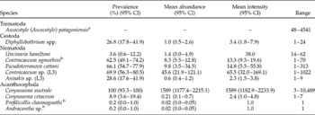
All South American sea lions harboured intestinal helminths; infection parameters are shown in table 1. A total of 97,325 helminth specimens were collected; 88,998 were acanthocephalans, 3684 were nematodes, 4589 were digeneans and 54 were cestodes. Adult specimens were ascribed to six species, namely, Ascocotyle (Ascocotyle) patagoniensis, Diphyllobothrium spp., Contracaecum ogmorhini s.s., Pseudoterranova cattani, Uncinaria hamiltoni and Corynosoma australe. In addition, five forms were found either as third-stage larvae (the nematodes Contracaecum sp. and Anisakis sp. type I) or juvenile individuals (Andracantha sp., Profilicollis chasmagnathi and Corynosoma cetaceum).
Infection parameters of helminths found in 56 South American sea lions (Otaria flavescens) from northern Patagonia, Argentina.

CI, confidence interval; L3, third-stage larvae.
a Infection parameters could not be calculated for the entire sample (see Materials and methods). The range corresponds to two hosts for which a complete census of specimens was obtained.
b New host record.
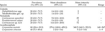
In the South American fur seals, helminths were collected from the four males; the female was uninfected (table 2). From a total of 1516 helminth individuals, 1408 specimens were acanthocephalans, 99 were nematodes and 9 were cestodes. Adult specimens corresponded to three species, namely, Diphyllobothrium spp., C. ogmorhini s.s. and C. australe. In addition, four forms were found either as third-stage larvae (Contracaecum sp.), fourth-stage larvae (P. cattani), juvenile (C. cetaceum) or sexually immature individuals (Tetrabothriidae gen. sp.).
Infection parameters of helminths found in five South American fur seals (Arctocephalus australis) from northern Patagonia, Argentina.

CI, confidence interval; L3, third-stage larvae.
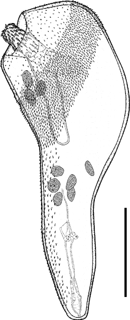
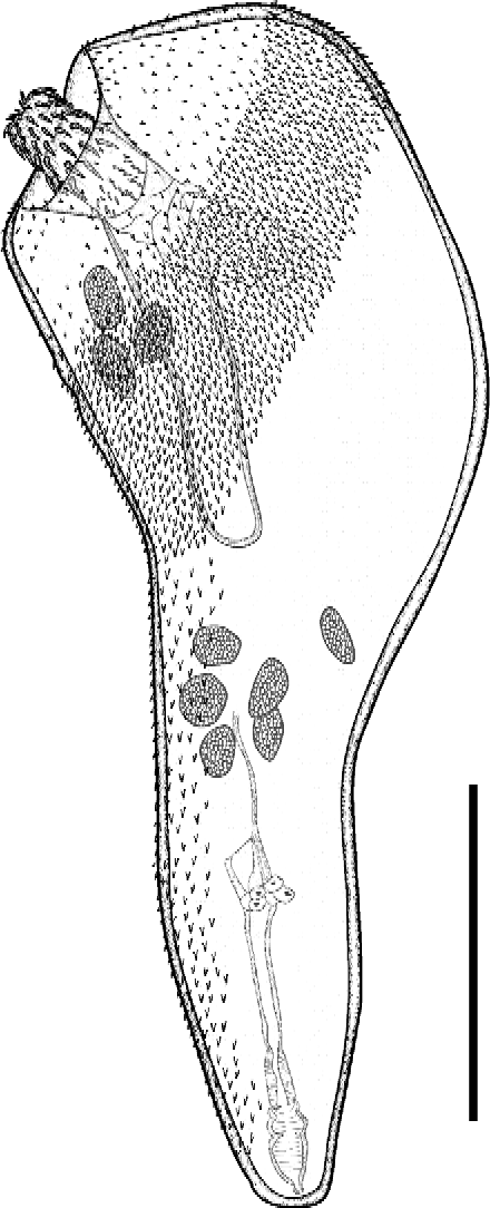
A single female specimen of an unidentified species of Andracantha was found in the rectum of one South American sea lion. The specimen, which was poorly preserved, had numerous ovarian balls. The pipe-shaped trunk was 2594 long and 1574 wide (disc diameter). The disc was covered with spines, except in a small circle on the anterior part where spines became scattered, or even disappeared in a small ventral area (fig. 1). A continuous field of faint spines was also observed along the hind trunk, but spines did not reach the vicinity of the genital pore (fig. 1). The proboscis was partly invaginated and its whole morphological features could only be ascertained by transparency. It had a conical shape, widest at its base, 670 long and 270 wide, with 12 hooks, the 4 basal ones being rootless.
Andracantha sp. from the intestine of the South American sea lion, Otaria flavescens from northern Patagonia, whole-mount lateral view. Scale bar: 1000 μm.
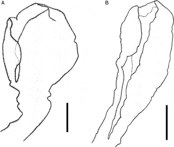
Specimens of Diphyllobothrium spp. collected from the South American fur seal had a rounded–ellipsoidal scolex in lateral view (n= 2; fig. 2A). Gravid proglottids were 1944 ± 214 (1667–2292, n= 13 from two specimens) long and 3391 ± 274 (3042–3833) wide. Width–length ratio of mature proglottids was 1:1.8 (1:1.3–2.2). Transverse tegumentary recesses and pits were observed extending ventrally from the anterior margin of proglottid to the anterior margin of genital atrium on the midline of proglottid. Eggs were 56.9 ± 2.6 (52.1–59.6) long and 39.6 ± 2.0 (36.2–43.3) wide (n= 25, from six proglottids of two specimens). No other structures were discernible.
Scolices of Diphyllobothrium spp. collected from the intestine of otariids from northern Patagonia, Argentina, lateral view. (A) Diphyllobothrium spp. from the South American fur seal, Arctocephalus australis. (B) Diphyllobothrium spp. from the South American sea lion, Otaria flavescens. Scale bars: 500 μm.

Specimens of Diphyllobothrium spp. from the South American sea lion had a lanceolate scolex (n= 54; fig. 2B). Gravid proglottids were 2874 ± 763 (1750–4042, n= 15 from two specimens) long and 2056 ± 263 (1667–2500, n= 15) wide. Width–length ratio of mature proglottids was 1:0.78 (1:0.42–1.33). Transverse tegumentary recesses or pits could not be observed in these specimens, probably because the tegument was poorly preserved. Eggs were 55.8 ± 2.2 (54.0–60.4) long and 35.2 ± 2.0 (33.3–37.5) wide (n= 18, from five proglottids of three specimens). No other structures were discernible.
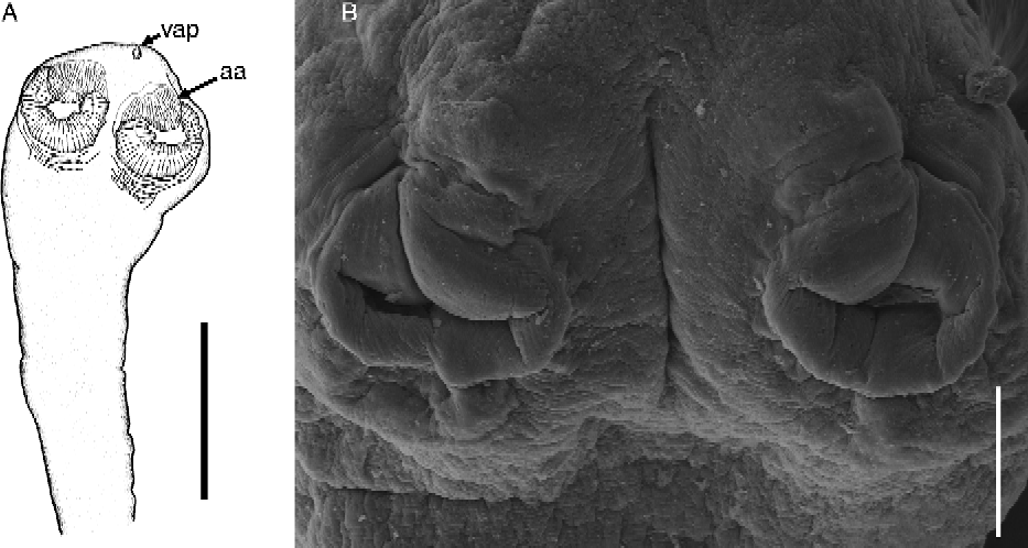
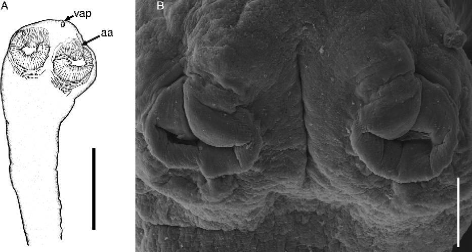
Specimens of Tetrabothriidae gen. sp. from the South American fur seal had a round scolex (n= 7; fig. 3A) with four round, sucker-like bothridia; 184 ± 51 (81–226, n= 10) long and 166 ± 36 (93–196) wide, each with a small appendage located in antero-lateral to anterio-medial position (fig. 3A and B). Remains of a sucker-like structure were observed at the apex of the scolex in two specimens (fig. 3A). Numerous fragments of strobili with non-mature proglottids were observed.
Tetrabothriidae gen. sp. from the South American fur seal, Arctocephalus australis. (A) Scolex, lateral view. (B) Scanning electron micrographs of the round, sucker-like bothridia, lateral view. Scale bars: 400 μm (A) and 60 μm (B). vap, vestiges of apical sucker; aa, anterior appendage.
The intestinal component community of helminths from the South American sea lion was composed of 11 species. At infracommunity level, species richness ranged from 1 to 6 species (mean ± SD: 3.1 ± 1.1 species). A total of 3 hosts (5.4%) were infected with 1 helminth species; 14 (25.0%) with 2; 20 (35.7%) with 3; 14 (25.0%) with 4; 4 (7.1%) with 5, and a single host (1.8%) with 6. In contrast, the intestinal component helminth community of the South American fur seal was composed of seven species. At infracommunity level, species richness ranged from 1 to 5 species (2.2 ± 1.9). One host (20%) was infected with 1 helminth species, 1 (20%) with 2, 1 (20%) with 3, and 1 host (20%) with 5. A single host (a 1-year-old female) was uninfected. There were no significant differences of infracommunity richness between sea lions and fur seals (Mann–Witney test, U= 91.5, P= 0.209).
Values of helminth species richness are strongly influenced by the taxa that were found only as larvae or juvenile individuals. When these taxa are excluded, the component community of the South American sea lion dropped to six species, and mean infracommunity species richness to 2.6 ± 0.9 species (range 1–5). A total of 5 hosts (8.9%) were infected with 1 helminth species; 20 (35.7%) with 2; 22 (39.3%) with 3; 8 (14.3%) with 4 and 1 (1.8%) with 5. In the South American fur seal, the component community excluding larvae and juveniles was composed of four species (mean infracommunity species richness 1.75 ± 1.0). A single host (20%) harboured 4 helminth species and 3 (60%) harboured 1.
Discussion
Parasite composition
Ascocotyle (A.) patagoniensis was recently described from the South American sea lion (Hernández-Orts et al., Reference Hernández-Orts, Montero, Crespo, García, Raga and Aznar2012). Species of the subgenus Ascocotyle infect birds and mammals of freshwater or brackish habitats (Font et al., Reference Font, Heard and Overstreet1984; Ostrowski de Nuñez, Reference Ostrowski de Núñez2001). Therefore, this is the first record of a species from this subgenus in a marine habitat and in a marine mammal. However, the life cycle of A. (A.) patagoniensis is yet to be elucidated; no metacercariae of this species were found in 542 individuals from 20 marine teleost species (Hernández-Orts et al., Reference Hernández-Orts, Montero, Crespo, García, Raga and Aznar2012), nor have specimens of Ascocotyle spp. been reported in marine fish from Patagonia (Timi & Poulin, Reference Timi and Poulin2003; Sardella & Timi, Reference Sardella and Timi2004; Vales et al., Reference Vales, García, Crespo and Timi2011, and references therein). Although the high parasite burdens indicated that it was not an accidental parasite, infection parameters of A. (A.) patagoniensis could not be determined reliably because we could not rule out that an indeterminate number of specimens had been lost during sieving, due to the small size of the parasite. It is therefore advisable that sieves < 0.2 mm mesh are used in future parasitological surveys of sea lions or other pinnipeds.
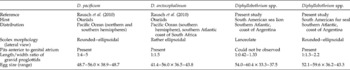
Species of Diphyllobothrium are common intestinal parasites of fur seals and sea lions (Rausch et al., Reference Rausch, Adams and Margolis2010). Two species have been reported frequently in otariids from the southern hemisphere, i.e. D. pacificum and D. arctocephalinum, in the Pacific and Atlantic Oceans, respectively (Rausch et al., Reference Rausch, Adams and Margolis2010). The specimens of Diphyllobothrium spp. collected in the present study were very poorly preserved, which prevents identification at the species level. Based on some morphological traits (e.g. scolex shape, proglottids and size of eggs) specimens of Diphyllobothrium spp. from South American fur seals resemble both D. pacificum and D. arctocephalinum (table 3). Similarities are less evident in specimens collected from South American sea lions (table 3). Molecular data and morphological examination based on suitable material will be necessary for a reliable identification of specimens of species of Diphyllobothrium infecting sea lions and fur seals in northern Patagonia.
Comparison of taxonomic data between Diphyllobothrium pacificum, D. arctocephalinum and unidentified species of Diphyllobothrium collected from sea lions and fur seals in northern Patagonia, Argentina. Measurements in micrometres.

Species of Tetrabothriidae occur as parasites of marine birds and marine mammals and are assigned to six genera based on features of the scolex and the reproductive system (Hoberg, Reference Hoberg, Khalil, Jones and Bray1994). No gravid proglottids were observed in any of the specimens collected from fur seals in our study. Nor did the structure of the scolex allow us to make a reliable generic allocation of our specimens. In tetrabothriids, the presence of vestiges of the larval apical sucker suggests that the ontogeny of the scolex is almost complete (Hoberg, Reference Hoberg1987; Hoberg et al., Reference Hoberg, Adams and Rausch1991; Hoberg & Measures, Reference Hoberg and Measures1995). Accordingly, our specimens probably represented a relatively recent infection and/or they could not progress development further. In any event, the structure of the scolex would preclude the assignation of our specimens to Tetrabothrius, which should exhibit clear, laterally directed auricular appendages at this stage of development (Hoberg, Reference Hoberg1987). The type of anterior appendages that we observed is reminiscent of, but not similar to, those from species of Anophryocephalus or Trygonocotyle (Hoberg, Reference Hoberg, Khalil, Jones and Bray1994, and references therein). New material should therefore be made available to perform further morphological and molecular analyses in order to identify these cestodes.
Species of Uncinaria have been reported in fur seals and sea lions worldwide, including the South American sea lion in the study area (Berón-Vera et al., Reference Berón-Vera, Crespo, Raga and Pedraza2004). Adults reproduce in the intestine of pups, and evidence suggests that pups become infected through the milk of mothers carrying third-stage larvae (Lyons et al., Reference Lyons, DeLong, Gulland, Melin, Tolliver and Spraker2000 and references therein). In our sample, individuals of U. hamiltoni were collected from the two youngest pups, 5 and 7 months old, but were not found in any of ten pups aged 8–10 months. The latter were probably in the weaning process (Cappozzo & Perrin, Reference Cappozzo, Perrin, Perrin, Würsig and Thewissen2009) and had no further opportunity of (re)infection. Therefore, these pups either had never been infected or had cleared previous infections. The latter possibility would confirm that most infections occurred soon after parturition, as has been suggested for species of Uncinaria infecting otariids in the northern hemisphere; in these species, the estimated life-span of the parasite in pups was 3–8 months (Lyons et al., Reference Lyons, DeLong, Gulland, Melin, Tolliver and Spraker2000).
Contracaecum ogmorhini s.s. and P. cattani have been reported in the stomach of pinnipeds in South America (George-Nascimento & Urrutia, Reference George-Nascimento and Urrutia2000; Timi et al., Reference Timi, Sardella and Mattiucci2003). In this study, fourth-stage larvae and adult specimens of both species were frequently found in the intestine of sea lions, and in 1 out of 5 fur seals. Most specimens, especially those collected from the middle and posterior intestine, were degraded, suggesting that they likely were senescent worms from the stomach that were passing throughout the gut. However, whether or not the upper intestine could be a suitable microhabitat for these nematodes is an open question.
Third-stage larvae of Contracaecum sp. were the most abundant nematodes collected from the intestine (tables 1 and 2). Larvae of Contracaecum sp. cannot be identified to the species level based on morphological traits. We believe that our material probably contains not only third-stage larvae from C. ogmorhini s.s., but also from sympatric species of Contracaecum that typically mature in marine birds or other pinnipeds, which come from digested fishes. Apart from C. ogmorhini s.s. at least four species of Contracaecum have been reported in northern Patagonia: C. pelagicum, from the Magellan penguin, Spheniscus magellanicus; C. chubutensis from the Imperial cormorant, Phalacrocorax atriceps; C. osculatum s.l. and C. miroungae from the elephant seal, Mirounga leonina (Mattiucci et al., Reference Mattiucci, Cianchi, Nascetti, Paggi, Sardella, Timi, Webb, Bastida, Rodríguez and Bullini2003; Garbin et al., Reference Garbin, Navone, Diaz and Cremonte2007, Reference Garbin, Diaz, Cremonte and Navone2008).
Species of Anisakis are typical parasites of cetaceans (Mattiucci & Nascetti, Reference Mattiucci and Nascetti2008). The morphology of the third-stage larvae of Anisakis sp. collected from South American sea lions conforms to Berland's (Reference Berland1961) type I, which corresponds to the larvae of A. simplex s.l. (Nascetti et al., Reference Nascetti, Paggi, Orecchia, Smith, Mattiucci and Bullini1986). These larvae are widespread in fishes from northern Patagonia (Timi & Poulin, Reference Timi and Poulin2003; Sardella & Timi, Reference Sardella and Timi2004; Timi & Lanfranchi, Reference Timi and Lanfranchi2009, and references therein), whereas adults have been recorded from species of toothed whales from the same locality (Dans et al., Reference Dans, Reyes, Pedraza, Raga and Crespo1999; Berón-Vera et al., Reference Berón-Vera, Crespo, Raga and Fernández2007, Reference Berón-Vera, Crespo and Raga2008).
Corynosoma australe was the most prevalent and abundant parasite from the intestine of the South American sea lion and the South American fur seal, accounting for >90% of all individual parasites found in both species. This acanthocephalan is a common parasite of pinnipeds (principally otariids) from the southern hemisphere, including sea lions (George-Nascimento & Marin, Reference George-Nascimento and Marin1992; Aznar et al., Reference Aznar, Hernández-Orts, Suárez, García-Varela, Raga and Cappozzo2012) and fur seals (Aznar et al., Reference Aznar, Cappozzo, Taddeo, Montero and Raga2004; Sardella et al., Reference Sardella, Mattiucci, Timi, Bastida, Rodríguez and Nascetti2005). In Uruguay, C. australe is known to readily infect the South American fur seal because the parasite seems to be ecologically ubiquitous, occurring in many species of both pelagic and demersal fish (Aznar et al., Reference Aznar, Cappozzo, Taddeo, Montero and Raga2004). The same situation appears to occur in northern Patagonia, where cystacanths of this species have been found in at least 17 pelagic, benthopelagic and benthic fish species (Hernández-Orts, unpublished data); there are additional records in other parasitological surveys (Timi & Poulin, Reference Timi and Poulin2003; Sardella & Timi, Reference Sardella and Timi2004; Vales et al., Reference Vales, García, Crespo and Timi2011, and references therein). Not surprisingly, C. australe appeared in all sea lions analysed, even in pups aged 4, 5 or 7 months.
Juvenile specimens of three additional acanthocephalan species, i.e. C. cetaceum, P. chasmagnathi and Andracantha sp. were found in this study. It has been suggested that C. cetaceum is specific to cetaceans, but juvenile specimens also occur in different pinniped species, including both species of otariids from South America (Aznar et al., Reference Aznar, Bush, Balbuena and Raga2001, Reference Aznar, Hernández-Orts, Suárez, García-Varela, Raga and Cappozzo2012). Our data confirm that C. cetaceum is apparently unable to mature in sea lions and fur seals. Definitive hosts for species of Profilicollis are marine fish-eating birds (Zdzitowiecki, Reference Zdzitowiecki, Wägele and Sieg1991). Profilicollis chasmagnathi is the only species from this genus that has been reported from the Atlantic coast of South America. There are records of cystacanths in grapsid crabs from Uruguay (Holcman-Spector et al., Reference Holcman-Spector, Mané-Garzón and Dei-Cas1977). Adult worms have been recorded in Olrog's gull, Larus atlanticus, from Bahía Blanca (La Sala & Martorelli, Reference La Sala and Martorelli2007) and in the kelp gull, Larus dominicanus, from northern Patagonia (Diaz et al., Reference Diaz, Cremonte and Navone2011).
Species of Andracantha also reproduce in marine birds, especially cormorants, worldwide (García-Varela et al., 2009). In Argentina, Andracantha sp. had only been reported from nototheniid fishes in the Beagle Channel (Laskowski & Zdzitowiecki, Reference Laskowski and Zdzitowiecki2009). We assigned our specimen to this genus based on the observed pattern of trunk spination, which conforms to the generic concept as defined by Zdzitowiecki (Reference Zdzitowiecki1989), i.e. the possession of conspicuous spines arranged in two circular fields separated from each other by either a bare zone or a zone covered with smaller spines. Unfortunately, we were unable to make a reliable specific assignation of our immature specimen because it, too, was in poor condition and had a partly inverted proboscis. Interestingly, our specimen closely resembles immature specimens of an unidentified species of Andracantha that was recently found in a Franciscana dolphin, Pontoporia blainvillei, in Buenos Aires Province (Aznar et al., Reference Aznar, Hernández-Orts, Suárez, García-Varela, Raga and Cappozzo2012). Overall, specimens of Andracantha collected from marine mammals in Argentina are most similar to A. phalacrocoracis, but the combination of a singular pattern of trunk armature and proboscis traits does not preclude the possibility that they might represent a new species (see Schmidt, Reference Schmidt1975; Zdzitowiecki, Reference Zdzitowiecki1989).
Parasite diversity
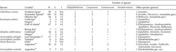
Otariids from northern Patagonia seem to be suitable hosts for 6–7 species (depending on whether there are one or two species of Diphyllobothrium), five of which belong to genera whose species infect otariids worldwide (table 4), i.e. Diphyllobothrium, Corynosoma, Pseudoterranova, Contracaecum and Uncinaria (Dailey, Reference Dailey1975; Lauckner, Reference Lauckner and Kinne1985; Nascetti et al., Reference Nascetti, Paggi, Orecchia, Smith, Mattiucci and Bullini1986; Aznar et al., Reference Aznar, Pérez-Ponce de León and Raga2006; Castinel et al., Reference Castinel, Duignan, Pomroy, Lyons, Nadler, Dailey, Wilkinson and Chilvers2006; Rausch et al., Reference Rausch, Adams and Margolis2010, and references therein). The global occurrence of this predictable group of species in pinnipeds suggests that associations might have been established prior to the splitting of the three major pinniped clades (Hoberg & Adams, Reference Hoberg and Adams2000); nevertheless, Uncinaria is almost exclusively associated with otariids (George-Nascimento et al., Reference George-Nascimento, Lima and Ortiz1992). However, the coevolutionary history between these five parasite taxa and otariids may be complex, including independent associations of the parasites with fur seals and sea lions during the expansion to the southern hemisphere (Hoberg & Adams, Reference Hoberg and Adams2000).
Richness and composition of intestinal heminth fauna of otariid species obtained from surveys worldwide. N, host sample size; C, number of helminth taxa in the total sample (component community); I, number of taxa per individual host (infracommunity) calculated as the sum of prevalences expressed on a per unit basis.

NA, Not available.
a Source: 1Machida (Reference Machida1969); 2Delyamure & Skrjabin (Reference Delyamure and Skrjabin1960); 3Krotov & Delyamure (Reference Krotov and Delyamure1952); 4Dailey & Hill (Reference Dailey and Hill1970); 5Stroud (Reference Stroud1978); 6Shults (Reference Shults1986); 7Sepulveda & Alcaino (Reference Sepulveda and Alcaino1993); 8Delyamure & Parukhin (Reference Delyamure and Parukhin1968); 9George-Nascimento & Carvajal (Reference George-Nascimento and Carvajal1981); 10Present study.
There are additional taxa that appear to have become associated with otariids at a more regional geographical scale. This might be the case of species of Anophryocephalus, Phocitrema and Stictodora in the northern hemisphere, and species of Ogmogaster and Ascocotyle in the southern hemisphere (see references in table 4). These associations are probably linked to a history of host-switching, with geographical restriction resulting, at least in part, from the temporal limits of the initial colonization event (Hoberg & Adams, Reference Hoberg and Adams2000; see also Hoberg, Reference Hoberg1995, for a detailed account of the history of Anophryocephalus spp.). Finally, there is another group of species whose consideration as actual community members is more doubtful. As many as six parasite taxa found in this study have cetaceans, marine birds and even fish as definitive hosts, pinnipeds being apparently unsuitable hosts for these taxa. This might also be the case at least for species of Anisakis, Tetrabothriidae, Bolbosoma and Nanophyetus reported in other otariids (table 4).
The exchange of parasites species is not particularly surprising in hosts that belong to the same trophic level, because many infective stages of parasites can end up in the ‘wrong’ hosts (Hoberg, Reference Hoberg1987, Reference Hoberg1996; Hoberg & Adams, Reference Hoberg and Adams2000; Raga et al., Reference Raga, Fernández, Balbuena, Aznar, Perrin, Würsig and Thewissen2009). This process is apparent in guilds of hosts with close phylogenetic affiliation; for instance, Antarctic pinnipeds share several species of Corynosoma, but not all species are able to mature in all pinniped species (Zdzitowiecki, Reference Zdzitowiecki, Wägele and Sieg1991). The point is, however, the extent to which species that do not reproduce in a given host can be qualified as members of its helminth community or assemblage with regard to taxonomic composition or diversity (e.g. species richness) of the assemblage, or the potential for interspecific interactions. For instance, the helminth component community of the South American sea lion is close to the upper limit of values reported for otariids worldwide (table 4). However, this does not result just from an artefact due to a high sampling effect, but also from the inclusion of parasites for which sea lions are putative non-hosts. Obviously, the category ‘non-host’ is based on the assumption that the absence of sexually mature worms is related to their inability to reproduce in sea lions and/or fur seals, rather than to the fact that these infections are recent (Mateu et al., Reference Mateu, Raga and Aznar2011; Aznar et al., Reference Aznar, Hernández-Orts, Suárez, García-Varela, Raga and Cappozzo2012). This assumption appears to be reasonably supported in parasite taxa that are typical in other definitive hosts, i.e. Andracantha sp. and P. chasmagnathi from birds, C. cetaceum and Anisakis sp. from cetaceans. However, the caveat remains that larger host sample sizes collected over longer time frames could be necessary to confirm categorization of sea lions and fur seals as ‘non-hosts’ for specific parasites.
Omitting the parasite taxa for which sea lion and fur seals are putative non-hosts can have a significant impact on the estimation of species richness, particularly at component community level, and can lead to more accurate comparisons among otariid species (table 4). Unfortunately, information about the reproductive status of helminths is frequently lacking from parasitological surveys of otariids, or it is merely assumed that any parasite found in a host is part of their helminth fauna (see references in table 4). However, data from our study highlight the quantitative difference of including or excluding parasites obtained from non-hosts (see Aznar et al., Reference Aznar, Hernández-Orts, Suárez, García-Varela, Raga and Cappozzo2012). We urge researchers to always provide appropriate information on maturity status of the parasites, if they are to be informative for parascript studies (e.g. Hoberg & Adams, Reference Hoberg and Adams2000) or meta-analyses (e.g. Poulin, Reference Poulin1995; Lindenfors et al., Reference Lindenfors, Nunn, Jones, Cunningham, Sechrest and Gittleman2007).
Acknowledgements
We are indebted to J.M. Carrillo, M.S. Leonardi and P. Mateu for technical assistance. We also thank to M. Ferrer-Montell, S. Sánchez-Quiñones, G. Shira and N. Fraija for their assistance with parasite collection. Thanks are also due to F. Grandi, R. Loizaga de Castro, M. Degrati and S. Dans of the Laboratorio de Mamíferos Marinos of the Centro Nacional Patagónico, and to A. Romero, G. Svendsen and R. González of the Instituto de Biología Marina y Pesquera Almirante Storni, for their assistance with stranded animals. The authors thank J.A. Balbuena for reviewing the paper. This study was supported by the following projects: Interacciones entre Pesquerías y Mamíferos Marinos en el Litoral Patagónico-Fueguino (ANPCYT/PICT9801-04025); Conservación de la Diversidad Biológica Marina y Prevención de la Contaminación en Patagonia (GEF/PNUD ARG 02/018); Estudio de las Amenazas para la Conservación de Mamíferos Marinos de Patagonia (BBVA, BIOCON 04); South American sea lion conservation programme of the Amnèville Zoo, France; CGL2007-6321 from the Ministry of Education and Science of Spain; and PROMETEO 2011-040 of the Valencian Government. Institutional support was given by Centro Nacional Patagónico (CONICET, Argentina). Permits were provided by Secretaría de Áreas Protegidas y Turismo, and Dirección de Fauna y Flora Silvestre, Chubut Province (Argentina). J.S.H.O. benefited from a PhD student grant from the Consejo Nacional de Ciencia y Tecnología (CONACYT) of the Mexican Government (scholarship number 197266).