Many organisms synthesize amazingly hard and tough functional materials by combining inorganic minerals with organic macromolecules in complex, hierarchical structures.[Reference Meyers, Chen, Lin and Seki1] For example, sea urchin teeth, which contain high magnesium content polycrystalline calcite (CaCO3) embedded in a protein-based matrix, are hard and tough enough to grind limestone rocks.[Reference Ma, Cohen, Addadi and Weiner2] Although biogenic calcite has been reported to be harder than geologic calcite,[Reference Wang, Addadi and Weiner3–Reference Griesshaber, Schmahl, Neuser, Pettke, Blum, Mutterlose and Brand8] the reasons for the increased hardness are not well understood. Changes in hardness have been speculated to arise from differences in magnesium[Reference Wang, Addadi and Weiner3–Reference Moureaux, Pérez-Huerta, Compère, Zhu, Leloup, Cusack and Dubois5, Reference Pérez-Huerta, Cusack, Zhu, England and Hughes7, Reference Griesshaber, Schmahl, Neuser, Pettke, Blum, Mutterlose and Brand8] and organic macromolecule content,[Reference Kunitake, Mangano, Peloquin, Baker and Estroff4, Reference Perez-Huerta, Cusack and Zhu6, Reference Pérez-Huerta, Cusack, Zhu, England and Hughes7, Reference Aizenberg and Hendler9] as well as microstructural features including crystallographic texture,[Reference Kunitake, Mangano, Peloquin, Baker and Estroff4, Reference Perez-Huerta, Cusack and Zhu6] crystallite size,[Reference Merkel, Deuschle, Griesshaber, Enders, Steinhauser, Hochleitner, Brand and Schmahl10] and inorganic-to-organic ratio.[Reference Wang, Addadi and Weiner3] However, it is not possible to quantify the relative importance of these compositional and microstructural differences from the existing data, because multiple parameters vary simultaneously between hardness measurements of different biologic systems and even within measurements of a single system. [Reference Moureaux, Pérez-Huerta, Compère, Zhu, Leloup, Cusack and Dubois5, Reference Griesshaber, Schmahl, Neuser, Pettke, Blum, Mutterlose and Brand8]
In previous work,[Reference Kunitake, Mangano, Peloquin, Baker and Estroff4] we showed that the hardness of single-crystal calcite is sensitive to the orientation of the crystal (both the crystal face indented and the azimuthal angle of the indenter with respect to that face), and that the combination of the mechanisms listed above leads to a hardness for single-crystal calcite from the outer shell of the mollusk Atrina rigida that is as much as 70% greater than the hardness of a relatively pure geologic single-crystal calcite, Iceland spar (at the same orientation). To provide more detailed insight into the effect of magnesium incorporation on the hardness of single-crystal calcite, we have produced synthetic samples with excellent control of both composition and microstructure, and investigated their mechanical properties using nanoindentation.
We created samples with a range of Mg concentrations by epitaxially overgrowing synthetic calcite seed crystals with a layer of calcite that contains a gradient in magnesium concentration. The equilibrium solubility of Mg in calcite is about 1 at.%.[Reference Anovitz and Essene11] To create a gradient in Mg concentration, a fixed volume solution of 1 mM:5 mM Mg:Ca was used.[Reference Raz, Weiner and Addadi12] As the thermodynamically favorable calcite is deposited, the ratio of magnesium to calcium ions remaining in solution increases, leading to increased magnesium incorporation in the overgrowth with time.
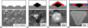
As seed crystals, we chose to use calcite grown in a 1 w/v% agarose hydrogel, which yields samples with uniform size and shape.[Reference Li and Estroff13] To orient the seed crystals with their c-axis perpendicular to the substrate, an array of ~50 µm deep impressions was made in a 1/16th-inch-thick aluminum sheet [Fig. 1(a)] using a home-built cubic zirconia indenter ground and polished to the shape of a calcite rhombohedron, generating an array of oriented sockets for the seed crystals. A single ~75 µm calcite seed was then carefully placed into each impression, letting gravity orient it such that the c-axis was aligned with the indentation axis [Fig. 1(b)]. Using a solution of 1 mM MgCl2 and 5 mM CaCl2,[Reference Falini, Gazzano and Ripamonti14] we then epitaxially overgrew the seed crystals by gas diffusion of NH4(CO3)2 leading to a ~20-μm-thick overgrowth of Mg-containing calcite [Fig. 1(c)]. After 10 days of overgrowth, the crystals were rinsed and dried, and cyanoacrylate resin was poured over the top to encapsulate the crystals. The seed crystals with Mg-containing calcite overgrowth were then polished to expose the {001} face for measurement [Fig. 1(d) and Supplemental Fig. S1].
Profile (schematic) and top (a–c: light and, d: electron microscope images) views of experimental setup. (a) Array of impressions in Al substrate made to hold seed crystals with their c-axis perpendicular to the substrate. (b) Seed crystal in impression. (c) Seed crystal with Mg-containing calcite overgrowth. (d) Secondary electron (SE) image of polished cross-section of crystal embedded within cyanoacrylate resin with black line overlay showing outline of seed crystal. Key: gray: Al substrate; black: calcite seed crystal; red: Mg-containing calcite overgrowth; blue: cyanoacrylate embedding resin.

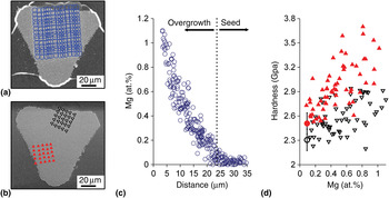
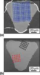
We then used wavelength dispersive spectroscopy (WDS) to measure the magnesium and calcium content in the overgrowth region for five different synthetic crystals [Fig. 2(a)]. The WDS data showed that the magnesium concentration was fairly uniform parallel to the growth front (Supplemental Fig. S2), and increased at a nonlinear rate as a function of distance from the seed crystal–overgrowth interface [Fig. 2(c)]. We also measured the Mg content in Iceland spar and A. rigida to be 0.107 ± 0.021 at.% (n = 32) and 0.38 ± 0.05 at.% (n = 48), respectively.
(a) Scanning electron microscope (SEM) image with overlay of blue circles showing location of WDS magnesium and calcium measurements (5 µm spot size). (b) SEM image with triangle array overlay showing location of nanoindentation measurements for 0° (open black) and 60° (filled red) azimuthal angles for one of five crystals. (c) Mg content as a function of distance from outer edge of crystals. Dotted line indicates approximate interface between seed-crystal and overgrowth region. For a diopside (MgCaSi2O6) standard, the Mg content was 18.2 at.% and the standard deviation as measured by counts per second was ±0.8 at.%. (d) Hardness as a function of Mg content in the overgrowth region. Triangles represent 0° (open black) and 60° (filled red) azimuthal angles while correspondingly colored circles represent the reference geologic values. For a standard fused silica sample, the hardness was 9.10 ± 0.15 GPa (n = 4). Plots in (c) and (d) are compilations of data from five different crystals.

We used nanoindentation to measure the hardness and plane strain indentation modulus[Reference Oliver and Pharr15] of the polished faces at two different azimuthal angles in the same five crystals [Fig. 2(b)]. The resulting indentations were ~200 nm deep. Surface topography scans were performed with the indenter tip before and after indenting. These scans are sensitive enough to detect differences in polishing between the seed crystal and the overgrowth region (Supplemental Fig. S1). To avoid any influence from the seed crystal or the sample edges, we included only indentations that were more than 2 µm away from these interfaces.
Hardness as a function of magnesium concentration was determined by using the distance from the overgrowth surface as a common parameter [Fig. 2(d)]. Hardness increases with increasing Mg content. Both 0° and 60° azimuthal orientations approach the values of geologic calcite with decreasing magnesium content, confirming the accuracy of this method. The plane strain indentation modulus of the overgrowth regions was similar to geologic calcite and consistent with DFT calculations[Reference Elstnerova, Friak, Fabritius, Lymperakis, Hickel, Petrov, Nikolov, Raabe, Ziegler, Hild and Neugebauer16] for small changes in composition (Supplemental Fig. S3).
We assume that the mechanism by which magnesium hardens calcite is solid solution hardening. X-ray diffraction,[Reference Pokroy, Fitch, Marin, Kapon, Adir and Zolotoyabko17] computational studies,[Reference Elstnerova, Friak, Fabritius, Lymperakis, Hickel, Petrov, Nikolov, Raabe, Ziegler, Hild and Neugebauer16, Reference Stashans and Chamba18] experimental phase diagrams,[Reference Anovitz and Essene11] transmission electron microscopy,[Reference Tsipursky and Buseck19, Reference Barber, Reeder and Smith20] atomic force microscopy,[Reference Davis, Dove, Wasylenki and De Yoreo21] and cathodoluminescence[Reference Paquette and Reeder22] all show that magnesium is incorporated substitutionally into the calcite lattice at low concentrations, ≤1 at.%. Since magnesium is a smaller cation than calcium, it is expected to create lattice distortions (a stress field)[Reference Elstnerova, Friak, Fabritius, Lymperakis, Hickel, Petrov, Nikolov, Raabe, Ziegler, Hild and Neugebauer16, Reference Stashans and Chamba18] that would hinder dislocation motion and increase the hardness. Theories for substitutional impurity strengthening suggest that the yield strength of single-crystal metals follows a square root dependence,[Reference Courtney23] and it has been proposed that a similar relationship holds for ionic single crystals.[Reference Evans and Langdon24] Therefore, we might expect a similar functional form for our hardness. We fit both linear and square root dependences to our data, but there was no difference in the quality of fit. In an attempt to extend our data set, we tried using a higher magnesium-to-calcium ratio (5 mM:5 mM) for the overgrowth solution.[Reference Long, Nasse, Ma and Qi25, Reference Long, Ma and Qi26] However, the overgrowth, while epitaxial, was no longer smooth and well defined, leading to difficulties in correlating hardness with magnesium content. Thus, our experiments were constrained to equilibrium magnesium contents of <1 at.%.
Given the relationship shown in Fig. 2(d), it is possible to evaluate the effect of magnesium in previous reports of the increased hardness of biogenic calcite. To a first-order approximation, we have assumed that the effect of magnesium on the hardness of calcite may be separated out from the contributions due to other impurities (e.g., biomacromolecules and microstructural features found in biogenic calcite). For calcite crystals (with 0.4 at.% Mg) from the mollusk, A. rigida, we reported a hardness between 3.5 and 4.2 GPa for the {001} plane depending on azimuthal angle.[Reference Kunitake, Mangano, Peloquin, Baker and Estroff4] From Fig. 2(d), a 0.4 at.% Mg-containing calcite crystal has a hardness of 2.4–2.9 GPa. Therefore, assuming that strengthening mechanisms in biogenic calcite are independent of each other, approximately 20% of the increased hardness found in calcite prisms from A. rigida could come from magnesium content. In another organism, the brachiopod Megerlia truncata, the magnesium content of the primary and secondary layers has been reported to range from 0.2 to 0.9%[Reference Griesshaber, Schmahl, Neuser, Pettke, Blum, Mutterlose and Brand8] and therefore all of the increased hardness of the dorsal valve, which has been measured at 2.85 ± 0.41 GPa, could come from magnesium solid solution hardening.[Reference Merkel, Deuschle, Griesshaber, Enders, Steinhauser, Hochleitner, Brand and Schmahl10]
Other species produce supersaturated solid-solution Mg-containing calcites (>1 at.%). For example, the spines from the sea urchin Paracentrotus lividus are comprised of calcite-containing 3–5% magnesium in solid solution, and the nanoindentation hardness on the {001} plane in the septa has been found to range from 3.6 to 4.1 GPa.[Reference Moureaux, Pérez-Huerta, Compère, Zhu, Leloup, Cusack and Dubois5] If we extrapolate from our data to the magnesium concentrations found in the sea urchin septa, it is possible that magnesium impurities could account for the increased hardness.
In summary, we have quantitatively determined the effect of magnesium incorporation on the hardness of calcite. Our results show that magnesium incorporation plays a significant role in hardening calcite, even at relatively low magnesium concentrations (in the equilibrium solid solubility regime <1 at.%). The results are consistent with a solid-solution strengthening mechanism. A significant fraction of the hardness of several biogenic calcites, compared with a nearly pure geologic reference, can be attributed to magnesium impurities. To elucidate other hardening mechanisms in biogenic calcites, a similar approach, in which synthetic samples with well-controlled composition and microstructure are prepared and tested (e.g., calcite with incorporated organic macromolecules), can be used.
Acknowledgments
We acknowledge support from NSF (DMR 0845212), the J.D. Watson Investigator Program (NYSTAR Contract No. C050017), the GI Bill (Chapter 33), and Hysitron Inc. We also acknowledge the Cornell Center for Materials Research (CCMR), a Materials Research Science and Engineering Center of the National Science Foundation (DMR 1120296), for both research support and use of the Electron Microscopy facility. We would also like to thank John Hunt for WDS measurements and Benjamin Shulman for help with sample preparation. Supporting Information is available online from MRS Communications or from the authors.
Supplementary materials
For supplementary material for this article, please visit http://dx.doi.org/10.1557/mrc.2012.20