Introduction
The most common chronic diseases of the paranasal sinuses are chronic paranasal sinusitis and fungal sinusitis. Clinically, blood-tinged post-nasal drip is seldom observed in these two disease entities.Reference Schwartz and Thiel1 The presence of this symptom might indicate a different diagnosis in patients with paranasal sinus disease. A larger amount of bleeding from paranasal sinus lesions would cause epistaxis, and exploration to investigate a possible sinonasal origin would be performed. However, if the bleeding was mild, blood-tinged post-nasal drip would be present and the differential diagnosis would be more challenging.
Although the presence of blood-tinged post-nasal drip would intuitively prompt suspicion of other sinonasal diseases or malignancies, no previously published study has assessed the significance of this symptom in patients with paranasal sinus disease. The purpose of this study was to analyse the clinical importance of this symptom in these patients.
Materials and methods
A prospective study was performed between August 2000 and April 2005. One hundred and ninety-three patients with paranasal sinus lesions who had undergone endoscopic sinus surgery for treatment or biopsy were recruited. Their ages ranged from seven to 73 years, with an average of 42 years. One hundred and thirty-six patients were male and 57 were female. Information on demographic features, clinical symptoms, computed tomography (CT) scan findings and pathological results were collected. Laterality, bony destruction, calcification and Lund–Mackay scoresReference Lund and Mackay2 for CT scan images were also documented. In addition to the usual symptoms of sinonasal diseases, a history of blood-tinged post-nasal drip was recorded. The definition of blood-tinged post-nasal drip was that the symptom was reported by patients during history-taking. In addition, patients had to describe the post-nasal drip sensation clearly, and there had to be no coughing or vomiting associated. This explicit definition aimed to exclude confusion with haemoptysis or haematemesis.
The frequency of blood-tinged post-nasal drip was analysed for different diseases. The distributions of diseases with and without blood-tinged post-nasal drip were compared. In addition, statistical interactions between the presence of blood-tinged post-nasal drip and the patient's age and sex were also analysed. We arbitrarily defined two age groups with a cut-off point of 40 years, to enable comparison. Other symptoms of sinusitis were also collected and analysed. In patients with chronic paranasal sinusitis, these analyses were performed separately. We investigated whether there was a difference in the frequency of blood-tinged post-nasal drip in chronic paranasal sinusitis patients with different local findings and different Lund–Mackay scores. The purpose was to find out if more severe disease was more likely to present with blood-tinged post-nasal drip.
Statistical analyses were performed with the Fisher's exact test. The significant threshold was set as a p value of less than 0.05 in two-tailed results.
Results and analysis
Of the total 193 patients, 12 (6.22 per cent) presented with blood-tinged post-nasal drip. Patients’ diagnoses included a variety of diseases; the distribution of different diseases with and without blood-tinged post-nasal drip is summarised in Table I. The most common diseases in these 193 patients were chronic paranasal sinusitis and fungal sinusitis. Only 10 patients were diagnosed with other conditions. In the 181 patients without blood-tinged post-nasal drip, 177 (97.8 per cent) had chronic paranasal sinusitis and fungal sinusitis. However, in the 12 patients who did present with blood-tinged post-nasal drip, six (50 per cent) were diagnosed with other conditions. This difference was statistically significant (Fisher's exact test, two tails, p < 0.001). Blood-tinged post-nasal drip was found in both patients with malignant disease, which included malignant melanoma of the maxillary sinus and nasal T cell lymphoma.
Distribution of diagnoses in patients with and without blood-tinged post-nasal drip

BTPND = blood-tinged post-nasal drip; AP = antrochoanal polyp; CG = cholesterol granuloma; CPS = chronic paranasal sinusitis; IP = inverted papilloma; MM = malignant melanoma; T cell = nasal T cell lymphoma
Among different diseases, the probability of patients presenting with blood-tinged post-nasal drip was significantly different (Figure 1). In patients with antrochoanal polyp, chronic paranasal sinusitis and fungal sinusitis, blood-tinged post-nasal drip were observed in 0 per cent (zero of one), 2.65 per cent (four of 151), and 6.25 per cent (two of 32), respectively. Nevertheless, all five patients with either cholesterol granuloma, malignant melanoma or nasal T cell lymphoma presented with blood-tinged post-nasal drip.
Frequency of blood-tinged post-nasal drip in different diseases. BTPND = blood-tinged post-nasal drip; AP = antrochoanal polyp; CG = cholesterol granuloma; CPS = chronic paranasal sinusitis; IP = inverted papilloma; MM = malignant melanoma; T cell = nasal T cell lymphoma

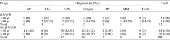
In the 181 patients with paranasal sinus diseases who did not have blood-tinged post-nasal drip, there was no difference in the distribution of diseases between older and younger age groups (Table II). However, in the 12 patients with paranasal sinus diseases who did have blood-tinged post-nasal drip, the older patient group included more diagnoses other than chronic paranasal sinusitis and fungal sinusitis, compared with the younger group (Table II). In addition, both patients with malignant diseases were older than 40 years.
Distribution of diagnoses in older and younger patients with and without blood-tinged post-nasal drip

Pt = patient; AP = antrochoanal polyp; CG = cholesterol granuloma; CPS = chronic paranasal sinusitis; IP = inverted papilloma; MM = malignant melanoma; T cell = nasal T cell lymphoma; BTPND = blood-tinged post-nasal drip
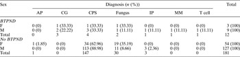
With regard to sex, fungal sinusitis was found more frequently in female patients without blood-tinged post-nasal drip (Table III). However, the proportion of diseases other than chronic paranasal sinusitis and fungal sinusitis was similar in males and females. In the 12 patients with blood-tinged post-nasal drip, the male:female ratio was increased, and the male patients tended to have diagnoses other than chronic paranasal sinusitis and fungal sinusitis (Table III). Both of the patients with malignant disease were male.
Distribution of diagnoses in female and male patients with and without blood-tinged post-nasal drip

AP = antrochoanal polyp; CG = cholesterol granuloma; CPS = chronic paranasal sinusitis; IP = inverted papilloma; MM = malignant melanoma; T cell = nasal T cell lymphoma; BTPND = blood-tinged post-nasal drip; F = female; M = male
Computed tomography images were analysed to assess whether they could provide clues for different diagnoses. In the 193 patients, bony destruction on CT scan images was observed in only three patients, including the patient with nasal T cell lymphoma and two patients with fungal sinusitis. Calcification was noted in seven patients, including six with fungal sinusitis and one with chronic paranasal sinusitis. The laterality of lesions could not aid the differential diagnosis. In other words, CT images alone could not assist the differential diagnosis for our patients. Other symptoms of sinusitis were also analysed; the results were similarly unhelpful in the differential diagnosis.
In the 151 chronic paranasal sinusitis patients, only four complained of blood-tinged post-nasal drip. The symptom was not associated with sex, age, or Lund–Mackay score. However, although not significant statistically, in patients with the local finding of sinonasal polyposis, the frequency of blood-tinged post-nasal drip was slightly higher (three of 53) than in patients with local findings of purulent discharge (one of 98) (Fisher's exact test, two tails, p = 0.1244).
The patient with cholesterol granuloma and the patient with malignant melanoma of the maxillary sinus had visited the chest department before presenting to our clinic. Chest plain X-rays and fibre-optic bronchoscopy were performed, with negative results.
The patient with malignant melanoma of the maxillary sinus presented with blood-tinged post-nasal drip as the only symptom. He was diagnosed with early stage (stage I) disease. Radical maxillectomy was performed, and no recurrence or metastasis was noted after one year's follow up.
Discussion
According to these results, blood-tinged post-nasal drip was not a frequent symptom in patients with paranasal sinus disease. Only 12 of the 193 patients complained of this symptom. However, most of these 12 patients had diagnoses other than chronic paranasal sinusitis or fungal sinusitis. In patients without blood-tinged post-nasal drip, 97.8 per cent had chronic paranasal sinusitis or fungal sinusitis. However, in patients with blood-tinged post-nasal drip, 50 per cent had other diagnoses, including malignancies of the sinonasal tract. These results show that, if a patient with a paranasal sinus lesion complains of blood-tinged post-nasal drip, the otolaryngologist should assess for diagnoses other than the usual sinusitis.
In addition, blood-tinged post-nasal drip was noted in both our patients with malignancies. Therefore, patients with this symptom, in addition to the usual sinonasal symptoms, should be assessed for malignant sinonasal disease.
Of our patients with blood-tinged post-nasal drip, older males had the highest chance of having diseases other than chronic paranasal sinusitis and fungal sinusitis (although this difference was not statistically significant). In addition, malignant disease was found in two such older males. Therefore, it should be a priority to establish definitive diagnoses in older men with paranasal sinus lesions who complain of blood-tinged post-nasal drip.
In this study, we found that CT image signs (including laterality, bony destruction and calcification) did not provide reliable additional clues in the differential diagnosis. The usual sinonasal symptoms were similarly unhelpful. Therefore, blood-tinged post-nasal drip is a very important clinical symptom, especially in older men, indicating a higher risk of diagnoses other than the usual sinusitis.
Blood-tinged post-nasal drip was noted in all five of our patients with cholesterol granuloma, malignant melanoma and nasal T cell lymphoma. In malignant disease, increased vascularity may induce bleeding. Upon physiological drainage, malignant diseases of the sinuses would present with blood-tinged post-nasal drip.Reference Lin, Yang and Lai3 On the other hand, the favoured hypotheses for the mechanism of formation of cholesterol granuloma of the sinuses involve obstruction of ventilation, haemorrhage and impaired drainage.Reference Chao4 This may explain why blood-tinged post-nasal drip presented with such frequency in these patients.
• Blood-tinged post-nasal drip is a rare manifestation of paranasal sinus disease
• This study assessed the clinical significance of this symptom in patients with paranasal sinus diseases
• The majority of patients without the symptom were diagnosed with chronic paranasal sinusitis and fungal sinusitis
• In patients with the symptom, the likelihood of diagnoses other than chronic paranasal sinusitis and fungal sinusitis increased significantly, especially in older males
• Although exploration for possible malignancies is necessary in all patients with paranasal sinus lesions, patients with blood-tinged post-nasal drip have a higher risk of more severe disease, especially older males
In our patients with chronic paranasal sinusitis and fungal sinusitis, blood-tinged post-nasal drip was noted in only 2.65 and 6.25 per cent, respectively. In these cases, the cause of bleeding was unknown, but might be due to an inflammatory response to the sinus lesions. Although the association between Lund–Mackay score and blood-tinged post-nasal drip was not statistically significant, the higher incidence of the symptom in patients with nasal polyposis, compared with those with purulent discharge, implies that more severe chronic paranasal sinusitis disease may be associated with a higher incidence of blood-tinged post-nasal drip. Another implication is that the pathogenesis of these two kinds of chronic paranasal sinusitis might be different. However, further study is necessary to clarify these hypotheses.
Clinically, blood-tinged post-nasal drip may be confused with blood-tinged sputum, which would intuitively be associated with a pulmonary or tracheal lesion. In this study, the one patient with cholesterol granuloma and the one patient with malignant melanoma of the maxillary sinus received fibre-optic bronchoscopy and chest plain X-rays before the final diagnoses were made. Because the symptoms of some sinus lesions are vague, patients presenting with blood-tinged post-nasal drip should be evaluated for possible sinus disease. Such patients should undergo sinoscopy, and CT scanning may be indicated, especially if a possible bleeding source from the sinuses is suspected.
Conclusion
These study findings show that blood-tinged post-nasal drip is an alerting symptom in patients with paranasal sinus lesions. The chance of diagnoses other than the usual sinusitis is increased significantly in such patients, especially in older males. In addition, exploration of sinus lesions should not be neglected in patients with blood-tinged post-nasal drip in whom a bleeding source in other sites of the upper aerodigestive tract has not been identified.