Ectopic pregnancy is defined as a pregnancy implanted outside the uterus, and >98% implant in the Fallopian tube (Refs Reference Jurkovic and Wilkinson1, Reference Sivalingam2). The aetiology of ectopic pregnancy is uncertain, but tubal implantation is probably due to retention of the embryo owing to impaired embryo–tubal transport and alterations in the tubal environment allowing implantation to occur (Refs Reference Shaw3, Reference Brown and Horne4). Once implanted in the Fallopian tube, partial development of the embryo and placenta proceeds for a time, but the thin-walled Fallopian tube is an unsatisfactory site for implantation. The vascular placental tissues penetrate into the thin tubal wall, and if breached, bleeding into the lumen of the tube and peritoneal cavity occurs.
Between 1 and 2% of all pregnancies in Europe and the USA are ectopic and, in the Western world, haemorrhage from an ectopic pregnancy remains the most common cause of maternal mortality in the first trimester of pregnancy (0.35/1000 ectopic pregnancies) (Refs Reference Jurkovic and Wilkinson1, Reference Sivalingam2, Reference Varma and Gupta5). In the developing world, the incidence of ectopic pregnancy is probably much higher, and it has been estimated that 10% of women admitted to hospital with a diagnosis of ectopic pregnancy ultimately die from the condition (Ref. Reference Leke6). In a World Health Organization analysis of maternal deaths, ectopic pregnancy was recorded as the cause in <1% of deaths in developing countries and ~5% in developed countries (Ref. Reference Khan7). Ectopic pregnancy is also a considerable cause of maternal morbidity, causing acute symptoms such as pelvic pain and vaginal bleeding, and long-term problems, such as infertility.
A diagnosis of ectopic pregnancy requires a differentiation to be made from a viable or nonviable intrauterine pregnancy. In severe cases, patients present with haemodynamic shock and the diagnosis is obvious. However, in most cases, early symptoms are either absent or subtle (abdominal pain, vaginal bleeding) and require investigation with transvaginal ultrasound and serial serum β-human chorionic gonadotropin (β-hCG) levels. Studies have shown that >50% of ectopic pregnancies remain undiagnosed at the patient's first presentation, and that the multiple visits and investigations currently necessary to diagnose and exclude ectopic pregnancy are a sizeable burden for health services (Refs Reference Robson and O'Shea8, Reference Wedderburn9). Data from our Scottish study indicate that health services spend between £139 ($232) and £941 ($1570) per patient diagnosing and excluding ectopic pregnancy (Ref. Reference Wedderburn9). This equates to an estimated £9 million ($15 million) per year in direct costs alone to health services per year throughout the UK.
Ectopic pregnancy can be treated expectantly, surgically or medically. Some ectopic pregnancies resolve without treatment (often without significant symptoms) and expectant management is therefore offered in selected cases: that is, if a woman is clinically stable or has falling serum levels of β-hCG, and there is the absence of fetal heart activity on ultrasound. The current surgical treatment of choice is laparoscopic salpingectomy (excision of the Fallopian tube) because of the theoretical risk of recurrence in a damaged Fallopian tube. Salpingotomy (when a linear incision is made in the Fallopian tube to remove the ectopic pregnancy, with the tube itself being retained) is generally performed when the contralateral tube is abnormal. The likelihood of subsequent intrauterine pregnancy seems to be similar after salpingectomy or salpingotomy (Ref. Reference Varma and Gupta5).
Salpingotomy by laparoscopy may lead to fewer complications and shorter recovery times compared with laparotomy, but may also be less likely to remove all the trophoblast (Ref. Reference Varma and Gupta5). Medical treatment relies on using a single (or multiple) intramuscular dose of the folic acid antagonist methotrexate (Refs Reference Jurkovic and Wilkinson1, Reference Sivalingam2). Although the exact mechanism of action is unknown, methotrexate is believed to cause either resorption or tubal abortion of the pregnancy. In addition to allowing preservation of the Fallopian tube, it is a much more cost-effective option when compared with surgery. Unfortunately, the efficacy of methotrexate falls with increasing size of the ectopic pregnancy. Most ectopic pregnancies are not suitable for medical management and require surgical excision. However, 90% present without clinical evidence of rupture, and potentially most of these stable ectopic pregnancies could be treated medically instead of surgically if an efficacious therapeutic existed.
This comprehensive review of the literature examines current understanding of the endocrinology of Fallopian tube biology and ectopic implantation, focusing on genes expressed in the Fallopian tube that are regulated by oestrogen and progesterone. We have not discussed the role of the placenta because we felt it was beyond the scope of this review, although we appreciate that it is not a passive player in ectopic pregnancy pathogenesis. Of note, much of the current Fallopian tube data are descriptive: there are a limited number of functional studies and no good animal models. However, the review concludes with a discussion of how recent advances in knowledge of this field are enabling the development of new disease biomarkers and could lead to the identification of novel medical treatments.
Our search strategy was as follows. We searched ‘Pubmed’ using the terms ‘Fallopian tube’ and ‘ectopic pregnancy’ in combination with ‘estrogen’ or ‘progesterone’ for studies published between 1999 and 2011. We mostly selected publications in the past 5 years, but did not exclude commonly referenced and important older publications. We also searched the reference lists of articles identified by this search strategy and selected those we judged as relevant. For a study to be included, it needed to be primarily focused on the effect of oestrogen or progesterone on Fallopian tube gene expression and function. Genes identified on this basis were then discussed in the context of ectopic pregnancy. Studies that were solely epidemiological in nature were not included.
Normal intrauterine embryo implantation
Successful intrauterine implantation is a complicated hormonally regulated process that requires synchronisation between the developing embryo and the differentiating uterine endometrium (Ref. Reference Norwitz, Schust and Fisher10).
Fertilisation occurs in the Fallopian tube within 24–48 h of ovulation. As the embryo migrates through the Fallopian tube, it develops from a fertilised ovum (zygote) to a mass of 12–16 cells (morula) and becomes encased in a nonadhesive protective coating (zona pellucida). Within 48–72 h after fertilisation, the morula enters the uterine cavity and becomes a blastocyst with the appearance of a fluid-filled inner cavity. The process of normal implantation occurs within 72 h of entering the uterus.
Normal intrauterine implantation only occurs if the endometrium of the uterus is receptive during a putative ‘window of implantation’ within the mid-luteal phase of the menstrual cycle (Ref. Reference Norwitz, Schust and Fisher10). Outside this time, the endometrium is resistant to embryo attachment. Cellular differentiation of the endometrium occurs with the appearance of apical epithelial protrusions (pinopodes), oedema of the endometrial stroma and vascular proliferation. At the molecular level, changes in the endometrial expression pattern of cell adhesion molecules, cytokines, HOX genes and growth factors are observed. Most of these changes are regulated by the sex steroid hormones oestrogen and progesterone (Ref. Reference Critchley and Saunders11). As many as 571 endometrial genes, representing 131 biochemical pathways, are considered as progesterone regulated (Ref. Reference Cloke12).
The embryo implants by attaching to the luminal epithelium and invading the stroma of the endometrium. Under the influence of the steroid hormones oestrogen and progesterone, the stromal cells surrounding the implanting embryo undergo a remarkable transformation event. This process, known as decidualisation, is an essential prerequisite for implantation.
The Fallopian tube and the endocrine environment
Mechanisms of sex steroid hormone action
Sex steroid hormones act through intracellular receptors to regulate the expression of downstream effector genes. Receptors for oestrogen and progesterone are localised predominantly to the nucleus of the cell. Binding of the ligand leads to receptor dimerisation and subsequent binding of steroid receptor complexes to hormone responsive elements on DNA that results in transcriptional activation or repression of target genes.
Two oestrogen receptor (ER) genes have been identified, ER-α (ESR1) and ER-β (ESR2), and splice variant isoforms of the latter are expressed in a variety of human tissues (Ref. Reference Critchley and Saunders11). ER-α and ER-β exhibit different functional properties in vitro and show distinct patterns of gene regulation, and it has been suggested that both ERβ1 and ERβ2 may have an impact on ER-mediated gene expression. The human progesterone receptor (PR) is expressed as two isoforms, PR-A (94 kDa) and PR-B (116 kDa), both encoded by a single gene (PGR) (Ref. Reference Critchley and Saunders11). Although both PR-A and PR-B bind progesterone, selective physiological roles for the two isoforms of PR have been documented. Generally, PR-B is transcriptionally the more active of the two isoforms (Ref. Reference Giangrande, Pollio and McDonnell13). Furthermore, PR-A can act as a dominant repressor of PR-B-dependent activation of progestin-sensitive reporter genes, and likewise inhibits the transcriptional activity of receptors for androgens, glucocorticoids and mineralocorticoids (Ref. Reference McDonnell14).
Sex steroid hormone expression in the Fallopian tube
Expression of sex steroid hormone receptors in the Fallopian tube does not simply mirror the changes seen in the endometrium. In the endometrium, the in vivo expression of PR and ER-α is reduced in the mid-luteal phase of the menstrual cycle under the influence of progesterone and seems to be closely associated with the onset of uterine receptivity (Ref. Reference Stavreus-Evers15). In the Fallopian tube, total expression of PR isoforms, PR-AB and PR-B, is significantly reduced during the mid-luteal phase of the cycle (Ref. Reference Horne16). By contrast, expression of ER-α and ER-β in the Fallopian tube remains constant across the menstrual cycle, that is ER-α is not downregulated in the mid-luteal phase, when the tissue is exposed to peak levels of circulating progesterone (Ref. Reference Horne16). Persistent expression of ER-α might explain why under normal circumstances the tubal epithelium is nonreceptive to the embryo as it passes through the Fallopian tube.
Sex steroid hormone-modulated morphological and cellular changes
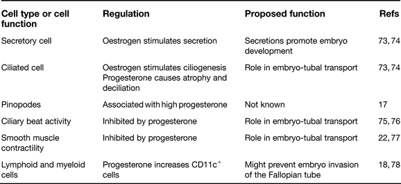
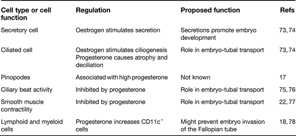
Like the endometrium, the morphology of the Fallopian tube epithelium undergoes cyclical changes under the influence of oestrogen and progesterone (Fig. 1, Table 1). Epithelial cells increase in height during the proliferative phase and reach maximal height and secretory activity at the time of ovulation. Subsequently, in the luteal phase, both cell types reduce in height and there is partial deciliation. Similar to the endometrium, pinopodes appear at the time of uterine receptivity (Ref. Reference Makrigiannakis17). The significance of their appearance is not known. Thus, oestrogen stimulates tubal epithelial cell hypertrophy, secretion and ciliogenesis, whereas atrophy, deciliation and pinopodes are associated with high circulating levels of serum progesterone (Ref. Reference Makrigiannakis17). In addition to epithelial cell proliferation, the sex steroid hormones control ciliary beat activity and smooth muscle contraction, important for the passage of the embryo through the Fallopian tube, and regulate Fallopian tube mucosa lymphoid and myeloid cell populations (Refs Reference Shaw3, Reference Shaw18).
The nonpregnant Fallopian tube. (a) Gross anatomy of the nonpregnant Fallopian tube. (b) Light microscopy image with haematoxylin and eosin staining of the Fallopian tube. The Fallopian tube is comprised of an outer serosa, with inner layers of smooth muscle arranged in a longitudinal and circular manner. Internal to the muscular layers is a mucosa comprised of a delicate vascular lamina propria containing lymphoid and myeloid cell populations. A single layer of columnar epithelium lines the inside of the lumen of the tube. (c) Scanning electron micrograph of the secretory and ciliated epithelial cells of the tubal epithelium. (d) Scanning electron micrograph showing pinopode formation within the Fallopian tube. (e) Progesterone and oestrogen-regulated genes in the Fallopian tube.

Sex steroid hormone-modulated morphological and cellular changes in the nonpregnant Fallopian tube
Progesterone-regulated gene expression
Prokineticins and their receptors
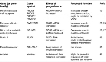
The prokineticins PROK1 and PROK2 are known for their effect on smooth muscle contractility. They are cognate ligands for two closely homologous G-protein-coupled receptors, PROKR1 and PROKR2, through which either prokineticin can signal. The PROKs and their receptor proteins are localised to the smooth muscle and epithelium of the Fallopian tube (Ref. Reference Shaw19). PROKR1 mRNA levels are upregulated in the Fallopian tube in the mid-luteal phase compared with the follicular phase, and increased PROKR1 expression is observed in tubal explant cultures treated with medroxy-progesterone acetate (Fig. 1, Table 2) (Ref. Reference Shaw19). Carefully controlled expression of PROKR1 in the Fallopian tube is likely to be important in regulating tubal smooth muscle contractility and in ensuring successful embryo-tubal transport. This mechanism is potentially mediated through the cyclo-oxygenases: PROK1 has been shown to upregulate COX-2 levels in the endometrium (Ref. Reference Evans20) and the human Fallopian tube expresses COX-2 (Ref. Reference Arbab21); some prostaglandins have recently been reported to increase Fallopian tube smooth muscle contractility (Ref. Reference Wanggren22).
Progesterone-regulated genes in the nonpregnant Fallopian tube

Endocannabinoid receptor
Genetic and pharmacological silencing of the endocannabinoid receptor CB1 (CNR1) has been shown to cause embryo retention within the Fallopian tube in mice (Ref. Reference Wang23). In the mouse oviduct, CNR1 mRNA and CB1 protein are expressed in the smooth muscle, and it is proposed that anandamide (AEA), the major ligand for CB1, controls Fallopian tube smooth muscle action (Ref. Reference Wang23). Interestingly, progesterone in the mouse uterus has been shown to regulate the production of fatty acid AEA hydrolase, an enzyme that controls the cellular uptake and degradation of AEA (Ref. Reference MacCarrone24). In humans, CB1 has been localised to the smooth muscle of the Fallopian tube and has been shown to be progesterone regulated (Fig. 1, Table 2) (Ref. Reference Horne25). Given the detrimental effects of high AEA levels on the embryo and the Fallopian tube, the human data further support the idea that there is an efficient system for its tight regulation under hormonal control (Ref. Reference Horne25).
Nitric oxide and nitric oxide synthetase
NO is produced by the Fallopian tube and has been shown to have a relaxing effect on tubal smooth muscle (Refs Reference Ekerhovd26, Reference Perez Martinez27). It has been reported that the three NOS isoforms are expressed by the human Fallopian tube and that they are regulated by the ovarian steroids in bovine oviduct, with expression higher around the time of oestrus (Refs Reference Ekerhovd28, Reference Lapointe29). In women, inducible NOS (NOS2) mRNA and protein levels are greater in the progesterone-dominant luteal phase of the menstrual cycle (Fig. 1, Table 2), supporting other studies in the rat, cow and pig also showing cyclic variation in the expression of iNOS throughout the oestrous cycle (Ref. Reference Al-Azemi30). These data suggest that NO could have a role in embryo-tubal transport.
Mucin-1
The antiadhesive glycoprotein mucin-1 (MUC1) is upregulated in luminal and glandular endometrial epithelial cells during the implantation phase (Ref. Reference Meseguer, Pellicer and Simon31), and in vitro studies suggest that the human embryo induces local removal of MUC1 to facilitate implantation (Ref. Reference Meseguer, Pellicer and Simon31). In the Fallopian tube, MUC1 is present at the apical surface of the epithelium throughout the menstrual cycle, but undergoes partial intracellular localisation in the luteal phase (Fig. 1, Table 2) (Ref. Reference Al-Azemi32). It has been suggested that MUC1 acts as a barrier, inhibiting premature embryo attachment at an anatomically inappropriate site such as the tubal epithelium (Ref. Reference Meseguer, Pellicer and Simon31).
Integrins
Endometrial expression of the integrin heterodimers α1β1, α4β1 and αvβ3 indicates receptivity to the presenting embryo, and functional blockade of αvβ3 results in reduced implantation in the mouse (Refs Reference Lessey33, Reference Illera34). A similar ‘window of implantation’ has been proposed to occur in the Fallopian tube, during which time the tubal epithelium is susceptible to ectopic implantation (Refs Reference Makrigiannakis17, Reference Sulz35). Two studies have reported increased immunohistochemical labelling intensity for the β3 subunit and more recently for the αvβ3 heterodimer in human tubal epithelium during the mid-luteal phase of the cycle. However, both these studies were based entirely on immunohistochemistry observations, and a more recent quantitative study from our group could find no evidence for changes in the expression or distribution of α1β1, α4β1 and αvβ3 during the window of implantation (Ref. Reference Brown36). Furthermore, in this study there was no evidence for cyclic redistribution of the integrin α(v)β(Reference Shaw3) ligand osteopontin within the Fallopian tube. Although we do not rule out the involvement of integrin endometrial receptivity markers in the establishment of ectopic pregnancy, these findings do not support their differential expression during a tubal implantation window.
Prolactin receptor
In contrast to the extensive studies of prolactin receptor (PRLR) expression and regulation in the human and mouse ovary and uterus, the mechanisms controlling the regulation of PRLR expression in the Fallopian tube are poorly understood. Nevertheless, there are published data to show that treatment with oestrogen or progesterone in vitro leads to decreases in PRLR expression in the Fallopian tube that are similar to PRL treatment (Ref. Reference Shao37). Oestrogen suppresses the short form of PRLR expression, whereas progesterone behaves like prolactin, regulating the long form of PRLR expression in the Fallopian tube. Taken together, the data provide evidence that the PRLR isoforms can be regulated independently or in combination in a tissue-specific manner by two different mechanisms, PRL or ovarian steroid hormones. Nevertheless, the physiological significance of PRLR isoforms in the human Fallopian tube in vivo remains unclear.
Activins
Several reports have demonstrated the expression of activins by tubal epithelial cells and the endometrium (Refs Reference Bahathiq38, Reference Florio39, Reference Mylonas40, Reference Refaat41). The expression of activins shows cyclical variation and is highest in the luteal phase (Fig. 1, Table 2) (Refs Reference Florio39, Reference Mylonas40). Activin A is also involved in the process of endometrial decidualisation as shown by in vitro and in vivo studies (Refs Reference Jones, Salamonsen and Findlay42, Reference Jones43). Additionally, activin A has been found to regulate trophoblast cell adhesive properties and facilitate implantation (Ref. Reference Stoikos44). More recently, clinical studies have suggested that measurement of serum activin A could differentiate between ectopic and viable intrauterine pregnancy (Ref. Reference Florio45), but this finding has not been replicated in other populations (Ref. Reference Kirk46).
Oestrogen-regulated gene expression
Adrenomedullin
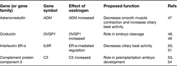
Adrenomedullin (ADM) is a peptide hormone that relaxes smooth muscle in various tissues and induces vasodilatation. A recent study in the human Fallopian tube demonstrated that higher oestradiol levels in the periovulatory phase, relative to that in the early follicular phase, induce ADM expression (Fig. 1, Table 3) (Ref. Reference Li47). It also showed that ADM increased ciliary beat activity in vitro, suggesting that dependent–dependent ADM expression was important for Fallopian tube function (Ref. Reference Li47).
Oestrogen-regulated genes in the nonpregnant Fallopian tube

Oviductin
Oviductin (OVGP1) is secreted specifically in the oviduct, with a maximal response at the time of ovulation and with the embryo as one of its purported targets (Fig. 1, Table 3). There is a significant positive correlation between serum oestradiol and luteinising hormone concentrations and oviductin mRNA expression, and a significant inverse correlation between serum progesterone concentrations and oviductin mRNA (Ref. Reference Briton-Jones48). Although oestrogen seems to be the main regulator of oviductin mRNA expression in oviduct mucosal cells, other factors, such as ER isoform and mature cell phenotype, have been proposed to mediate the regulatory process (Ref. Reference Briton-Jones49). This implies a putative role of oviductin in facilitating embryo cleavage and successful implantation.
Interleukin 6R-α
Treatment with interleukin-6 (IL-6), a proinflammatory and immunoregulatory cytokine, inhibits cilia activity in the human Fallopian tube in vitro (Ref. Reference Papathanasiou50). One of two receptors for IL-6, IL-6R-α is restricted to the cilia of epithelial cells and oestrogen selectively downregulates IL-6R-α expression mediated by ER-α (Fig. 1, Table 3) (Ref. Reference Shao51). These data suggest that oestrogenic regulation of tubal IL-6R is associated with the oestrogen-induced embryo transport process in vivo.
Complement protein component-3
Fallopian-tube-derived embryotrophic factors (ETFs) stimulate the development of mouse preimplantation embryos (Ref. Reference Liu52). The most abundant ETF is ETF-3, which contains mainly complement protein component-3 (C3) and its derivatives, C3b and iC3b. Sex steroids, particularly oestradiol, and preimplantation embryos have been shown to regulate the expression pattern of C3 in the Fallopian tube (Fig. 1, Table 3) (Ref. Reference Lee53). These data, together with a recent finding that C3 is converted to embryotrophic iC3b in the presence of preimplantation embryos (Ref. Reference Tse54), support a physiological role for C3 in preimplantation embryo development.
Ectopic implantation in the Fallopian tube
Morphological and cellular changes
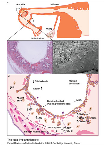
The Fallopian tube epithelium containing an ectopic pregnancy demonstrates a marked reduction in the number of ciliated cells in comparison with the Fallopian tube from an intrauterine pregnancy of the same gestational age, and marked deciliation is sometimes seen (Fig. 2). These changes have also been noted in biopsies from women undergoing tubal surgery who later develop an ectopic pregnancy, so it is plausible that they are causative.
The tubal implantation site. (a) Gross anatomical representation of a tubal ectopic pregnancy. (b) Light microscopy image with haematoxylin and eosin staining of a tubal implantation site. (c) Scanning electron micrograph of a tubal implantation site. Note marked deciliation. (d) Progesterone and oestrogen-regulated genes at the tubal implantation site.

Within the Fallopian tube mucosa, CD45+ leukocytes, CD68+ cells (which are likely to be macrophages) and CD11c+ cells are significantly increased in women with ectopic pregnancy compared with the mid-luteal phase nonpregnant Fallopian tube (Fig. 2) (Ref. Reference Shaw18). These cells may simply be a reflection of the increased progesterone levels present during pregnancy. However, it is likely that invasion of the tubal wall by trophoblast results in inflammatory signals causing an influx of immune cells to the Fallopian tube.
Changes in sex steroid hormone receptor expression
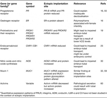
Levels of PR-B and PR protein are reduced in Fallopian tubes from women with ectopic pregnancy (Refs Reference Horne16, Reference Land and Arends55), and this observation may offer an explanation for the reduction in tubal decidualisation observed with tubal implantation. Mice deficient in PR fail to mount a decidual response, and a recent study in humans has shown that PRs regulate distinct gene networks and cellular functions in decidualising endometrium (Ref. Reference Cloke12). In addition, ER-α protein is absent in the Fallopian tube of women with ectopic pregnancy (Ref. Reference Horne16). ER-α gene polymorphisms have been associated with female infertility (Ref. Reference Corbo56), and ER-α has been shown to serve as a dominant regulator in Fallopian tube development in the rat (Ref. Reference Mowa and Iwanaga57). Furthermore, a recent study in mice has identified a molecular mechanism for ER-α-mediated tubal protein synthesis and secretion that appears to be important for successful embryonic development (Ref. Reference Shao58). Thus, low ER-α may promote low PR activity preventing decidualisation leading to ectopic pregnancy with abnormal implantation.
Changes in expression of progesterone-regulated genes
Three of the genes that appear to be progesterone regulated in the nonpregnant Fallopian tube are altered with tubal implantation in a pattern reflective of the high circulating progesterone levels and the absence of PR associated with ectopic pregnancy (Fig. 2, Table 4). NOS2 mRNA and protein levels are greater in the Fallopian tubes of women with ectopic pregnancy compared with pseudopregnant women (Ref. Reference Al-Azemi30). MUC1 expression is reduced, and its glycosylation pattern altered, in the Fallopian tubes of women with ectopic pregnancy (Refs Reference Al-Azemi32, Reference Savaris59). In addition, activin and activin receptor protein levels are increased in Fallopian tubes from women with tubal ectopic pregnancy compared with Fallopian tubes from pseudopregnant women (Refs Reference Refaat60). However, the levels of activin and activin receptor mRNA were decreased, and the authors suggest that this might indicate pathological secretion of activins and their receptors as causal in the development of tubal ectopic pregnancy.
Gene expression at the ectopic implantation site

aQuantitative expression patterns of PRLR, integrins, ADM, oviductin, IL6R-α and C3 have not been studied in the context of ectopic implantation.
However, two progesterone-regulated genes, PROKR and CB1, do not exhibit expression patterns reflective of progesterone levels (Fig. 2, Table 4). PROKR levels are lower in Fallopian tubes from ectopic pregnancies than in mid-luteal phase Fallopian tubes collected from nonpregnant women (Ref. Reference Shaw19). This could be the result of lower maternal serum β-human chorionic gonadotrophin (hCG) levels observed in ectopic pregnancy. hCG increases PROK1 expression in the endometrium (Ref. Reference Evans61), and, generally, ectopic pregnancies have lower hCG levels than normal intrauterine pregnancies. CB1 expression is also lower in Fallopian tubes from women with ectopic pregnancy compared with Fallopian tube tissue from nonpregnant women (Ref. Reference Horne25). The coordinated downregulation of embryonic CB1 and uterine AEA levels is important for both embryo activation and uterine receptivity (two events critical for successful intrauterine implantation), so it is not inconceivable that CB1 expression could be under embryonic control (Ref. Reference Wang62).
There is also an increasing body of evidence to suggest that the changes in PROKR and CB1 expression observed with ectopic pregnancy have occurred prior to the implantation event and represent pathological processes predisposing to ectopic pregnancy (Refs Reference Horne25, Reference Shaw63, Reference Shaw64). The signalling downstream of PROKR and CB1, and any interaction between these two genes, in the Fallopian tube warrants further investigation.
Quantitative expression patterns of PRLR and the integrins have not been studied in the context of ectopic implantation.
Changes in expression of oestrogen-regulated genes
Similar to PRLR and integrins, quantitative expression patterns of ADM, oviductin, IL6R-α and C3 have not been studied in the context of ectopic implantation.
Ectopic implantation and the endometrium
When an embryo implants in the Fallopian tube, maternal cells at the implantation site usually only display very limited, if any, decidual differentiation, but intrauterine endometrial decidualisation is maintained. Nevertheless, similarities in the cellular composition of decidualised endometrium in ectopic and intrauterine pregnancies have been described. Specifically, the relative numbers of leukocyte subtypes in decidualised endometrium from ectopic pregnancies are reported to be the same as those found in decidua from an intrauterine pregnancy (Ref. Reference von Rango65). We have demonstrated, however, that there are differences at a morphological level, and in the expression of markers of decidualisation (prolactin and IGFBP-1) and inhibins/activins in the decidualised endometrium of ectopic compared with intrauterine pregnancies of similar gestations (Refs Reference Horne66, Reference Duncan67). The most obvious difference in the decidualised endometrium from an ectopic and an intrauterine pregnancy is the absence of trophoblast. There are no published data to our knowledge to suggest that these genes are regulated by trophoblast signals. Nevertheless, recent in vitro studies, using a functional genomics approach, have shown that conditioned media from trophoblasts alter the local immune environment of the decidua to facilitate embryo implantation by inducing proinflammatory cytokines and cells (Ref. Reference Hess68). This provides an interesting potential mechanism for the changes observed in these studies.
Research in progress and outstanding research questions
There are clearly deficiencies in diagnostic tools for ectopic pregnancy. A one-by-one scientific approach has revealed over 20 candidate biomarkers that could be used as a test to diagnose ectopic pregnancy, although at present their clinical utility is very limited (Ref. Reference Cartwright69). Our review of the literature has shown that these biomarkers can be clustered into themes: uterine markers of normal implantation, markers of abnormal embryo/trophoblast growth, markers of abnormal corpus luteum function, markers of a growing pregnancy in the Fallopian tube, and markers of inflammation and peritoneal irritation. We have used this ‘thematic’ approach to identify two new biomarkers.
Firstly, we identified a new uterine marker of normal implantation, activin B. Using microarray technology, we showed that inhibin/activin β B subunit expression was lower in the decidualised endometrium from ectopic and intrauterine pregnancies, and that this was reflected in circulating levels of activin B (Ref. Reference Horne66). Secondly, we searched for markers of abnormal embryo/trophoblast growth and showed reduced expression of the proangiogenic factor, placental growth factor (PIGF), at the ectopic compared with the normal intrauterine implantation site (Ref. Reference Horne70). Again, this difference was demonstrated in circulating levels.
Our group and others are also using the 3D biomarker discovery strategy consisting of abundant protein immunodepletion, Sodium dodecyl sulfate (SDS) gels, Liquid chromatography–mass spectroscopy (LC-MS)/MS and label-free quantitation of MS signal intensities to identify candidate biomarkers (Ref. Reference Beer71). In addition, emerging data highlight the medically relevant potential of determining microRNA levels in serum to use as biomarkers (Ref. Reference Gilad72). Candidates identified from these novel approaches, activin B and PIGF, in combination with known markers of abnormal corpus luteum function, markers of a growing pregnancy in the Fallopian tube, and markers of inflammation and peritoneal irritation, are currently being explored in large-scale studies as a diagnostic test for early pregnancy location and outcome.
There is also a need for new or adjuvant medical treatments for ectopic pregnancy. Currently, methotrexate can resolve selected ectopic pregnancies that fulfil strict clinical criteria suggesting they are small (serum hCG <3000 IU/l and no fetal cardiac activity) and unruptured (haemodynamically stable, no abdominal distension and no free fluid in the Pouch of Douglas on ultrasound). Unfortunately, the efficacy of methotrexate falls with increasing ectopic size, and this means that only 25% of ectopic pregnancies are suitable for medical management (Ref. Reference Varma and Gupta5). We believe that the development of relevant animal and new in vitro models to further understand the endocrinology of ectopic pregnancy will lead to the identification of specific inhibitors that could replace or be used in combination with methotrexate to efficiently treat larger cases of ectopic pregnancy.
Acknowledgements and funding
We are grateful to the peer reviewers for their helpful comments, Professor Jane Norman and Dr Jeremy Brown for their contributions to the manuscript, Dr Alistair Williams for his advice on images, Mrs Sheila Milne for secretarial support, and Mr Ronnie Grant and Mr Ted Pinner for graphical support. A.W.H. receives grant support from UK Medical Research Council (MRC G0802808 2009-13), IKTF (2009–2011), and an Albert McKern Bequest (2010–11).
Conflict of interest statement
A.W.H. and H.O.D.C. hold a UK patent for a diagnostic biomarker for ectopic pregnancy (no. 0712801.0). A.W.H. is Medical Advisor to the Ectopic Pregnancy Trust.