Introduction
Menopause occurs at an average age of 51 years.Reference McKinlay, Brambilla and Posner 1 With increasing life expectancy, a progressively larger portion of a woman’s lifetime is postmenopausal, giving rise to new factors influencing women’s health after menopause. Menopause not only marks the end of a women’s fertile lifespan, but a younger age at menopause is also associated with an increased risk of cardiovascular disease, osteoporosis and overall mortality.Reference Daan and Fauser 2 With women worldwide delaying child bearing until their 30s, a young age at menopause and the inevitable decade of subfertility preceding, also causes a significant amount of emotional distress and relational problems as well as an increasing demand on medically assisted reproductive treatments.
The total number of follicles is established at around 5 months gestational age and is therefore finite at ~7 million.Reference Faddy and Gosden 3 , Reference Wallace and Kelsey 4 When a woman reaches menarche this number has already been reduced to ~400,000.Reference Baker 5 The onset of menopause is thought to occur when the ovarian follicle count reaches ~1000 follicles.Reference Wallace and Kelsey 4 Therefore, early life could be a particularly relevant period in determining follicular endowment and age at natural menopause. Suboptimal conditions during early development including maternal undernutrition or placental insufficiency could influence the total amount of follicles that are formed. Alternatively, adverse prenatal or postnatal conditions or accelerated general aging may increase oocyte attrition. Several studies have established that early life exposure to an adverse environment can lead to impaired adult health, including an elevated risk of cardiovascular disease and metabolic syndrome.Reference Whincup, Kaye and Owen 6 – Reference Newnham and Ross 9
There is also substantial evidence from animal studies and epidemiological studies supporting the hypothesis that poor intrauterine and early life nutrition cause early aging and decreased longevity.Reference Tarry-Adkins and Ozanne 10 – Reference Ozanne and Hales 14 Furthermore, aging of the reproductive system precedes somatic aging.Reference Li, Geng and Zheng 15 Nutritional stress during gestation and infancy could affect the ovarian germ cell population resulting in a diminished ovarian reserve and a younger age at menopause. Throughout human history periods of famine have been a source of nutritional stress. In periods of nutritional abundance, a low birth weight and impaired early childhood growth are commonly used indicators for intrauterine and early childhood nutritional stress.
Here, we systematically examined the existing literature investigating the effect of adverse intrauterine conditions, early childhood growth and famine exposure on the age of natural menopause.
Material and methods
Search strategies
This review was conducted according to PRISMA and MOOSE guidelines. The protocol of this study is registered at PROSPERO under registration number CRD42016049239. A comprehensive search was performed in the bibliographic databases PubMed, EMBASE.com, CINAHL (via EBSCO) and Cochrane library (via Wiley) with no language or date restrictions from inception to 1 January 2017. Additionally the reference lists of relevant studies, review articles and opinion papers were checked by snowball search to identify any secondary references. Search terms included controlled terms (MeSH in Pubmed, EMtree in Embase.com), as well as free text terms. We used free text terms only in the Cochrane Library. The following terms were used (including synonyms and closely related words) as index terms or free-text words for ‘birth weight’ or ‘gestational age’ or ‘famine’ or developmental origins of health and disease’ and the outcome variable ‘menopause’ or ‘premature menopause’ with aid of a clinical librarian (L.J.S.). Detailed information on the search strategy is listed in the Supplementary Appendix 1.
Study selection and eligibility
After identifying and excluding duplicate studies, two independent reviewers (S.S. and M.V.) evaluated titles and abstracts for suitability using a customized in-exclusion chart. Any disagreement between reviewers was resolved through consensus. If disagreement was not resolved, full text articles were reviewed. Studies were included if they were (i) cross-sectional, case–control, cohort or intervention studies, (ii) were conducted in adult humans, (iii) reported age at menopause, (iv) reported birth weight, gestational age, childhood growth or famine exposure, (v) the reported birth parameters and childhood influences were related to women whose menopausal age was reported. Full texts were retrieved for studies that could satisfy all selection criteria and independently reviewed by two authors (S.S. and M.V.) for eligibility using the same in-exclusion chart. Any disagreement between reviewers was resolved through consensus, if consensus could not be reached, a third author was consulted (R.C.P.).
Quality assessment and data extraction
Selected studies were assessed independently for methodological validity by reviewers S.S. and M.V., using the Newcastle–Ottawa scale with a maximum of nine stars.Reference Wells, Shea and O’Connell 16 Included studies were classified as low quality (0–4), moderate quality (5–6) and high quality (7–9). Any disagreement between reviewers was resolved through consensus if consensus could not be reached, a third author was consulted (R.C.P.). A piloted data extraction form was used to extract relevant information. The form included questions on publication year, study design, baseline population, number of participants, definition of outcome variable, type of exposure and reported risk estimates.
Data synthesis and analysis
Providing the available data were suitable they were entered in 2×2 tables, data were transformed to Review Manager (RevMan 5.3) for meta-analysis, to estimate hazards ratios (HR) and odds ratios. We assessed heterogeneity by eyeball test and the I2 statistic, the later was distinguished as high heterogeneity, ergo not fit to be pooled, when I2⩾75–100%.
Results
Identification of relevant studies
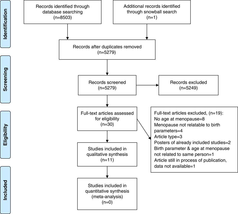
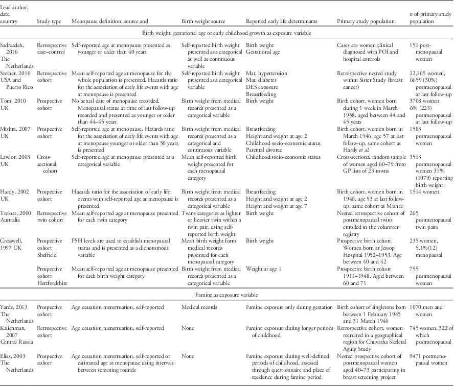
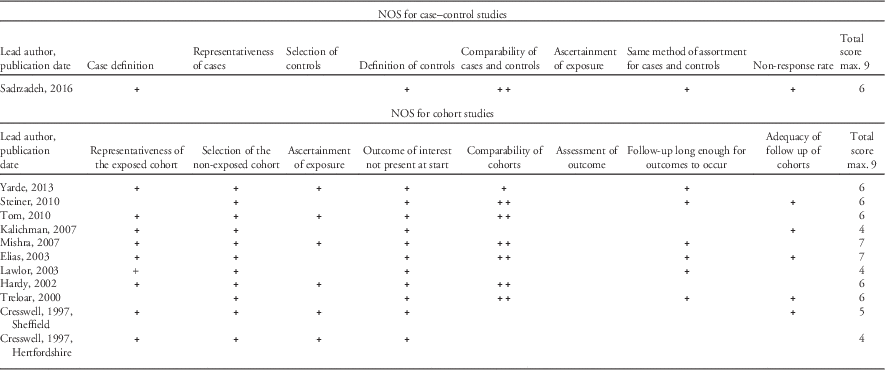
The search strategy resulted in 5278 unique citations and one additional study through snowball search. In total, 11 studies were deemed eligible and included. Nine of the final 11 studies were cohort studies, 1 case–control study and 1 twin study. The study described by Mishra et al. is a follow-up study of the same cohort investigated by Hardy et al. but included more participants and additional outcome variables. The selection procedure is visualized in Fig. 1 and characteristics of the final 11 articles are listed in Table 1. Table 2 shows the results of quality assessment using the Newcastle–Ottawa scale.
Flow chart of study selection process.

General characteristics of all 11 included studies listed by exposure variable and publication dates

POI, premature ovarian insufficiencies; DES, diethylstilbestrol; FSH, follicle stimulating hormone.
Quality assessment of included studies in the meta-analysis using Newcastle–Ottawa Scale (NOS)

The reported exposure variables as well as reported risk estimates differed significantly. Although, some studies reported mean birth weights others reported birth weight in categories. Some, but not all studies reported gestational age. We also could not find a common nominator for famine exposure, studies reported on intrauterine exposure, early childhood exposure or exposure at puberty and the duration of exposure varied greatly. The outcome variable menopause was also reported in various ways. Some dichotomized menopause defining a different menopausal age as breaking point. Others reported mean age at menopause. There was also a great heterogeneity in reported risk estimates, from reporting only raw data or descriptive statistics to HR, odds ratios or only P values. Due to this diversity of reported data and risk estimates we were unable to pool data or performed meta-analysis on pooled data.
Birth weight, neonatal adiposity and age at menopause
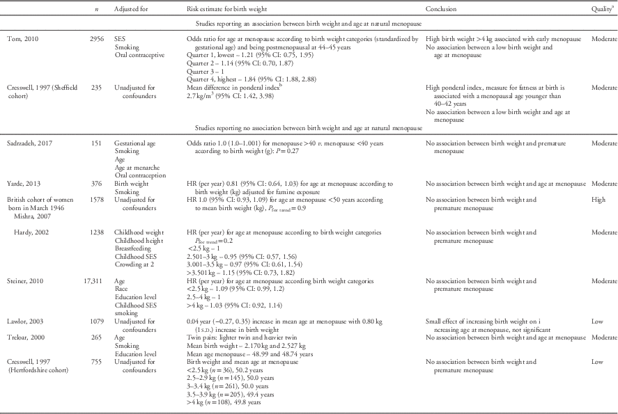
From the 11 included studies, nineReference Sadrzadeh, Painter, van Kasteren, Braat and Lambalk 17 – Reference Cresswell, Egger and Fall 26 reported how birth weight was associated with age at natural menopause. None of the studies reported a statistically significant association between low birth weight and age at natural menopause. Two studiesReference Tom, Cooper and Kuh 19 , Reference Cresswell, Egger and Fall 26 reported on the association between a higher birth weight and age at natural menopause. Tom et al. found a statistically significant association, with an odds ratio (95% CI) of 1.84 (1.88, 2.88) for the highest birth weight quartile to enter menopause at a younger age the other reported association was not statistical significant (Table 3). Cresswell et al. (Sheffield cohort) reported an association between a higher ponderal index [birth weight/height (m3)] a marker of neonatal adiposity and a menopausal age under 40–42 years.
Studies reporting age at menopause in relation to birth weight. The study of Cresswell et al. is included twice as it reports on two different cohorts

SES, socio-economic status; HR, hazard ratio.
a Low quality (0–4), moderate quality (5–6) and high quality (7–9).
b Ponderal index=birth weight(kg)/height (m3).
Gestational age or prematurity and age at menopause
One case–control studyReference Sadrzadeh, Painter, van Kasteren, Braat and Lambalk 17 reported an association between premature birth before 37 weeks of gestation, and age at natural menopause before the age of 40 with an odds ratio of 4.66 (95% CI: 1.3, 16.7).
Exposure to famine and age at menopause
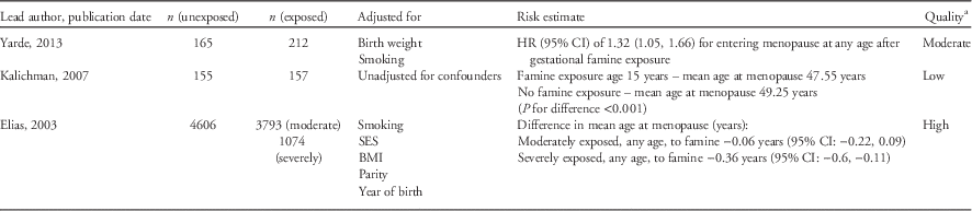
Three studies reported on well-defined periods of famine exposure in relation to a younger age at menopause.Reference Yarde, Broekmans and van der Pal-de Bruin 18 , Reference Kalichman, Malkin and Kobyliansky 20 , Reference Elias, van Noord, Peeters, den Tonkelaar and Grobbee 22 In these studies, the age at which women had been exposed to famine varied: Yarde et al. reported on famine during gestation. Exposure to famine was associated with a 32% increase in the HR of natural menopause at any age [HR (95% CI): 1.32 (1.05, 1.66)] after adjusting for smoking and birth weight. Kalichman et al. assessed the effects of famine exposure during puberty, estimating that menopause occurred 1.7 years earlier (95% CI: 1.12, 2.27) compared with famine unexposed women. Elias et al. reported on the effect of famine exposure at any age with extra focus on exposure during childhood. Famine exposure at 2–7 years had the largest effect with severely famine exposed girls entering menopause 1.83 years (95% CI: −3.03, −0.63) earlier compared with unexposed women (Table 4).
Studies reporting age at menopause in relation to famine exposure

SES, socio-economic status; BMI, body mass index – weight (kg)/height (m2).
a Low quality (0–4), moderate quality (5–6) and high quality (7–9).
Childhood growth and age at menopause
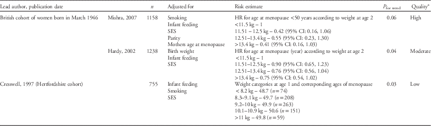
Three studies detected an association between low childhood weight (at age 1 or 2 years old) and a younger age at menopause. Cresswell et al. (Hertfordshire cohort), Mishra et al. and Hardy et al. all concluded that a lower weight in infancy was associated with a younger age at menopause (Table 5).
Studies reporting age at menopause in relation to childhood growth

HR, hazards ratio; SES, socio-economic status.
a Low quality (0–4), moderate quality (5–6) and high quality (7–9).
Additional childhood factors
A few of the included studies also explored other childhood exposures and their effect on the age of menopause. Although these findings are not in the scope of our review question, we have summarized the main positive associations. An association between a low socio-economic status during childhood and an early age at menopause was reported by Lawlor et al., P for trend<0.001; Hardy et al. P for trend=0.06 and Mishra et al. P for trend=0.03. All studies used multiple indicators for childhood socio-economic status. Mishra et al. also reported that parental divorce was associated with a younger age at menopause. Women experiencing parental divorce during childhood had a HR (95% CI) of 6.52 (2.0, 21.3) to enter menopause before the age of 50 compared with other women. Steiner et al. report an association between gestational DES exposure and a younger age at menopause, HR 1.45 (95% CI: 1.27, 1.65). Hardy et al. and Mishra et al. documented that breastfed infants had a later age at menopause, HR (95% CI) and P for trend are 0.74 years (0.57, 0.97), P for trend =0.06 and 0.81 (0.65–0.99), P for trend=0.05, respectively.
Quality
The score on the Newcastle–Ottawa scale for studies included in our systematic review ranged from 4 to 7. Two studies were categorized as high, three as low and seven as moderate. The two cohorts reported on by Cresswell et al. were assessed separately. There were no striking differences in quality of studies reporting no effect compared with studies reporting positive effects. Quality of the studies is reported in the Table 2.
Conclusion
Our systematic review indicates that prenatal or postnatal famine exposure and slow childhood growth might both be associated with a younger age at menopause. These effects seem to be independent of birth weight. All six included studies investigating the relation between childhood growth or childhood exposure to famine report a positive relation between an impaired childhood growth or famine exposure and a younger age at menopause. The association between birth weight, as a summary measure of poor intrauterine conditions, and age at natural menopause were largely null: nine studies report on the association between birth weight and age at menopause but only one reports a significant association with a higher weight at birth and one of a significant association with increased adiposity at birth. Low weight at the ages one and two as well as exposure to famine, however, were consistently associated with younger age at menopause.
Our finding is most likely a reflection of a premature or accelerated senescence related to early life nutritional stress. Increased oxidative stress has been opted as a possible pathway linking nutritional stress and cellular aging through an accelerated shortening of telomeres.Reference Diamanti-Kandarakis, Papalou, Kandaraki and Kassi 27 Telomeres, a unique nucleotide repeat sequence at the distal ends of chromosomes, shorten after each mitotic division and when they reach a critical length the cell seizes to divide.Reference Wong and Collins 28 Animal studies detect increased oxidative stress levels as well as an impaired telomere length in intrauterine nutritionally deprived rodents and non-human primates.Reference Aiken, Tarry-Adkins and Ozanne 29 – Reference Tarry-Adkins, Martin-Gronert, Chen, Cripps and Ozanne 31 The enzyme telomerase has the ability to elongate the telomeric DNA. Telomerase activity declines with age and there is evidence of declined activity in women with ovarian insufficiency.Reference Butts, Riethman and Ratcliffe 32 , Reference Kinugawa, Murakami, Okamura and Yajima 33 To summarize, nutritional stress during gestation and infancy could affect the ovarian germ cell population mediated through increased oxidative stress and reduced telomerase activity resulting in a diminished ovarian reserve and a younger age at menopause.
By systematically searching and reviewing all published data on this subject we minimized the chance of citation bias favoring a specific outcome. However our study has some weaknesses. Due to the differences in definition of the outcome variable and the reported risk estimates we were unable to pool data from different studies for statistical analysis. Not all included studies were initially designed to assess the effect of early life events on age at menopause this might have contributed to the heterogeneity between studies. Our conclusions therefore, could not be based on quantitative analysis but were based on a detected trend in reported study results.
The number of studies we were able to include were also limited. Although we have no direct evidence we cannot exclude the possibility of publication bias with an under representation of smaller studies reporting no association between early life circumstances and age at natural menopause. Other famine cohorts have published on the effects of famine on other health parameters but none of them has published on the relation between age at natural menopause and famine exposure.Reference Thurner, Klimek and Szell 34 – Reference Kannisto, Christensen and Vaupel 38 This might be due to lack of data on the subject but it could also reflect the fact that studies yielding negative or no associations are less likely to be submitted or to be accepted for publication.
Another aspect to consider is that premature ovarian insufficiency, with menopausal ages younger than 40 years, might have a different etiology than age at natural menopause and not just be an extreme variation. While an early age at menopause might be the reflection of a premature senescence it has been postulated that a much younger menopausal age has a different pathophysiology.Reference Nelson, Covington and Rebar 39 , Reference Pal and Santoro 40 Most included studies however, did not differentiate between an early age at natural menopause and premature age at menopause. To investigate early life influences on age at natural menopause future studies should focus on collecting data on periconceptional, prenatal and postnatal nutritional status of mother and child in a prospective setting. We advise to put more emphasis on gestational age, early childhood growth as well as periconceptional and gestational nutritional status of mother and child rather than birth weight when testing the Developmental Origins of Health and Disease concept on age at natural menopause. Another interesting field of investigation, already pursued in many Western countries, is the effect of nutritional abundance and the overload of high carbohydrate and high protein nutrients in our modern diet on the development of our fertility.
The impact of these issues on fertility are currently not well understood, but is likely to be detrimental. To determine age at menopause we advise to follow the 2011 STRAW criteria, defining menopause retrospectively after 1 year of amenorrhea without a history of ovarian disease or surgery or genetic abnormalities. Although there are currently no reliable hormonal markers of menopausal status, follicle stimulating hormone should be considered as a potential marker of reproductive aging.Reference Harlow, Gass and Hall 41
We conclude that famine during gestation and childhood as well as impaired childhood growth seems to be related to a younger age at menopause. This could lead to a shorter reproductive lifespan in populations with compromised early life nutrition, and may add to the increased burden of cardio-metabolic disease in postmenopausal women.
Acknowledgments
The authors would like to thank all authors who elaborated on their data after email contact. The authors also thank Peter van de Ven, consultant epidemiology and biostatistics, for advising us on the evaluation of data and calculating additional risk estimates.
Financial Support
This piece of work received no specific grant from any funding agency, commercial or not-for-profit sectors.
Conflicts of Interest
None.
Supplementary material
To view supplementary material for this article, please visit https://doi.org/10.1017/S2040174417000952