Introduction
Current reproductive biotechnologies, e.g. in vitro fertilization, cloning using nuclear transfer or transgenesis, depend on a rich source of oocytes. As oocytes matured in vivo cannot be easily obtained in a large enough quantity for these technologies, oocytes matured in vitro are often used for these purposes.
The maturing oocyte underwent germinal vesicle (GV) breakdown and then passed through the stages of metaphase I (MI), anaphase I (AI) and telophase I (TI). Maturation is completed at the metaphase II (MII) stage when oocyte meiosis is blocked spontaneously (Wassarman, Reference Wassarman, Knobil and Neil1988). Exit from this meiotic block depends on the activating stimulus, which induces destruction or inactivation of the molecules responsible for the maintenance of meiotic blockage. During fertilization, this activating stimulus is brought to the oocyte by the sperm (Yanagimachi, Reference Yanagimachi, Knobil and Neil1988).
In vitro matured oocytes, however, could be of an inadequate quality.This problem is sometimes tackled using prolonged in vitro culture of the oocytes. Oocytes cultured in vitro after reaching the MII stage were used for in vitro fertilization (Chian et al., Reference Chian, Nakahara, Niwa and Funahashi1992; Pavlok et al., Reference Pavlok, Kalab and Bobak1997), cloning (Stice et al., Reference Stice, Keefer and Matthews1994; Tanaka & Kanagawa, Reference Tanaka and Kanagawa1997; Iwamoto et al., Reference Iwamoto, Onishi, Fuchimoto, Somfai, Suzuki, Yazaki, Hashimoto, Takeda, Tagami, Hanada, Noguchi, Kaneko, Nagai and Kikuchi2005; Hall et al., Reference Hall, Compton, Stojkovic, Nesbitt, Herbert, Murdoch and Stojkovic2007) or production of parthenogenetic embryos (Jolliff & Prather Reference Jolliff and Prather1997; Iwamoto et al., Reference Iwamoto, Onishi, Fuchimoto, Somfai, Suzuki, Yazaki, Hashimoto, Takeda, Tagami, Hanada, Noguchi, Kaneko, Nagai and Kikuchi2005).
Prolonged in vitro culture of mammalian oocytes is accompanied by a complex process designated ‘ageing’. Developmental competence could be reduced significantly in aged oocytes (Nagano et al., Reference Nagano, Katagiri and Takahashi2006). The aged pig oocytes exhibited changes in their cytoskeletal arrangement (Kim et al., Reference Kim, Moon, Prather and Day1996; Suzuki et al., Reference Suzuki, Takashima and Toyokawa2002) or exhibited changes in the activities of various kinases (Kikuchi et al., Reference Kikuchi, Naito, Noguchi, Shimada, Kaneko, Yamashita, Tojo and Toyoda1999, Reference Kikuchi, Naito, Noguchi, Shimada, Kaneko, Yamashita, Aoki, Tojo and Toyoda2000). Ageing can result in spontaneous parthenogenetic activation, fragmentation or lysis in pig oocytes (Petrova et al., Reference Petrova, Sedmikova, Chmelikova, Svestkova and Rajmon2004, Reference Petrova, Rajmon, Sedmikova, Kuthanova, Jilek and Rozinek2005). All these changes deteriorate the quality or viability of the oocytes and, therefore, interfere with their success in reproductive biotechnologies. The nature of these processes is not completely understood. Programmed cell death or apoptosis is involved in these processes (Austin, Reference Austin1970; Fujino et al., Reference Fujino, Ozaki, Yamamasu, Ito, Matsuoka, Hayashi, Nakamura, Ogita, Sato and Inoue1996; Sasaki & Chiba, Reference Sasaki and Chiba2001; Yüce & Sadler, Reference Yüce and Sadler2001) and apoptosis is exerted in the oocyte as fragmentation (Takase et al., Reference Takase, Ishikawa and Hoshiai1995; Fujino et al., Reference Fujino, Ozaki, Yamamasu, Ito, Matsuoka, Hayashi, Nakamura, Ogita, Sato and Inoue1996; Tarin et al., Reference Tarin, Perez-Albala and Cano2001). Apoptosis is triggered and accomplished by a wide spectrum of specific bioactive molecules, including caspases, proteases responsible for the dismantling of the cell and many pro- or antiapoptotic factors from the Bcl-2 gene family (Adams & Cory, Reference Adams and Cory1998).
Many studies on somatic cells have proved the significant role of histone acetylation in apoptosis (e.g. Drexler & Euler Reference Drexler and Euler2005; Subramanian et al., Reference Subramanian, Opipari, Castle and Kwok2005; Wang et al., Reference Wang, Kim, Lee, Kim, Seo, Yu and Lee2005; Sonnemann et al., Reference Sonnemann, Hartwig, Plath, Kumar, Muller and Beck2006; Takai et al., Reference Takai, Ueda, Nishida, Nasu, Matsuda, Kusumoto and Narahara2006). Histones represent an essential component of nucleosome in eucaryotic cells. The DNA sequence containing 147 base pairs is bound to an octamer composed of doublets of histone proteins H2A, H2B, H3 and H4. Amino termini of these histones are on the surface of the nucleosome and they could be modified posttranslationally by reversible acetylation (Allfrey et al., Reference Allfrey, Faulkner and Mirsky1964). The extent of histone acetylation results from the activities of two families of enzymes – histone acetyltransferases (HATs) and histone deacetylases (HDACs) (Bertos et al., Reference Bertos, Wang and Yang2001; Marmorstein & Roth, Reference Marmorstein and Roth2001). Histone acetylation also plays an important role in the meiotic maturation of oocytes (Kim et al., Reference Kim, Liu, Tazaki, Nagata and Aoki2003; Wang et al., Reference Wang, Wang, Ai, Xiong, Yin, Hou, Chen, Schatten and Sun2006a, Reference Wang, Yin, Ai, Liang, Hou, Chen, Schatten and Sunb). This role of histone acetylation was also shown in pig oocytes (Endo et al., Reference Endo, Naito, Aoki, Kume and Tojo2005; Wang et al., Reference Wang, Wang, Ai, Xiong, Yin, Hou, Chen, Schatten and Sun2006a, Bui et al., Reference Bui, Thuan, Kishigami, Wakayama, Hikichi, Ohta, Mizutani, Yamaoka, Wakayama and Miyano2007).
Very limited data are available on the role of histone acetylation in ageing mammalian oocytes. The aim of this study was to investigate the role of histone acetylation in the ageing pig oocytes matured in vitro. We used an inhibitor of HDACs, trichostatin A (TSA), for this purpose.
Materials and Methods
Isolation and culture of oocytes
Pig ovaries were obtained from a local slaughterhouse from gilts at an unknown stage of the estrous cycle and transported to the laboratory in a saline solution (0.9% natrium chloride) at 39 °C within 1 h. Fully grown oocytes were collected from follicles by aspirating follicles that were 2–5 mm in diameter with a 20-gauge needle. Only oocytes with compact cumuli were chosen for further study. Before culture, the oocytes were washed three times in a maturation culture medium.
The oocytes were cultured in a modified M199 medium (Gibco BRL, Life Technologies) containing sodium bicarbonate (0.039 ml of a 7.0% solution per ml of medium), calcium lactate (0.6 mg/ml), sodium pyruvate (0.25 mg/ml), gentamicin (0.025 mg/ml), HEPES (1.5 mg/ml), 13.5 IU eCG: 6.6 IU hCG/ml (P.G.600 Intervet) and 10% fetal calf serum (Gibco BRL, Life Technologies).
The oocytes were cultured for 48 h in 3.5-cm diameter Petri dishes (Nunc) containing 3.0 ml of the culture medium at 39 °C in a mixture of 5.0% CO2 in air.
Evaluation of oocytes
At the end of the culture, the oocytes or embryos were mounted on slides, fixed with acetic alcohol (1 : 3 v/v) for at least 24 h and stained with 1.0% orcein. The oocytes were examined under a phase contrast microscope. Activation was considered to have occurred if the oocytes were at the pronuclear stage. Oocytes remaining at MII or arrested at AII or at TII stages were not considered as activated. In cleaved parthenogenetic embryos, the number of nuclei was counted at the end of the culture.
Oocyte activation and culture of embryos
Oocytes were activated using the method described by Jilek et al. (Reference Jilek, Huttelova, Petr, Holubova and Rozinek2001). Briefly, oocytes matured in vitro were denuded from their cumulus cells and were subjected to a 5 min treatment with 25 μM calcium ionophore A23185. The calcium ionophore was diluted in a modified M199 medium without fetal calf serum and without bovine serum albumin. After the treatment, the oocytes were washed in a M199 medium supplemented with bovine serum albumin and cultured for 2 h in a NCSU23 culture medium (Peters & Wells, Reference Peters and Wells1993) supplemented with 2 mM 6-dimethylaminopurine (DMAP). The oocytes were then washed carefully and they were cultured in a DMAP-free NCSU23 medium in 4-well Petri dishes (Nunc), each well containing 1.0 ml of the culture medium. The oocytes were cultured at 39 °C in a mixture of 5.0% CO2 in air for 7 days.
Experimental arrangement
Experiment 1 had the purpose of investigating the fates of pig oocytes aged in vitro. In vitro matured oocytes were denuded of cumulus cells, placed in a M199 culture medium and cultured for another 1, 2, 3, 4 or 5 days.
Experiment 2 was performed to evaluate the effect of histone deacetylase (HDATs) inhibition on the maturation of pig oocytes. Oocytes were matured in vitro for 24 or 48 h in the medium supplemented with the specific inhibitor of HDATs trichostatin A (100 or 1000 nM). Control experiments were performed on oocytes matured for 24 or 48 h in an inhibitor-free medium.
The aim of Experiment 3 was to study the ageing in oocytes matured under the effect of trichostatin A. Oocytes were matured in a medium supplemented with trichostatin A (100 or 1000 nM) and then they were denuded of cumulus cells, placed in a M199 culture medium and cultured for another 3 days.
Experiment 4 was performed to investigate the effects of histone deacetylase (HDATs) inhibition on the ageing of in vitro matured pig oocytes. The oocytes were matured in vitro for 48 h and were cultured for another 3 days in the medium supplemented with the specific inhibitor of HDATs trichostatin A (100 or 1000 nM). Control experiments were performed on oocytes aged for 3 days in an inhibitor-free medium.
Experiment 5 had the purpose of investigating the developmental capacity of oocytes aged under the effect of trichostatin A. Oocytes matured in vitro were cultured for another day in the medium supplemented with the specific inhibitor of HDATs trichostatin A (100 or 1000 nM). The oocytes were then activated by a combined treatment with calcium ionophore and 6-DMAP and cultured in a NCSU23 medium as described above. Control oocytes were activated either after 1 day ageing in a inhibitor-free medium or were activated immediately after 48 h maturation.
At the end of each experiment, the oocytes were evaluated as described above.
Statistical analysis
Data from all experiments were subjected to statistical analysis. Experiments were performed four times. The results were pooled for presentation and evaluated by chi-square analysis (Snedecor & Cochran, Reference Snedecor and Cochran1980). The mean percentage of oocytes or embryos reaching the given stage of maturation or development in all trials did not vary from the pooled percentage by >2.5%. A p value of less than 0.05 was considered significant.
Results
Oocytes cultured in vitro for 48 h reached the stage of MII in 96%. The remaining oocytes were at the MI, AI or TI stages.
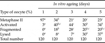
The course of oocyte ageing during their culture in vitro was investigated in Experiment 1 and its results are shown in Table 1. During the first day of ageing, the oocytes remained mainly at the MII stage. During the second day of ageing, there was a significant decrease in oocytes remaining at the MII stage. Concomitantly, there was a significant increase in the ratio of oocytes, which underwent spontaneous parthenogenetic activation or fragmentation. During the third day of oocyte ageing, the ratio of oocytes at the MII stage was further diminished and the ratio of fragmented oocytes further increased. On the fourth day of ageing, the ratio of parthenogenetically activated oocytes decreased and the ratio of fragmented oocytes increased. There were no significant changes in oocytes on the fifth day of ageing.
The fates of pig oocytes ageing in vitro

Oocytes matured in vitro for 48 h and then further cultured for another 1, 2, 3, 4 or 5 days.
a–c Statistically significant differences between oocytes of different age within the same type of oocyte (i.e. differences within the row) are indicated by different superscripts.
The most significant changes were observed on the third day of ageing, when the ratio of MII oocytes reached its minimum and the ratios of fragmented and parthenogenetically activated oocytes reached their maximum. The ratio of lysed oocytes is not yet significantly elevated. For these reasons we used oocytes aged for 3 days in the following experiments.
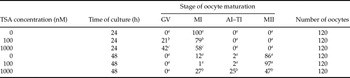
In Experiment 2, we observed delayed maturation of pig oocytes cultured in TSA-supplemented medium. After isolation from the ovary, all checked oocytes were at the GV stage. All oocytes cultured in a TSA-free medium for 24 h were at the MI stage. Oocytes cultured in TSA for the same time had a lower percentage of maturing oocytes (71% of oocytes at MI for 100 nM TSA, 58% of MI for 1000 nM TSA). After 48 h culture, the maturation was the same for oocytes matured either with 100 nM TSA or a TSA-free medium. This finding indicates that HDAC inhibition did not block, but only delayed, oocyte maturation. The extent of this delay depends on the TSA concentration. A significantly lower portion of oocytes matured to MI when cultured for 24 h with 1000 nM TSA. After 48 h culture with 1000 nM TSA, the portion of oocytes at the MII stage was also significantly reduced. Results are shown in Table 2.
Effect of TSA on the maturation of pig oocytes

Oocytes were matured in vitro either for 24 or 48 h with various concentration of TSA (0, 100 or 1000 nM) and then checked for their nuclear maturation.
a–c Statistically significant differences between oocytes at the same stage of maturation and exposed to TSA for the same time interval (24 or 48 h) are indicated by different superscripts.
In Experiment 3, we followed the ageing of oocytes matured under the influence of TSA. We did not observe any positive effect of maturation of oocytes with TSA on the subsequent 3 day ageing. Results are shown in Table 3.
Ageing of pig oocytes matured under the influence of TSA

Oocytes were cultured for 48 h in TSA-supplemented medium (0, 100 or 1000 nM TSA) a then they were cultured for 3 days in TSA-free medium.
a There were no statistically significant differences between oocytes, which were at the same state at the end of ageing after different treatments during ageing (i.e. there were insignificant differences within columns).
In Experiment 4, we investigated ageing of oocytes in a TSA-supplemented medium. TSA significantly lowered the percentage of fragmented oocytes. Results are shown in Table 4.
Effect of TSA on ageing of in vitro matured pig oocytes

Oocytes were cultured in vitro in TSA-free medium for 48 h and then they were cultured for another 3 days with respective concentration of TSA (0, 100 or 1000 nM).
a,b Statistically significant differences between oocytes that were at the same state at the end of ageing after different treatments during ageing (i.e. differences within columns) are indicated by different superscripts.
In Experiment 5, we investigated the developmental competence of oocytes matured in vitro and subsequently aged for 24 h in TSA. Ageing in 100 nM TSA had no effect on the oocytes. All oocytes (n = 120) were at the MII stage at the end of 24 h ageing. Ageing in 1000 nM of TSA significantly increased the rate of spontaneously activated oocytes (23%, n = 120). The rate of fragmented (8%) or lysed (2%) oocytes did not differ significantly in oocytes ageing for 24 h in a TSA-free medium or a medium supplemented with 100 nM of TSA.
Developmental competence in oocytes aged for 24 h was tested after their parthenogenetic activation using a combined treatment with calcium ionophore and 6-dimethylaminopurine (25 μM of calcium ionophore A23187 for 5 min followed by 2 h culture in 2 mM 6-DMAP). This artificially induced parthenogenetic activation resulted at the same activation rate in oocytes aged in a TSA-free medium or oocytes aged in a medium supplemented with 100 or 1000 nM of TSA. Twenty-four hours after activation treatment, the activation rate was 87, 94 or 93%, respectively. Control oocytes undergoing 24 h ageing without artificial activating treatment and without TSA treatment had a significantly lower activation rate (0%, n = 120). Twenty three per cent of oocytes were spontaneously activated when they aged for 24 h with 100 nM TSA without artificial activation treatment (n = 120). Significantly more (36%, n = 120) oocytes underwent spontaneous activation after 24 h ageing in 1000 nM TSA without artificial activation treatment.
Parthenogenetic development induced by artificial treatment was significantly higher in oocytes treated after 24 h ageing in 100 nM of TSA (see Table 5). Although the cleavage rate was the same in all treatments (ageing in 0, 100 or 1000 nM of TSA), the oocytes aged in 1000 nM TSA did not develop beyond the 8-cell stage. Control oocytes artificially activated immediately after 48 h maturation (i.e. without ageing) had a significantly higher cleavage rate (89%, n = 120) after 7 day culture in a NCSU23 medium. the rate of morula (15%) or blastocyst (29%) did not differ significantly, however, from the development of oocytes artificially activated after 24 h ageing in 100 nM TSA.
Parthenogenetic development in oocytes activated by artificial stimulus after 24 h ageing with TSA

In vitro matured oocytes were aged for 24 h in respective concentration of TSA, then subjected to artificial activation treatment (25 μM of calcium ionophore A23187 for 5 min followed by 2 h culture in 2 mM 6-DMAP) and subsequently cultured for 7 days in a NCSU23 culture medium.
a–c Statistically significant differences between oocytes at the same stage of parthenogenetic development after ageing under different treatments (i.e. differences within columns) are indicated by different superscripts.
Discussion
In the present study we demonstrated the positive effect of the HDAC inhibitor trichostatin A on in vitro ageing of pig oocytes. Fragmentation was significantly reduced in oocytes aged under the effects of trichostatin A. Moreover, despite the ageing these oocytes maintained developmental competence comparable to that of oocytes that were not aged in vitro.
Histone acetylation plays an important role in the mitotic or meiotic cell cycle (Kruhlak et al., Reference Kruhlak, Hendzel, Fischle, Bertos, Hameed, Yang, Verdin and Bazett-Jones2001; Endo et al., Reference Endo, Naito, Aoki, Kume and Tojo2005; Akiyama et al., Reference Akiyama, Nagata and Aoki2006; Ivanovska & Orr-Weaver, Reference Ivanovska and Orr-Weaver2006; Wang et al., Reference Wang, Wang, Ai, Xiong, Yin, Hou, Chen, Schatten and Sun2006a, Reference Wang, Yin, Ai, Liang, Hou, Chen, Schatten and Sunb). During the maturation of the mouse oocyte (Kim et al., Reference Kim, Liu, Tazaki, Nagata and Aoki2003; Akiyama et al., Reference Akiyama, Kim, Nagata and Aoki2004; Spinaci et al., Reference Spinaci, Seren and Mattioli2004) or pig oocytes (Endo et al., Reference Endo, Naito, Aoki, Kume and Tojo2005; Wang et al., Reference Wang, Wang, Ai, Xiong, Yin, Hou, Chen, Schatten and Sun2006a; Bui et al., Reference Bui, Thuan, Kishigami, Wakayama, Hikichi, Ohta, Mizutani, Yamaoka, Wakayama and Miyano2007) There is a high level of acetylation in immature oocytes generally at the GV stage. The level of histone acetylation decreases at GV breakdown and during the oocyte entry into the MI stage. At stages AI and TI, histone acetylation increases and acetylation decreases again when maturation is completed at the stage of MII. There are numerous exceptions from this general model that result from the interspecies differences. It is also clear that some lysines could be acetylated specifically on histones during meiotic metaphase (Wang et al., Reference Wang, Wang, Ai, Xiong, Yin, Hou, Chen, Schatten and Sun2006a, Reference Wang, Yin, Ai, Liang, Hou, Chen, Schatten and Sunb).
Inhibition of HDAC results in hyperacetylation of histones, but this inhibition has no effect on the meiotic maturation of pig oocytes under in vitro conditions (Endo et al., Reference Endo, Naito, Aoki, Kume and Tojo2005). Other authors, however, have observed a certain delay in maturation occurring under the effects of TSA (Wang et al., Reference Wang, Wang, Ai, Xiong, Yin, Hou, Chen, Schatten and Sun2006a). This finding is in agreement with our observation and with the general observation on somatic cells, which were arrested at the G1 or G2/M stages of the mitotic cell cycle after inhibition of HDACs (Yoshida & Beppu, Reference Yoshida and Beppu1988; Yamashita et al., Reference Yamashita, Shimada, Harimoto, Rikimaru, Shirabe, Tanaka and Sugimachi2003; Toth et al., Reference Toth, Knoch, Wachsmuth, Frank-Stohr, Stohr, Bacher, Muller and Rippe2004).
In our study, the maturation of oocytes under the affects of the HDAC inhibitor did not influence spontaneous parthenogenetic activation, fragmentation or lysis during the subsequent oocyte ageing under in vitro conditions. The inhibition of HDAC during oocyte maturation could even have a negative effect on oocyte quality, because there are data indicating impaired meiosis and aneuploidy in oocytes with hyperacetylated histones (Akiyama et al., Reference Akiyama, Nagata and Aoki2006, Wang et al., Reference Wang, Wang, Ai, Xiong, Yin, Hou, Chen, Schatten and Sun2006a, Reference Wang, Yin, Ai, Liang, Hou, Chen, Schatten and Sunb). Akiyama et al. (Reference Akiyama, Nagata and Aoki2006) even demonstrated relationships between the abnormal histone acetylation during maturation of oocyte from aged mouse females and more frequent aneuploidy in these oocytes. For these reasons, the application of TSA during oocyte maturation could be questionable, as the oocytes resulting from such culture could be impaired.
The inhibition of HDAC during oocyte ageing, however, had a beneficial effect under our conditions. We observed a decreased ratio of fragmented oocytes.
Oocyte fragmentation is one of the symptoms of apoptotic cell death (Perez et al., Reference Perez, Tao and Tilly1999). Jurisicova et al. (Reference Jurisicova, Lee, D'Estaing, Tilly and Perez2006) demonstrated that oocytes are protected from fragmentation by a treatment that also protects oocytes from spontaneous parthenogenetic activation. This situation is not the case in our study, however, because the ratio of spontaneously activated oocytes did not decrease significantly after oocyte treatment with TSA. Moreover, the proapoptotic effect of HDAC inhibitors in somatic cells was described (Sonnemann et al., Reference Sonnemann, Hartwig, Plath, Kumar, Muller and Beck2006; Takai et al., Reference Takai, Ueda, Nishida, Nasu, Matsuda, Kusumoto and Narahara2006). A similar effect on apoptosis in somatic cells was also seen in TSA (Wetzel et al., Reference Wetzel, Premkumar, Arnold and Pollack2005). The different effect of inhibitors of HDAC on somatic cells and aged pig oocytes could be caused by different pathways that trigger apoptosis in oocytes and somatic cells. In somatic cells, TSA activates caspase 3 (Wetzel et al., Reference Wetzel, Premkumar, Arnold and Pollack2005), caspase 8 but not caspase 9 (Komata et al., Reference Komata, Kanzawa, Nashimoto, Aoki, Endo, Kon, Takahashi, Kondo and Tanaka2005). Apoptosis in antral follicles depends on caspase 3 (Nicholas et al., Reference Nicholas, Alberio, Fouladi-Nashta and Webb2005) and this caspase could be also activated in oocytes (Tatemoto et al., Reference Tatemoto, Sakurai and Muto2000). On the other hand, the oocyte apoptosis could be independent of caspase 3, because oocyte apoptosis was observed even in the mouse with knockout of gene for caspase 3 (Matikainen et al., Reference Matikainen, Perez, Zheng, Kluzak, Rueda, Flavell and Tilly2001). Increased histone acetylation induced by TSA in ageing oocytes could affect the chromatin configuration and its susceptibility to DNA degradation (Ivanovska & Orr-Weaver, Reference Ivanovska and Orr-Weaver2006).
There was a significant increase in the developmental competence of oocytes ageing under the effects of TSA. Parthenogenetic embryos developing from aged oocytes had diminished developmental competence, caused by abnormal gene expression during early embryonic development (Hall et al., Reference Hall, Compton, Stojkovic, Nesbitt, Herbert, Murdoch and Stojkovic2007). Histone hyperacetylation induced by TSA treatment surprisingly had a positive effect on the embryonic development of embryos originated from the cloning using the transfer of nuclei from somatic cells (Rybouchkin et al., Reference Rybouchkin, Kato and Tsunodaz2006). Low developmental competence of cloned embryos is also caused by the misexpressed genes. Mechanisms through which TSA could correct gene expression in embryos, however, remain unclear (Rybouchkin et al., Reference Rybouchkin, Kato and Tsunodaz2006). We can speculate that the decreased developmental competence in parthenogenetic embryos could be the consequence of gene misexpression and because TSA exerts a similar correcting effect as to the one it performs in embryos that have originated from nuclear transfer. As embryos that originated from mouse oocytes and treated with TSA have increased aneuploidy, elevated embryonic mortality in utero and increased mortality early after delivery, we stress that the quality of embryos that originate from pig oocytes treated with TSA deserves particular attention.
Based on our data, we can conclude that the treatment of in vitro matured pig oocytes with the HDAC inhibitor reduced symptoms of oocyte ageing significantly and increased the developmental capacity of embryos that had originated from these oocytes after artificial parthenogenetic activation.
Acknowledgement
We thank Mrs Lucy Westcott for editorial assistance with this manuscript. The project was supported by grants NAZV QG 50052, MZE 0002701401, MSM 6046070901.