Implications
Livestock production aims to provide meats of high and consistent eating quality. Intramuscular fat (IMF) content positively influences sensory quality traits, for instance, taste and flavour. Visible IMF of meat is also considered as a positive or a negative quality criterion depending on the countries. In any case, muscle biochemistry and genomics can help to better understand the biological mechanisms determining IMF content. This also helps to identify biological markers of IMF to predict the ability of farm animals to deposit IMF early in age in order to satisfy consumers’ expectations and ensure the competitiveness of meat and fish production.
Introduction
During the past century, livestock production has progressively shifted from providing large amounts of high-value proteins to nourish populations, into promoting secure and highly convenient meats of consistent eating quality. The amount of intramuscular fat (IMF) and its fatty acid composition play major roles in the quality attributes of meats, including sensory properties and healthy considerations. It is generally assumed that IMF content positively influences sensory quality traits, including flavour, juiciness and tenderness of meat or firmness of fish, whereas a low amount of fat induces a less tasty meat. The amount of visible fat is also regarded as a quality criterion of beef in many developed countries as it is judged positively in Asia and North America, whereas an excess of visible fat is mainly unpopular in European countries. Indeed, consumers are becoming more and more aware of the relationships between meat consumption (in particular of red meat) and the amount of saturated fatty acids (SFA) in human diet contributing to raise total and low-density lipoprotein cholesterol (for review, see Valsta et al., Reference Valsta, Tapanainen and Mannisto2005). Recent guidelines from the World Health Organization (WHO, 2003) and the Food and Agriculture Organisation did emphasize the importance of maintaining a balanced diet to reduce the incidence of various diseases such as obesity, type-2 diabetes, cancer and cardiovascular pathologies. It is then recommended that total fat should contribute to less than 15% to 30% of total energy intake, including precise recommendations concerning SFA, n-6 polyunsaturated fatty acids (PUFA), n-3 PUFA and trans fatty acids. Therefore, reductions in the intake of total fat, especially of SFA from animal products, are considered as important issues nowadays, because a great part of the SFAs in the human diet are obtained from meat, poultry, fish and dairy products (Dupont et al., Reference Dupont, White and Feldman1991) in addition to the ‘manufactured foods’, such as sausages, biscuits, cakes, etc. In parallel, it is recommended to consume at least two portions of fish per week in order to meet n-3 PUFA supply.
Meat cuts include not only IMF but also subcutaneous and intermuscular fat depots (Culioli et al., Reference Culioli, Bérri and Mourot2003) that contribute to the total amount of fat of meats purchased at retail. Furthermore, oil and fat added for cooking also participate to the amount of fat really eaten by the consumers (Geay et al., Reference Geay, Bauchart, Hocquette and Culioli2002). Despite these considerations, a better understanding of the biological mechanisms determining the amount and composition of IMF remains a hotspot of research conducted in most countries in order to satisfy consumers’ expectations and ensure the competitiveness of meat production all over the world.
This study will deal with biological markers of IMF in farm animals in order to predict early in age and without adverse consequences, the ability of those animals to deposit IMF during growth. A biomarker is ‘a characteristic that is objectively measured and evaluated as an indicator of normal biologic processes, pathogenic processes, or pharmaceutical responses to therapeutic intervention’ (for review, see Hocquette et al., Reference Hocquette, Cassar-Malek, Scalbert and Guillou2009). Biomarkers may be phenotypic traits or gene characteristics. In addition, reproducibility of the procedures to assess the marker must be guaranteed. The identification of biomarkers relies on existing scientific knowledge for the regulation of IMF content by physiological and nutritional factors, which will be therefore detailed in the different sections of this study. We will also discuss biomarkers both for genetic improvement of farm animals, and to predict the consequences of feed intake and diet composition.
IMF and marbling: definition and role in palatability of meat. Variability between muscle types and cuts
What is IMF?
IMF corresponds to the amount of fat within muscles. It is then different from intermuscular fat that refers to the fat located between different muscles in a same cut. The amount of IMF can be measured on muscle samples by various analytical methods (e.g. Folch et al., Reference Folch, Lees and Slone-Stanley1957) or estimated in vivo by real-time ultrasound (e.g. Hassen et al., Reference Hassen, Wilson, Amin, Rouse and Hays2001; Newcom et al., Reference Newcom, Baas and Lampe2002) and H1-NMR (e.g. Boesch et al., Reference Boesch, Slotboom, Hoppeler and Kreis1997; Toussaint et al., Reference Toussaint, Médale, Davenel, Akoka, Fauconneau and Haffray2002) techniques. Chemically, IMF covers the sum of phospholipids (mainly found in cell membranes), triglycerides (which are the main forms of energy reserves) and cholesterol. In muscles of mammals and avian species, triglycerides are mainly stored not only within intramuscular adipocytes (about 80% at least, Essen-Gustavsson et al., Reference Essen-Gustavsson, Karlsson, Lundström and Enfält1994; Gondret et al., Reference Gondret, Mourot and Bonneau1998), but also within myofibres cytoplasm in droplets in close vicinity to mitochondria (5% to 20% of total triglycerides). In fish, IMF is deposited differently depending on fibre type. Thus, in the red muscle, it is stored as droplets within the fibres, whereas in the white muscle, lipids accumulate within adipocytes mainly located in the myosepta, (i.e. the connective tissue surrounding the fibres; Jafri, Reference Jafri1973; Henderson and Tocher, Reference Henderson and Tocher1987; Zhou et al., Reference Zhou, Ackman and Morrison1996; Nanton et al., Reference Nanton, Vegusdal, Bencze Rørå, Ruyter, Baeverfjord and Torstensen2007).
Marbling is the term used in the beef industry to refer to the appearance of white flecks or streaks of IMF between the bundles of muscle fibres. Under the microscope, marbling fat appears as a specific adipose depot, with adipocytes embedded in a connective tissue matrix in close proximity to a blood capillary. Among bovine breeds, differences exist not only in the amount of marbling but also in structure, and distribution of the marbling flecks in muscles (Albrecht et al., Reference Albrecht, Teuscher, Ender and Wegner2006). Marbling is an integral part of beef grading in the United States, especially because a segment of consumers is willing to pay a premium for guaranteed quality beef. The best grades are obtained in heavy feedlot cattle with a high carcass fat content (ca 25% to 30%), because a 5% increase in carcass fat generally corresponds to an average of 1% increase in IMF content (Goutefongea and Dumont, Reference Goutefongea and Dumont1990). Marbling in beef is not regarded in the SEUROP classification system of carcasses or in European grading systems. Marbling is less visible in pork than in beef, because extractable lipids in pork loins range only from 0.76% to 8% (Rincker et al., Reference Rincker, Killefer, Ellis, Brewer and McKeith2008). However, consumers are generally able to visually differentiate among pork chops with low (1%), medium (2.3%) or high IMF (3.5%) contents (Brewer et al., Reference Brewer, Zhu and McKeith2001), likely because chops with high IMF content are judged to be lighter even when they are trimmed from external fats.
Relationship between IMF level and meat quality
It is generally accepted that IMF positively influences flavour, juiciness, tenderness and/or firmness and the overall acceptability of meat in different species (Hodgson et al., Reference Hodgson, Davis, Smith, Savelli and Cross1991; Hovenier et al., Reference Hovenier, Kanis and Verhoeven1993; Fernandez et al., Reference Fernandez, Monin, Talmant, Mourot and Lebret1999; Wood et al., Reference Wood, Enser, Fisher, Nute, Sheard, Richardson, Hughes and Whittington2008), although research results are quite controversial. There is a general agreement that very low levels of IMF lead to dry and less-tasty meat. However, significant relationships with sensory quality traits are often observed only in case of high variations in IMF content. For instance, Chartrin et al. (Reference Chartrin, Meteau, Juin, Bernadet, Guy, Larzul, Remignon, Mourot, Duclos and Baeza2006a) showed that increasing lipid levels from 1.7% to 8.5% in breast muscle increased lightness, yellowness, cooking loss, tenderness and flavour of duck meat. The major changes in scores for sensory attributes recorded by trained panelists were observed for lipid levels around 3%. The minimum amount of IMF to achieve acceptable consumer satisfaction is about 3% to 4% for beef (Savell and Cross, Reference Savell and Cross1986), and 5% for sheep meat (Hopkins et al., Reference Hopkins, Hegarty, Walker and Pethick2006). In fresh pork meat, Fernandez et al. (Reference Fernandez, Monin, Talmant, Mourot and Lebret1999) reported that flavour and juiciness were significantly enhanced when IMF levels increased above approximately 2.5%. Barton-Gade and Bejerholm (Reference Barton-Gade and Bejerholm1985) also reported that IMF levels in pork meat had to reach values above 2% before any noticeable effects on sensory qualities could be detected. The importance of muscle lipid content on the sensory quality was also demonstrated in fish. By studying fillets of Atlantic salmon with IMF levels ranging from 2.9% to 10.7%, Robb et al. (Reference Robb, Kestin, Warriss and Nute2002) showed the correlation of the changes in IMF with attributes of flavour and texture of fillets. Oily and fishy flavours increased with IMF, whereas firmness decreased. As regards consumers’ preference, there is no general consensus. However, low IMF is generally preferred for cooked fillet and high IMF for smoked fillet.
The relationship between IMF and sensory quality traits could also depend of confounding effects such as breed and other sources of variations in eating quality. For instance, the relationship between IMF content and Warner-Bratzler shear force (i.e. a method to assess texture of meat) was linear in high-fat Duroc pig breed, whereas it was not significant in low-fat Landrace and Berkshire breeds (van Laack et al., Reference Van Laack, Stevens and Stalder2001). In commercial genetic line of pigs that displayed a high propensity to marble, Rincker et al. (Reference Rincker, Killefer, Ellis, Brewer and McKeith2008) indicated that no threshold level of marbling was necessary to ensure a positive experience because increasing IMF content from 1% to 8% improved the palatability of meat by only one-point taste panel. Furthermore, IMF has only small effects on perceived pork tenderness and texture (Lonergan et al., Reference Lonergan, Stalder, Huff-Lonergan, Knight, Goodwin, Prusa and Beitz2007) within defined pH classification (i.e. another source of variation in meat quality). Similarly, it has been shown in beef that the contribution of marbling to the variation in palatability may explain only 10% to 15% of the variance in palatability (Dikeman, Reference Dikeman1987; Jeremiah et al., Reference Jeremiah, Dugan, Aalhus and Gibson2003a). However, when variations in tenderness are controlled, the contribution of marbling to palatability is more important due to its specific contribution to juiciness (about 38.4% of the variation; Jeremiah et al., Reference Jeremiah, Dugan, Aalhus and Gibson2003b) and flavour (Thompson, Reference Thompson2004).
Indeed, IMF affects directly juiciness and flavour, but indirectly tenderness (Jeremiah et al., Reference Jeremiah, Dugan, Aalhus and Gibson2003a). IMF deposited between fibres fascicules disrupted the structure of the endomysium, thus separating and diluting perimysial collagen fibres and disorganizing the structure of intramuscular connective tissue that contributes to increase meat tenderness. This mechanism occurs, however, only in heavily marbled muscles (Nishimura et al., Reference Nishimura, Hattori and Takahashi1999). This is especially important in beef, in which collagen is a source of variation in tenderness. Meat from older animals contains high IMF content (see below), which has a diluting and consequently a tenderizing effect on meat; they have also more cross-links of collagen that are unfavourable to tenderness (Webb and O’Neill, Reference Webb and O’Neill2008). In that context, some beef producers who are specialized in the production from young bulls of late-maturing breeds (Belgian Blue, Blonde d’Aquitaine or even Limousine) would consider that IMF content is too low (<3%) to ensure taste of the meat.
The perception of meat quality by consumers depends greatly on their socio-demographic backgrounds and hence on cultural factors and health expectation. Therefore, livestock animals are reared and fed to different levels of fatness in different countries. It follows that not only carcass composition data but also IMF content vary between countries. For instance, beef contains more IMF in the United States (8% to 11% for the best grades) and in Japan (up to 20%) than in France (up to 6%). Health professionals need to take note of these differences, although the emphasis has, however, shifted away from fat quantity to fat quality (i.e. composition in fatty acids) to reduce saturated fatty acid dietary intake, total cholesterol and the risk of cardiovascular diseases. Recent reviews have been published on that aspect (e.g. Valsta et al., Reference Valsta, Tapanainen and Mannisto2005; Wood et al., Reference Wood, Enser, Fisher, Nute, Sheard, Richardson, Hughes and Whittington2008), which will be not under consideration in this study.
Variability in IMF level in meats
The chemical composition of muscles is relatively constant (about 75% of water, 19% to 25% of proteins, and 1% to 2% of minerals and glycogen). However, their lipidic part is highly variable, both between species, between individuals in a given species and between muscles and cuts. Meat from pork, rabbit and poultry is generally poorer in fat than beef and lamb. Among poultry, chicken and turkey have especially low IMF level (<1% in breast meat) As in mammals and poultry, huge differences in IMF are observed among fish species depending on the ability of the muscle to store energy as fat. This trait is used to classify fish species: ‘lean’ species such as cod contain less than 3%, ‘fatty’ species such as Atlantic salmon contain more than 10% and species with a muscle lipid level between 4% and 10%, such as Rainbow trout, are considered as intermediate (Médale et al., Reference Médale, Lefèvre and Corraze2003).
IMF content varies from 3% to 11% in different muscles within a group of 25 Canada Aberdeen beef carcasses (Jeremiah et al., Reference Jeremiah, Dugan, Aalhus and Gibson2003a). This may be explained at least by both intrinsic biological patterns (relative importance of oxidative and glycolytic metabolisms in muscle fibres; Hocquette et al., Reference Hocquette, Ortigues-Marty, Damon, Herpin and Geay2000) and rearing factors (commercial slaughter weight regarding to sexual maturity; Renand et al., Reference Renand, Larzul, Le Bihan-Duval and Le Roy2003). In fish, the structure of muscle differs from that of land and avian species. Muscle mass is made of a large proportion of ‘white (or light) muscle’ mainly composed of glycolytic fibres used for burst and high-speed swimming and a low proportion (less than 10% except in some tuna species) of ‘red (or dark) muscle’ mainly composed of oxidative fibres that ensure slow swimming. Red muscle is higher in fat content than white muscle; muscle fat content generally increases from the head to the tail, and dorsal white muscle contains less fat than ventral muscle (Katikou et al., Reference Katikou, Hughes and Robb2001).
There are also great variations in IMF content among species and cuts after cooking. For example, after cooking, the proportion of lipids varies from 2.5% (veal escalope) to 17.3% (grilled lamb cutlet) with intermediary values of 3.6% (grilled rumpsteak or boiled shin with vegetables) and 11.8% (grilled steak cut from the ribs; Geay et al., Reference Geay, Bauchart, Hocquette and Culioli2001). It is important to state that the contribution of meat consumption to the total intake of fat in the human diets remains quite low (e.g. about 5% for beef), although the contribution of fat to lean meat is generally higher for processed products such as low fat sausages (10%) to salami (40% to 50%, Culioli et al., Reference Culioli, Bérri and Mourot2003). The industry is now facing the challenge to produce meat with enough IMF to satisfy eating experiences (and recent studies confirm that there is a chemical perception of dietary fat in the oral cavity), but without any excess of fat to satisfy health concerns and good appearance of the meat products.
Development of IMF and its relation with muscle growth
From a developmental point of view, marbling is the last adipose tissue to be deposited in finishing animals, although adipose tissue starts to accumulate in the early weaning periods (Hauser et al., Reference Hauser, Mourot, De Clercq, Genart and Remacle1997; Harper and Pethick, Reference Harper and Pethick2004). At the metabolic level, IMF content of species such as cattle and pigs that synthesize fat in the muscle results from the balance between uptake, synthesis and degradation of triacylglycerols, which involve many metabolic pathways in both intramuscular adipocytes and myofibres. In avian and most fish species, lipid synthesis is negligible, if any, in the muscle (Rollin et al., Reference Rollin, Médale, Gutières, Blanc and Kaushik2003). Endogenous lipids are mainly synthesized in liver, and then exported to extrahepatic tissues including the muscles by the blood stream. In these species, IMF thus results from the balance among dietary fat supply, de novo synthesis by the liver, uptake by muscle of blood non esterified fatty acids of after lipoprotein lipase (LPL) action, of fatty acids from plasma triglycerides and the subsequent partitioning of fatty acids towards oxidation for energy production and storage in the muscle tissue.
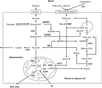
Therefore, the accretion rate of IMF content depends not only on the variation during the development of number and intrinsic metabolic activity of adipocytes inside the muscle tissue (Figure 1), but also on the muscle growth rate and metabolic activity of other organs. For instance, animals having a high muscularity with a high glycolytic activity (e.g. double-muscled cattle) or tissue with a high glycolytic activity (e.g. fish white muscle) generally display a reduced development of IMF. This suggests that muscle cells and adipocytes interplay during growth. In addition, early events that influence adipogenesis inside the muscle (i.e. proliferation and differentiation of adipose cells; type of the connective structure embedding adipocytes) might be involved in interindividual differences in IMF content. Increasing muscularity will also dilute the final fat content of muscle.
Overview of energy metabolism in muscular or adipose cells. Lipogenesis (right) is a major metabolic pathway in adipose cells. Mitochondrial oxidation is a major pathway in oxidative muscle fibres. ACC = acetyl-CoA carboxylase; COX = cytochrome-c oxydase; CPT-I = carnitine palmitoyl-transferase-I; CS = citrate synthase; ME: malic enzyme; H- or A-FABP = fatty acid-binding protein (H-FABP: heart and muscle isoform; A-FABP: adipocyte-isoform); FAS = fatty-acid synthase; G3PDH = glycerol-3 phosphate dehydrogenase; G6PDH = glucose-6-phosphate dehydrogenase; ICDH = isocitrate dehydrogenase; HAD = hydroxyacyl-CoA dehydrogenase; LDH = lactate dehydrogenase; LPL = lipoprotein lipase; PDH = pyruvate dehydrogenase; PFK = phosphofructokinase; TG = triglycerides.

How might marbling begin?
In the past decades, it has been demonstrated that tissue characteristics in postnatal life (and especially those of the muscle tissue; Picard et al., Reference Picard, Lefaucheur, Berri and Duclos2002) have their origins early in life. The foetal programming process determines not only the muscle mass but also its physiological properties, such as total fibre number, and likely the amount of IMF. The earliest development that is directly relevant to meat quality and quantity is then the formation of muscle fibres, but differences in early expression of fatty acid metabolism genes has been also revealed as important in relation to IMF content at slaughter (e.g. Cagnazzo et al., Reference Cagnazzo, te Pas, Priem, de Wit, Pool, Davoli and Russo2006).
The source of cells that will develop subsequently in intramuscular adipocytes is stem cells (reviewed by Harper and Pethick, Reference Harper and Pethick2004). Stem cells derived either from mesenchymal stem cells or from satellite cells have received specific attention (Tajbakhsh, Reference Tajbakhsh2005). However, the mechanisms involved in deriving different lineages to obtain adipose, muscle and fibroblastic cells remain largely elusive. Interaction between adipocytes and myoblasts in the very early stages of growth is likely to occur and influences the respective differentiation of both cellular types. As an example, impairment of insulin signalling has been demonstrated in muscle cells by in vitro coculture with human adipocytes (Dietze et al., Reference Dietze, Koenen, Röhrig, Horikoshi, Hauner and Eckel2002). Then, the activity of adipocytes in secreting adipokines (such as leptin; Kokta et al., Reference Kokta, Dodson, Gertler and Hill2004) as well as that of muscle fibres secreting myokines (such as myostatin, Artaza et al., Reference Artaza, Bhasin, Magee, Reisz-Porszasz, Shen, Groome, Meerasahib and Gonzalez-Cadavid2005; Hirai et al., Reference Hirai, Matsumoto, Hino, Kawachi, Matsui and Yano2007) are among the key putative mechanisms of this interaction. For instance, mutations in the myostatin gene or growth differentiation factor-8 in beef cattle increase the muscle mass in the double-muscled phenotype and leads to smaller adipocytes and fewer fat islands in muscle (Wegner et al., Reference Wegner, Albrecht and Ender1998; Cassar-Malek et al., Reference Cassar-Malek, Passelaigue, Bernard, Léger and Hocquette2007a). The exact mechanism by which myostatin regulates fat metabolism is unknown, and both direct (i.e. by a control on some genes involved in adipogenesis) and indirect effects (i.e. the anabolic effects of the myostatin mutations on skeletal muscle tissue that shift energy metabolites in such a manner as to prevent fat accumulation) have been suspected.
Another complexity in the regulation of intramuscular adipocyte development is added by the possible existence of interconversion of muscle satellite cells into adipocytes as shown in vitro (Kook et al., Reference Kook, Choi, Son, Lee, Hwang, Lee, Chang, Choi and Lee2006; Singh et al., Reference Singh, Chae, Hwang, Yoo, Ahn, Lee, Lee, Park and Chung2007; for review, see Chung and Johnson, Reference Chung and Johnson2008). It remains, however, to be determined whether this phenomenon also exists in vivo, and may account for fat infiltration into muscles observed following denervation (Dulor et al., Reference Dulor, Cambon, Vigneron, Reyne, Nougues, Casteilla and Bacou1998) or sarcopenia (for review, see Pahor and Kritchevsky, Reference Pahor and Kritchevsky1998). However, although some scientists have provided evidence for transdifferentiation of muscle cells to preadipocytes, the localization of marbling adipocytes to the perimysium in most cases would support the idea that marbling arises primarily from fibroblasts associated with perimysial connective tissue (for review, see Smith et al., Reference Smith, Kawachi, Choi, Choi, Wu and Sawyer2009). Taken together, much research is now needed to identify molecular markers related to stem cell differentiation or satellite cells’ interconversion to better understand the origin of intramuscular adipocytes for the subsequent development of IMF.
Foetal and postnatal growth
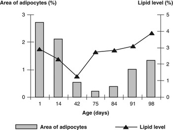
Although the different steps of ontogenesis have been described for both muscle (e.g. Picard et al., Reference Picard, Gagniere, Geay, Hocquette and Robelin1995 and Reference Picard, Lefaucheur, Berri and Duclos2002) and adipose tissues (e.g. reviews from Boone et al., Reference Boone, Mourot, Gregoire and Remacle2000; Hausman et al., Reference Hausman, Dodson, Ajuwon, Azain, Barnes, Guan, Jiang, Poulos, Sainz, Smith, Spurlock, Novakofski, Fernyhough and Bergen2009) in different species, the precise pattern of IMF ontogenesis and its regulation remains still poorly characterized. At the cellular level, triglycerides are generally initially stored within muscle fibres in mammals. A noticeable specificity of birds is that muscle lipid is high at hatching and mainly stored in intramuscular adipocytes (Chartrin et al., Reference Chartrin, Bernadet, Guy, Mourot, Hocquette, Rideau, Duclos and Baéza2007; Figure 2). After hatching, it is likely that the lipids stored in the adipocytes during embryonic life are used by the muscle fibres for growth in this species in which early energy metabolism is based on the use of lipids stored in egg yolk (Noble and Cocchi, Reference Noble and Cocchi1990; Klasing, Reference Klasing1998). For this reason, duck muscle progressively stores lipids first in fibres then in adipocytes as in mammals (Figure 2). Thereafter (e.g. from the first postnatal month onwards in pigs; Hauser et al., Reference Hauser, Mourot, De Clercq, Genart and Remacle1997), intramuscular adipocytes will increase both in size and number as shown, for instance, in rabbits (Gondret et al., Reference Gondret, Mourot and Bonneau1998) and pigs (Gondret and Lebret, Reference Gondret and Lebret2002). In the case of beef, marbling flecks become larger and new, small, round marbling flecks appear. These results suggest that hyperplasia of adipocytes, not initially visible (and hence early differentiated), plays an important role in marbling during growth (Albrecht et al., Reference Albrecht, Teuscher, Ender and Wegner2006). In practice, prenatal restriction (sufficient to reduce calf birth weight) has minimal effects on IMF level (Greenwood et al., Reference Greenwood, Cafe, Hearnshaw, Hennessy, Thompson and Morris2006; Greenwood and Cafe, Reference Greenwood and Cafe2007) unlike postnatal restriction that will clearly reduce IMF level.
Evolution with age of lipid level and the relative area occupied by adipocytes on cross-sections of breast muscle from mule duck (Chartrin et al., Reference Chartrin, Bernadet, Guy, Mourot, Hocquette, Rideau, Duclos and Baéza2007).

Because fat is deposited at a lower rate than muscle growth during the first periods of postnatal life and at a greater rate than lean tissues when animals get older, the concentration of fat in muscle (i.e. IMF content) will inevitably increase later in an animal’s life. This does not mean that the rate of fat accretion in intramuscular adipocytes is late maturing relative to other depots. Taken together, expression of IMF level within muscle homogenates generally follows a curve with an S shape that includes a period at the beginning of life in which IMF level does not increase, a period of linear development and sometimes (at least for early-maturing species) a plateau that represents a maximum of fat deposition capacity in the muscle at mature body size. In this context, the level of IMF at the start of the growth period (even if it is low) is likely a key determinant of the final level of IMF after finishing at least in cattle (reviews from Pethick et al., Reference Pethick, Barendse, Hocquette, Thompson and Wang2004, Reference Pethick, Harper, Hocquette and Wang2006 and Reference Pethick, Harper and Oddy2007). As assessing IMF content at this period of time is difficult, this emphasizes the usefulness of early predicting markers. As for non-muscular adipose tissue differentiation programme (Figure 1), it is then likely that a variety of genes involved in adipocyte development (such as insulin-responsiveness glucose transport GLUT4, LPL, lipogenic enzymes), and the master regulators of adipogenesis (peroxisome proliferator-activated receptor gamma (PPARG) and sterol-regulatory element-binding protein-1(SREBP1); for review, see Hausman et al., Reference Hausman, Dodson, Ajuwon, Azain, Barnes, Guan, Jiang, Poulos, Sainz, Smith, Spurlock, Novakofski, Fernyhough and Bergen2009) would be of great potential for predicting subsequent IMF development. Whether they could serve as early biomarkers should be discussed later in the text. Furthermore, any markers related to muscle differentiation growth may be also likely relevant to predict the ability of an animal to deposit IMF; however, this remains to be assessed.
Relationship of IMF level with carcass fatness and with muscle fibre types
It is generally accepted that a high muscle mass is associated with a low fat mass in the carcass and less IMF. This is obviously true with extreme animals such as double-muscled bovines (Hocquette et al., Reference Hocquette, Bas, Bauchart, Vermorel and Geay1998 and Reference Hocquette, Ortigues-Marty, Pethick, Herpin and Fernandez1999). This suggests that muscle and non-muscular adipose tissue interplay during the process of IMF accumulation, through hormonal changes such as thyroid hormones (Cassar-Malek et al., Reference Cassar-Malek, Picard, Kahl and Hocquette2007b) and the insulin-glucose axis (Hocquette et al., Reference Hocquette, Sauerwein, Higashiyama, Picard and Abe2006), which have been shown to be important in the differentiation of bovine muscles for instance. Attempts have been made to predict IMF level from blood parameters (Adachi et al., Reference Adachi, Kawano, Tsuno, Nomura, Yamamoto, Arikawa, Tsuji, Adachi, Onimaru and Ohwada1999). For instance, double-muscled bovines with low IMF level are characterized also by lower triiodothyronine, insulin and glucose plasma concentrations (Hocquette et al., Reference Hocquette, Bas, Bauchart, Vermorel and Geay1999) than normal animals of the same breed. Serum leptin has been correlated with fat deposition traits in ruminants (Geary et al., Reference Geary, McFadin, MacNeil, Grings, Short, Funston and Keisler2003), but not with IMF level (Altmann et al., Reference Altmann, Sauerwein and Von Borell2006). When different bovine breeds that differ in their ability to deposit fat are compared, no clear relationships between muscle growth potential and fat deposition are observed (Albertí et al., Reference Albertí, Panea, Sañudo, Olleta, Ripoll, Ertbjerg, Christensen, Gigli, Failla, Concetti, Hocquette, Jailler, Rudel, Renand, Nute, Richardson and Williams2008). Differences in muscle characteristics across bovine breeds can be associated not only with their fatness score but also with their biological type (rustic, beef or dairy) or with their geographical origin.
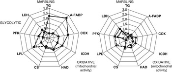
Within a given muscle, it is also known that oxidative fibres contain more phospholipids and more triglycerides (Figure 3). As a consequence, differences in IMF content between species and between muscles are often parallel to differences in muscle fibre types. For instance, breast muscle of chickens that is only composed of type IIb glycolytic fibres, has lower lipid content (1% to 2%) than breast muscle of ducks (2% to 3% of lipids), which contains only 15% type IIb glycolytic fibres (Baéza, Reference Baéza1995). Similarly, muscles from thigh and drumstick in chicken have higher IMF levels than breast muscles of the same species (4% to 5% v. 1% to 2%, Rabot, Reference Rabot1998). The fat breeds in cattle tend to have also more oxidative muscles than the lean breeds (Hocquette et al., Reference Hocquette, Jurie, Picard, Alberti, Panea, Christensen, Failla, Gigli, Levéziel, Olleta, Sañudo, Ertbjerg, Nute and Williams2007a). However, there is no strict association between IMF content and oxidative metabolism of the muscle fibres. This is evident, for instance, when bisons with oxidative muscles and low IMF are compared to Bos Taurus with less oxidative muscles but more IMF (Agabriel et al., Reference Agabriel, Bony and Micol1998). Within a given muscle, many studies in pigs have shown that the red part of semitendinosus (48% of type I fibres) displays a lower IMF content that the white part (20% of type I fibres). Indeed, the main part of IMF variability depends on differences in the number of intramuscular adipocytes located between fibre bundles. Adipocyte number has poor relationships with myofibre type composition as shown in pigs (Damon et al., Reference Damon, Louveau, Lefaucheur, Lebret, Vincent, Leroy, Sanchez, Herpin and Gondret2006). Similarly, Pekin ducks have higher IMF levels and a higher relative muscle surface occupied by adipocytes than Muscovy ducks, despite the typology of muscle fibres does not depend on species (Chartrin et al., Reference Chartrin, Schiavone, Bernadet, Guy, Mourot, Duclos and Baeza2005).
Relative levels of muscle triglyceride contents, of oxidative and glycolytic metabolic enzymes in muscles fibres and of A-FABP protein expression in rectus abdominis muscle (left) and semitendinosus muscle (right) in Limousine steers (dot lines) and cross-bred Angus × Japanese Black steers (full lines). It is noteworthy that the red oxidative muscle (r. abdominis) has more A-FABP and contains more IMF than the fast-glycolytic muscle (semitendinosus). Differences between genotypes are higher for the red oxidative muscle (r. abdominis) than for the fast-glycolytic one (semitendinosus). Adapted from Jurie et al. (Reference Jurie, Cassar-Malek, Bonnet, Leroux, Bauchart, Boulesteix, Pethick and Hocquette2007). COX = cytochrome-c oxydase; CS = citrate synthase; A-FABP = fatty acid-binding protein (A-FABP: adipocyte-isoform); ICDH = isocitrate dehydrogenase; HAD = hydroxyacyl-CoA dehydrogenase; LDH = lactate dehydrogenase; LPL = lipoprotein lipase; PFK = phosphofructokinase; TG = triglycerides.

We can conclude from all these observations that indicators of carcass fatness and muscle types are poorly markers of the ability of animals to deposit IMF. Of course, high muscle breeds will grow for longer and IMF will always be low at any given carcass weight. Similarly, the more red muscle is probably a prerequisite for IMF development, as intramuscular adipocytes need blood vessels. But, redness does not guarantee fat development. Indicators of adipocyte number are probably more relevant. The best marker of adipocyte number that can be assessed is probably the content in adipocyte-type fatty acid-binding protein (A-FABP, encoded by FABP4 gene) as shown in pigs (Damon et al., Reference Damon, Louveau, Lefaucheur, Lebret, Vincent, Leroy, Sanchez, Herpin and Gondret2006) and cattle (Jurie et al., Reference Jurie, Cassar-Malek, Bonnet, Leroux, Bauchart, Boulesteix, Pethick and Hocquette2007; Figure 3).
Metabolic indicators of the ability of animals to deposit IMF
IMF accumulation during growth results not only from an increase in the number of adipocytes within muscles but also from an increase in their volume. This is especially true when considering nutritional challenge to modulate IMF accumulation (e.g. Gondret and Lebret, Reference Gondret and Lebret2002) in pigs. Leptin expression at the mRNA level in muscles is then a good indicator of marbling (Bonnet et al., Reference Bonnet, Faulconnier, Leroux, Jurie, Cassar-Malek, Bauchart, Boulesteix, Pethick, Hocquette and Chilliard2007); this is likely because leptin is secreted by enlarged adipocytes.
Taken together, the activity of various lipogenic enzymes (Figure 1) such as acetyl-CoA carboxylase as one key lipogenic enzyme and of NADH-producing enzymes such as glucose-6-phosphate dehydrogenase or malic enzyme have been related to IMF level (Mourot and Kouba, Reference Mourot and Kouba1999 in pigs; Chartrin et al., Reference Chartrin, Bernadet, Guy, Mourot, Hocquette, Rideau, Duclos and Baeza2006b in ducks; Bonnet et al., Reference Bonnet, Faulconnier, Leroux, Jurie, Cassar-Malek, Bauchart, Boulesteix, Pethick, Hocquette and Chilliard2007 in cattle). Furthermore, the uptake of fatty acids by muscle LPL is found generally higher in muscles with a high fat content (Chartrin et al., Reference Chartrin, Bernadet, Guy, Mourot, Hocquette, Rideau, Duclos and Baeza2006b in ducks), especially in species that are characterized by a low lipogenic activity (Leveille et al., Reference Leveille, O’Hea and Chakbabarty1968; Henderson and Sargent, Reference Henderson and Sargent1981). Thus, the capacity for lipid uptake by muscle tissue is a major determinant of the control of IMF level in these species. In rainbow trout, a better correlation with IMF has been, however, observed for very low density lipoproteins (VLDL) receptors or for fatty acid transporter FAT/CD36 than for LPL (Kolditz et al., Reference Kolditz, Plagnes-Juan, Quillet, Lefèvre and Médale2009). However, in some studies, neither LPL and lipogenic enzymes in cattle (Bonnet et al., Reference Bonnet, Faulconnier, Leroux, Jurie, Cassar-Malek, Bauchart, Boulesteix, Pethick, Hocquette and Chilliard2007), nor oxidative and lipogenic enzymes in pigs (Damon et al., Reference Damon, Louveau, Lefaucheur, Lebret, Vincent, Leroy, Sanchez, Herpin and Gondret2006) were shown to be related to IMF variation. First, IMF accumulation during growth is the result of a balance between fatty acids synthesis into adipocytes and their oxidation within muscle fibres, rather than upregulation of a single pathway in mammals (Gondret et al. (Reference Gondret, Hocquette and Herpin2004a) in rabbits, Kolditz et al. (Reference Kolditz, Plagnes-Juan, Quillet, Lefèvre and Médale2009) in fish); the turnover of fatty acids within the muscle must be considered. This assumption has been recently confirmed with the demonstration that muscle with a high IMF content displays a low adenosine monophosphate-activated protein kinase (AMP-K) activity compared with low-fat muscle. Because AMP-kinase inhibits lipogenesis while promoting fatty-acid oxidation, a low AMP-K activity is expected to promote fat accumulation (Underwood et al., Reference Underwood, Means, Zhu, Ford, Hess and Du2008). Furthermore, to unravel discrepancies between studies, one has also to go back to the characteristics of experimental designs, considering the fact that different breeds, different muscles, different ages, different feeding regimen or different individuals are compared. Then, the relationships between IMF content and metabolic markers could depend on the source of variation in IMF. This is the reason why some authors have preferred to combine both blood indicators and metabolic enzyme activities to predict IMF variability, but the equations of prediction differ between muscles (Gondret et al., Reference Gondret, Hocquette and Herpin2004a). Furthermore, because enzymes of the same metabolic pathway (for instance, in mitochondria) are sometimes largely unrelated (Gondret et al., Reference Gondret, Damon, Jadhao, Houdebine, Herpin and Hocquette2004b), it is important to combine different metabolic markers.
Genetic and genomic markers of IMF content
Genetic selection
Heritability of IMF content is relatively high (h2 = 0.26 to 0.86, mean of 0.50) in pigs (Sellier, Reference Sellier1998; Newcom et al., Reference Newcom, Baas, Schwab and Stalder2005) and cattle as well as in fish (0.2 to 0.5; Rye and Gjerde, Reference Rye and Gjerde1996). IMF content shows a moderate but positive genetic correlation with carcass fatness (r G close to 0.30, Sellier, Reference Sellier1998). The genetic correlation of IMF content is on average positive with tenderness (r G = 0.41) or negative with Warner-Bratzler shear force (r G = −0.50) in cattle. As marbling score in these animals is genetically highly correlated with muscle lipid content (r G = 0.91), selection based on the former may lead to a correlated improvement in tenderness (for review, see Hocquette et al., Reference Hocquette, Sauerwein, Higashiyama, Picard and Abe2006). This relationship explains most of the efforts dedicated to the development of live scanning for IMF content as a selection tool in the United States and Australia (Reverter et al., Reference Reverter, Johnston, Graser, Wolcot and Upton2000; Hassen et al., Reference Hassen, Wilson, Amin, Rouse and Hays2001; Sapp et al., Reference Sapp, Bertrand, Pringle and Wilson2002). However, as marbling is positively genetically correlated to the carcass fatness, the undesirable side effects of selection on the basis of IMF content or marbling are often the increase in carcass fatness. Conversely, selection against body fat content has generally induced a decrease in IMF level (e.g. Baéza et al., Reference Baéza, de Carville, Salichon, Marche and Leclercq1997 and Reference Baéza, Dessay, Wacrenier, Marche and Listrat2002 in ducks for abdominal fat selection). However, the genetic dissociation between carcass fat content and IMF content by selection is possible. Examples are given in chickens in which two lines divergently selected for abdominal fat level (1.4% v. 3.93%, Berri et al., Reference Berri, Le Bihan-Duval, Baeza, Chartrin, Millet and Bordeau2005) did not display any differences in IMF level in thigh and breast muscles (Ricard et al., Reference Ricard, Leclercq and Touraille1983; Berri et al., Reference Berri, Le Bihan-Duval, Baeza, Chartrin, Millet and Bordeau2005). Another example is the divergent selection for IMF content in trout that lead to a lean muscle line with IMF lower than 5% and a fatty muscle line with IMF up to 10% without any significant difference in whole body fat content (Kolditz et al., 2008).
Considerable efforts have been made during the past decades to identify quantitative trait loci (QTL) for IMF (de Koning et al., 1999 and Sanchez et al., 2007; in pigs, and Jennen et al., 2005; in chickens). In cattle, research was conducted by the US Meat Animal Research Center, Nebraska and also in US Universities of Colorado, Texas, Louisiana and Florida (reviewed by Burrow et al., Reference Burrow, Moore, Johnston, Barense and Bindon2001), other reports by Kim et al. (Reference Kim, Davis, Sanders, Turner, Miller, Savell, Smith and Taylor1998) and Riley et al. (Reference Riley, Chase, Hamond, West, Johnson, Olson and Coleman2003) for steers intensively fattened in feedlots. Other results were obtained by the Cooperative Research Center for the cattle and beef industry in Australia (Reverter et al., Reference Reverter, Johnston, Ferguson, Pery, Godard, Burow, Oddy, Thompson and Bindon2000 and Reference Reverter, Johnston, Graser, Wolcot and Upton2003; Johnston et al., Reference Johnston, Reverter, Ferguson, Thompson and Burow2003) for steers and heifers of temperate and tropically adapted breeds.
Finally, the existence of a major gene with a great effect on IMF content has been postulated by segregation analysis (Janss et al., Reference Janss, Van Arendonk and Brascamp1997). The gene, named MI, has a recessive allele (imf) that increases IMF content and originates from the Chinese Meishan pig breed. However, the same authors have also located several small QTL with reduced individual effects afterwards. In Duroc pig population, Sanchez et al. (Reference Sanchez, Iannuccelli, Basso, Bidanel, Billon, Gandemer, Gilbert, Larzul, Legault, Riquet, Milan and Le Roy2007) further indicated that the putative major gene corresponded in fact to two QTL.
Genetic markers
Various genetic markers associated to IMF deposition or marbling have been reported; they include the polymorphic microsatellite loci CSSM34 and ETH10, which are associated with marbling scores in the Angus, Shorthorn and Wagyu cattle (Barendse, Reference Barendse2002). They also include the thyroglobulin (TG) gene in cattle (Barendse, Reference Barendse2002), the diacylglycerol acyltransferase 1 or 2 (DGAT1; DGAT2) genes in cattle (Thaller et al., Reference Thaller, Kühn, Winter, Ewald, Bellmann, Wegner, Zühlke and Fries2003), pigs (Nonneman and Rohrer, Reference Nonneman and Rohrer2002), chicken (Bourneuf et al., Reference Bourneuf, Herault, Chicault, Carre, Assaf, Monnier, Mottier, Lagarrigue, Douaire, Mosser and Diot2006) and sheep (Xu et al., Reference Xu, Chen, Ma and Xue2009), and the splicing factor serine-arginine-rich protein (SFRS18) gene in pigs (Wang et al., Reference Wang, Xue, Wang, Liu, Xu, Zhao, Jiang, Dodson and Chen2009b). However, the results are often inconsistent. For instance, the thyroglobulin marker (commercialized as the GeneSTAR MVPs for Marbling, Pfizer Animal Health, Kalamazoo, MI, USA) has been reported to have no effect in Simmental steers (Rincker et al., Reference Rincker, Pyatt, Berger and Faulkner2006). In addition, recent Beef CRC work in Australia has proved the current markers (including TG) used by Catipult Genetics in Australia as ineffective (Graser, Reference Graser2008).
From scientific knowledge on how IMF starts to accumulate in the muscle, it is then obvious that genes belonging to adipogenic process within muscles could be also good candidates for predicting IMF content. Single-nucleotide polymorphisms (SNPs) in various adipogenic genes have been then described. Zhao et al. (Reference Zhao, Liu, Li, Jiang, DU and Zhu2007) found an association between an SNP of the adipose differentiation-related protein (ADRP) gene and IMF level in chicken. Wu et al. (Reference Wu, Liu, Hou, Wang and Kong2008) found an association between an intronic SNP of PPARG gene and IMF level in four meat-type duck populations. Lipogenic genes have been postulated as good markers for IMF content or its composition, such as fatty acid synthase, SREBP1 and stearoyl-CoA desaturase-1 (SCD1; e.g. Bourneuf et al., Reference Bourneuf, Herault, Chicault, Carre, Assaf, Monnier, Mottier, Lagarrigue, Douaire, Mosser and Diot2006; Hoashi et al., Reference Hoashi, Ashida, Ohsaki, Utsugi, Sasazaki, Taniguchi, Oyama, Mukai and Mannen2007 and Reference Hoashi, Hinenoya, Tanaka, Ohsaki, Sasazaki, Taniguchi, Oyama, Mukai and Mannen2008 in cattle; Hérault et al., Reference Hérault, Robert, Al Mohammad, Chartrin, Baéza and Diot2008 in chickens). Genes involved in intracellular fatty-acid transport within skeletal muscles have been also notably proposed. Especially, studies put light on DNA markers or expression levels of FABP4 in pigs (Gerbens et al., Reference Gerbens, Jansen, Van Erp, Harders, Meuwissen, Rettenberger, Veerkamp and Te Pas1998 and Reference Gerbens, Verburg, Van Moerkerk, Engel, Buist, Veerkamp and te Pas2001; Damon et al., Reference Damon, Louveau, Lefaucheur, Lebret, Vincent, Leroy, Sanchez, Herpin and Gondret2006), cattle (Jurie et al., Reference Jurie, Cassar-Malek, Bonnet, Leroux, Bauchart, Boulesteix, Pethick and Hocquette2007; Barendse et al., Reference Barendse, Bunch, Thomas and Harrison2009) and avian species (Luo et al., Reference Luo, Chen, Wen, Zhao, Zheng and Sun2006 in chicken; Saez et al., Reference Saez, Gentes, Hocquette and Baéza2008 in ducks) to be associated to variations in IMF content. One study reported a specific allele of the PNAS-4 gene (i.e. known to be involved in early muscle development) prevalent in Chinese indigenous cattle breeds that are much fatter than the other studied genotypes (Mo et al., Reference Mo, Zhu, te Pas, Li, Yang, Wang, Wang and Li2008).
This list of genetic markers associated with IMF or marbling is not exhaustive (for a recent review, see Gao et al., Reference Gao, Zhang, Hu and Li2007). The associations of these gene markers are, however, not always positive depending on the species, the breed and the trait, which is under study (IMF content, marbling or other ones). For instance, Cheong et al. (Reference Cheong, Yoon, Park, Kim, Bae, Namgoong, Lee, Han, Kim, Cheong and Shin2008) in cattle reported some associations with marbling score of polymorphic sites in genes related to tenderness; this may be, however, a false-positive association due the relationships between IMF content and tenderness. Then, none of these markers are omnipotent. This may be not a problem anymore due to the advent of genomic selection (Meuwissen et al., Reference Meuwissen, Hayes and Goddard2001). This approach called genomic selection is based on markers that cover the whole genome, and has an increasing application due to the advent of chips for genotyping. In theory, the whole genetic variance is potentially explained by all the markers analysed simultaneously and the whole genetic value of each animal is well estimated. There is a huge challenge in this area for the immediate future. This is likely to have major effects on the agendas of research and commercial organizations, as genomic selection will probably redesign animal breeding and management programmes. But, this type of approach emphasizes the need for (i) powerful SNP panels and also (ii) large phenotypic databases for discovery and validation (Visscher, Reference Visscher2008). In the case of phenotypes, we need an ontology system to better define and standardize the measurements of phenotypes. An international undertaking entitled ‘Animal Trait Ontology’ has been initiated with the goal to make the comparison of phenotypic information between species more easy, including laboratory models and agronomical species (Hughes et al., Reference Hughes, Bao, Hu, Honavar and Reecy2008).
Genomic principles and main findings
Nowadays, scientists have access to gene networks and interaction, thanks to the development of transcriptomics and proteomics that allow the high-throughput detection of genes and proteins differentially expressed between conditions without any positional or functional a priori. Several studies dealing with functional genomics in livestock animals have been published so far (reviewed by Lehnert et al., Reference Lehnert, Wang, Tan and Reverter2006; Hocquette et al., Reference Hocquette, Lehnert, Barendse, Cassar-Malek and Picard2007b; Cassar-Malek et al., Reference Cassar-Malek, Picard, Bernard and Hocquette2008 for cattle). The global nature of genomics technology could be then an advantage for elucidating the complex physiological control of IMF fat content, which is likely mediated through multiple biochemical and molecular mechanisms in both adipocytes and myofibres. Differential-display reverse transcription polymerase chain reaction (ddRT-PCR; Sasaki et al., Reference Sasaki, Nagai, Nagata, Doronbekov, Nishimura, Yoshioka, Fujita, Shiga, Miyake, Taniguchi and Yamada2006; Lee et al., Reference Lee, Park, Cho, Kim, Lee, Jeon, Lee, Im, Oh, Thompson and Yoon2007), microarrays (Wang et al., Reference Wang, Byrne, Reverter, Harper, Taniguchi, McWilliam, Mannen, Oyama and Lehnert2005a and Reference Wang, Reverter, Mannen, Taniguchi, Harper, Oyama, Byrne, Oka, Tsuji and Lehnert2005b, Kolditz et al., 2008; Liu et al., Reference Liu, Damon, Guitton, Guisle, Ecolan, Vincent, Cherel and Gondret2009) eventually using muscle-dedicated (Sudre et al., Reference Sudre, Leroux, Cassar-Malek, Hocquette and Martin2005) or fat-enriched arrays (Lehnert et al., Reference Lehnert, Wang, Tan and Reverter2006) and proteomics (Kolditz et al., 2008; Liu et al., Reference Liu, Damon, Guitton, Guisle, Ecolan, Vincent, Cherel and Gondret2009) have been recently used in muscles of livestock animals and fishes to examine gene and protein expressions at different developmental stages during fattening period, between two breeds or two selected lines with a different propensity to accumulate IMF fat, and (or) between individuals with contrasting levels for this trait. In some studies, it cannot be excluded that the differences seen in gene expressions are due to breed characteristics rather than to variations in IMF per se.
Taken together, this allowed a great number of differentially expressed genes associated with IMF level to be identified. For instance, ddRT-PCR lead to the identification of NAT1 (i.e. a translational suppressor) by comparing bovine muscles with different IMF contents from different finishing periods on high-grain regimen (Childs et al., Reference Childs, Goad, Allan, Pomp, Krehbiel, Geisert, Morgan and Malayer2002). Putative functional genes were also found to be differentially expressed (e.g. ATP citrate lyase) or, surprisingly, not differentially expressed (e.g. PPARG) between animals extremes for IMF content (Childs et al., Reference Childs, Goad, Allan, Pomp, Krehbiel, Geisert, Morgan and Malayer2002). Transcriptomic studies identified other genes (e.g. 12-lipoxygenase, prostaglandin-D synthase), as key candidates involved in the control of fat accumulation in ruminants (Cho et al., Reference Cho, Han, Kang, Lee and Choi2002). In various studies, expression approaches also identified various biological categories of genes to be potentially involved in IMF accumulation. Wang et al. (Reference Wang, Byrne, Reverter, Harper, Taniguchi, McWilliam, Mannen, Oyama and Lehnert2005a) showed that the genes, which are more expressed in muscles from Japanese Black cattle (which produce marbled beef), compared with Holsteins, are associated with the thyroid hormone pathway, unsaturated fatty-acid synthesis and fat deposition including FABP4. Some genes involved in muscle structure have been also related to changes in IMF content and deposition (Luo et al., Reference Luo, Chen, Wen, Zhao, Zheng and Sun2006 in chicken; Lee et al., Reference Lee, Park, Cho, Kim, Lee, Jeon, Lee, Im, Oh, Thompson and Yoon2007; Liu et al., Reference Liu, Damon, Guitton, Guisle, Ecolan, Vincent, Cherel and Gondret2009) suggesting that components of extracellular matrix could exert effects on adipocyte-lineage cells and their development. Transcriptomic studies were also performed during muscle growth in a time when animals had none visible differences in IMF level. One study comparing different genotypes of cattle from 3 to 25 months of age indicated that adipogenesis- and lipogenesis-related genes (such as FABP4 again, AdipQ, and c/EBPbeta) were more expressed at 7 months of age in muscles of genotypes with a potential higher propensity to deposit IMF (Wang et al., Reference Wang, Bower, Reverter, Tan, De Jager, Wang, McWilliam, Cafe, Greenwood and Lehnert2009a). This is a great importance because the adequate timing to reveal early markers of IMF content remains elusive in much species.
Only a few studies have so far been published concerning the detection of diffentially expressed proteins in meats and flesh poor or rich in fats (Kim et al., Reference Kim, Cho, Lee, Park, Lee, Cho, Choy, Yoon, Im and Park2008 in cattle; Kolditz et al., 2008 in trout; Liu et al., Reference Liu, Damon, Guitton, Guisle, Ecolan, Vincent, Cherel and Gondret2009 in pigs). For instance, it was shown that some structural proteins (α-actin, T-complex protein-1) are differentially expressed between Korean beef, which is rich in fat, and another breed with low IMF, and the heat-shock protein-27 previously identified as a biomarker for tenderness (Morzel et al., Reference Morzel, Terlouw, Chambon, Micol and Picard2008) correlates therefore well with IMF content (Kim et al., Reference Kim, Cho, Lee, Park, Lee, Cho, Choy, Yoon, Im and Park2008). In this latter study, a new protein (inositol 1,4,5-triphosphate receptor IP3R1) involved in calcium signalling pathway is also shown to be associated with low IMF level (Kim et al., Reference Kim, Cho, Lee, Park, Lee, Cho, Choy, Yoon, Im and Park2008).
A recent combination of transcriptome and proteome analyses (Kolditz et al., 2008) of the liver of the two rainbow trout lines selected for IMF revealed that major changes induced by the selection procedure were not only related to lipid metabolism. Besides changes in abundance of markers of lipid synthesis and transport, such as H-FABP, G3PDH and 6-PGD, most of the alterations induced by the divergent selection for IMF occurred in protein and amino-acid metabolism. In particular, levels of mitochondrial aspartate aminotransferase, glutamate dehydrogenase and alanine: glyoxylate aminotransferase, three key enzymes that play a major role in amino-acid catabolism, were increased in the liver of the ‘fatty muscle’ fish. Transcripts and proteins that functioned in amino-acid bioconversion (betaine-homocysteine S-methyltransferase and 4-aminobutyrate aminotransferase) and in proteasome-dependent proteolysis were also enhanced in this line. All these findings suggest an increase in hepatic flux for energy production through amino-acid metabolism in the fatty muscle line compared to the lean muscle line. This difference in nutrient utilization could be at least in part orchestrated by PPARα (the expression of which was concomitantly decreased), exerting opposing controls of fatty-acid oxidation and amino-acid catabolism.
Outlook for the future
It is clear that animal phenotype is not determined solely by gene and protein expression changes. Different general technical issues have to be solved in genomics to better understand how IMF is determined. They include progress in proteomics to better assess translational regulation and post-translational modifications, and also the functional validation of potential markers of IMF deposition by inhibition or overexpression of the involved genes in cell lines and animal models such as rodent or zebrafish. For instance, Roorda et al. (Reference Roorda, Hesselink, Schaart, Moonen-Kornips, Martinez-Martinez, Losen, De Baets, Mensink and Schrauwen2005) have indicated that overexpression of DGAT1 protein in muscle using DNA electroporation was able to induce intramyocellular triglyceride storage in rats.
It is also clear that the associations of the genomic markers for IMF are not always consistent depending on the species, the breed or the muscle type; this problem may be resolved by assessing all the markers within a single genomic tool such a DNA chip dedicated to IMF deposition.
Another challenge for gene expression researchers is to turn the current knowledge into practical biological assays useful for the meat industry. Unlike geneticists (which have developed commercial DNA tests), biochemists missed this challenge. Some scientists do not believe in RNA-based methods to develop into robust quantitative assays (Lehnert et al., Reference Lehnert, Wang, Tan and Reverter2006), although those methods have been standardized before proteomics tools. Alternatives are to develop protein-based assays (Guillemin et al., Reference Guillemin, Meunier, Jurie, Cassar-Malek, Hocquette, Levéziel and Picard2009), or to detect the biomarkers in peripheral blood instead of within muscle samples. In addition, it is probable that expression profiling will become integrated with genotyping outputs in the next future. The combination of linkage genetics and expression profiling (genomics) is called ‘genetical genomics’ and is forecasted to become important (Kadarmideen et al., Reference Kadarmideen, Von Rohr and Janss2006).
Nutritional regulation of IMF level and potential outputs
Metabolic specificities of intramuscular adipose tissue and practical outputs
Much evidence is accumulating to indicate that IMF is not just an ectopic location of subcutaneous lipids, but is characterized by specific metabolic features. First, the proliferative potential is lower in bovine intramuscular preadipocytes compared to bovine subcutaneous preadipocytes (Wan et al., Reference Wan, Du, Ren and Meng2009). Similarly, the adipogenic response to antidiabetic agents thiazolidinediones is lower in stroma–vascular cell cultures derived from IMF than in adipose stroma–vascular cell cultures (Hausman and Poulos, Reference Hausman and Poulos2004; Poulos and Hausman, Reference Poulos and Hausman2006). Second, inherent differences in the capacity to differentiate exist between cells from IMF and subcutaneous adipose tissue (Grant et al., Reference Grant, Ortiz-Colon, Doumit, Tempelman and Buskirk2008 in cattle). Third, another evidence of the specificities of intramuscular adipocytes is provided by the relative low activity levels of enzymes of lipogenesis in those cells compared to subcutaneous adipocytes in pigs (Gardan et al., Reference Gardan, Gondret and Louveau2006) and in cattle (Bonnet et al., Reference Bonnet, Faulconnier, Leroux, Jurie, Cassar-Malek, Bauchart, Boulesteix, Pethick, Hocquette and Chilliard2007). One key metabolic problem of IMF adipocytes is indeed their low capacity for synthesis and indeed degradation of fatty acids. Recent proteomic investigation reveals that not only lipogenesis, but also indicators of lipolysis, fatty-acid oxidation and basal-energy metabolism are lower in abundance in adipocytes isolated from pig muscle than in fat cells isolated from other body fat depots (Gondret et al., Reference Gondret, Guitton, Guillerm-Regost and Louveau2008). Differences in gene expression between IMF depot and subcutaneous adipose tissue have been also reported in cattle (Ross et al., Reference Ross, Smith, Krehbiel, Malayer, DeSilva, Morgan, White, Hersom, Horn and Geisert2005). An additional evidence of intramuscular specific metabolism is given by cattle in which marbling adipocytes preferentially use glucose/lactate carbon, whereas subcutaneous adipose tissue uses mainly acetate as a source of acetyl units for lipogenesis (reviewed by Smith et al., Reference Smith, Kawachi, Choi, Choi, Wu and Sawyer2009). Then, a higher level of GLUT4 expression and higher activities of metabolic enzymes involved in the conversion of glucose into long-chain fatty acids (namely phosphofructokinase and ATP-citrate lyase) were detected in intramuscular adipose tissue compared to subcutaneous fat in these species (Hocquette et al., Reference Hocquette, Jurie, Bonnet and Pethick2005). More recently, it was shown that the rate of glucose incorporation into fatty acid was more than twice as high as the rate of acetate incorporation in IMF adipocytes, whereas they are similar in subcutaneous adipose tissue (Rhoades et al., Reference Rhoades, Sawyer, Chung, Schell, Lunt and Smith2007). Thus, when fat synthesis is limited (such as within IMF adipocytes), glucose provides a greater proportion of acetyl units for lipogenesis than acetate (for review, see Smith et al., Reference Smith, Kawachi, Choi, Choi, Wu and Sawyer2009).
Although these studies did not provide any biomarkers, they could be used to propose new feeding strategies. For instance, it should be proposed that feeding a hay-based diet (providing mainly acetate) may alter intramuscular adipose tissue metabolism with less effect on subcutaneous adipose tissue accretion in ruminants. A recent experiment indicated that feeding a corn-based diet (providing a great amount of propionate, a glucogenic precursor) enhances glucose uptake in IMF depot, whereas feeding a hay-based diet reduces insulin action without altering acetate incorporation in fatty acids (Rhoades et al., Reference Rhoades, Sawyer, Chung, Schell, Lunt and Smith2007). Because subcutaneous fat used acetate more effectively than intramuscular adipose tissue, feeding a hay-based diet would then promote subcutaneous fat deposition over intramuscular adipose tissue accretion. Conversely, greater absorption of the gluconeogenic precursor propionate earlier in the growth period might result in increased IMF deposition and greater marbling scores as observed in winter-wheat fed steers (Choat et al., Reference Choat, Krehbiel, Duff, Kirksey, Lauriault, Rivera, Capitan, Walker, Donart and Goad2003). Generally speaking, diets that promote glucose supply to the muscle might increase IMF deposition in ruminants. This can be achieved by the administration of glucogenic precursors, such as propylene glycol, which induces elevated propionate in the rumen and higher levels of blood insulin and glucose (Myung and Sun, Reference Myung and Sun2007). This can also be achieved by maximizing fermentation in the rumen or by increasing starch digestion in the small intestine (Rowe et al., Reference Rowe, Choct and Pethick1999). Diets with a high glycaemic index (i.e. allowing rapid glucose absorption and concomitant high insulin levels) will deliver increased levels of net energy for lipogenesis; this explains why grain feeding promotes more IMF development compared with grass finishing (Pethick et al., Reference Pethick, Harper and Oddy2004). We must keep in mind, however, that it is not so easy to favour IMF development as the expense of subcutaneous adipose tissue through nutrition factors only, but when combined with genetics, nutritional factors may be very useful.
Manipulating dietary protein, energy and micronutrients
The majority of studies have indicated that marbling scores are less affected by differences in diet than backfat thickness or total carcass fat. It is also easier to manipulate the muscle lipid composition by nutritional challenge than IMF content.
In fish with carnivorous (piscivorous) feeding habits, such as salmonids, synthesis of fat from dietary digestible carbohydrates is weak (Brauge et al., Reference Brauge, Corraze and Médale1995). The most efficient nutritional way to manage IMF content is thus the dietary energy content through dietary fat. Indeed, high fat diets enhance muscle lipid uptake mediated by muscle VLDL-R and FAT/CD-36 protein (Kolditz et al., Reference Kolditz, Plagnes-Juan, Quillet, Lefèvre and Médale2009).
In all species, a feed restriction generally causes a decrease in IMF level (Gondret et al., Reference Gondret, Lebas and Bonneau2000 in rabbits; Rasmussen et al., Reference Rasmussen, Ostenfeld, Ronsholdt and McLean2000 in fish, Santoso, Reference Santoso2001; Pethick et al., Reference Pethick, Davidson, Hopkins, Jacob, D’Souza, Thompson and Walker2005 in lambs; Gondret and Lebret, Reference Gondret and Lebret2002 and Reference Gondret and Lebret2007 in pigs; Ljubic et al., Reference Ljubic, Milinkovic-Tur, Pirsljin and Zdelar-Tuk2007 in avian species). In rabbits as in other species, energy restriction decreases activities of some lipogenic enzymes without any change in muscle fibre type composition (Gondret et al., Reference Gondret, Lebas and Bonneau2000), confirming again the idea that IMF is poorly related to fibre types. Fasting was also shown to strongly affect both muscle structure and IMF levels in fish so that the current practice to reduce IMF at slaughter is a finishing period of feeding with a low-fat diet that enables to manage IMF without affecting other muscle components (Rasmussen et al., Reference Rasmussen, Ostenfeld, Ronsholdt and McLean2000; Rasmussen, Reference Rasmussen2001).
A way to manipulate IMF content could be also a restriction/refeeding strategy. Mild nutritional restriction followed by ad libitum feeding have major effects on metabolic enzyme activities, but they do not increase IMF level (Cassar-Malek et al., Reference Cassar-Malek, Hocquette, Jurie, Listrat, Jailler, Bauchart, Briand and Picard2004 in cattle, Gondret and Lebret, Reference Gondret and Lebret2007 in pigs). The effect of a severe undernutrition period followed by refeeding has been examined using microarray technology in cattle to get a broader view of the changes that occur. Major underexpression of genes encoding muscle structural proteins, extracellular matrix and muscle metabolic enzymes (especially those belonging to the metabolic glycolytic pathway) have been observed (Lehnert et al., Reference Lehnert, Wang, Tan and Reverter2006), and the expression of most of the genes was then restored after refeeding.
In various species, overfeeding induces an increased IMF content (Guillerm-Regost et al., Reference Guillerm-Regost, Louveau, Sebert, Damon, Champ and Gondret2006 in pigs; Chartrin et al., Reference Chartrin, Bernadet, Guy, Mourot, Hocquette, Rideau, Duclos and Baéza2007 in ducks). As the amount of feed ingested by ducks or geese is much higher during the overfeeding period than that ingested by birds fed ad libitum (Chartrin et al., Reference Chartrin, Bernadet, Guy, Mourot, Duclos and Baéza2006c), the effect of overfeeding on IMF level, and also on intramuscular adipocyte characteristics and overall muscle energy metabolism is then stronger in these species than in other species. However, such strategies generally induce a greater increase in fat stores in organs other than muscles (i.e. liver in fatty goose or duck, subcutaneous fat).
The most successful nutritional strategy in non-ruminants to dissociate fat accumulation in muscle to that in other parts of the body has been obtained with subtle protein-deficient diets (Gondret and Lebret, Reference Gondret and Lebret2002; Doran et al., Reference Doran, Moule, Teye, Whittington, Hallett and Wood2006; D’Souza et al., Reference D’Souza, Pethick, Dunshea, Pluske and Mullan2008 in pigs). The basis of these strategies was to restrict muscle development, but the mechanisms involved in the observed increase in IMF content remain elusive. One of the possible explanations could be that lower dietary protein levels stimulate the expression of SCD1, which catalyses the cellular biosynthesis of monounsaturated fatty acids (Doran et al., Reference Doran, Moule, Teye, Whittington, Hallett and Wood2006). Indeed, oleic and palmitoleic fatty acids, as the main products of SCD, are the main monounsaturated fatty acids in fat depots and membrane phospholipids, and a different pattern of SCD gene isoforms may exist between muscle and subcutaneous fat tissues. Similarly, Da Costa et al. (Reference Da Costa, McGillivray, Bai, Wood, Evans and Chang2004) have shown that a low-lysine (low protein) diet increases SCD transcriptional rate in pig muscles. The picture is, however, somewhat different in fish where fat deposition is increased by feeding diets with high protein levels (Reinitz, Reference Reinitz1983). When protein intake exceeds the level that fish is able to use to synthesize body protein, carbons from dietary protein are partly stored as fat. These strategies are less conclusive in cattle. Two studies indicated that diets containing more or less proteins than recommended amounts did not lead to significant differences in marbling or IMF (Oddy et al., Reference Oddy, Smith, Dobos, Harper and Allingham2000; Pethick et al., Reference Pethick, McIntyre and Tudor2000).
In cattle, it is also known that vitamin A deficiency is associated with an elevated IMF content (reviews from Harper and Pethick, Reference Harper and Pethick2004 and Pethick et al., Reference Pethick, Harper, Hocquette and Wang2006). For instance, a low intake of ß-carotene or diet deficient in vitamin A increase marbling score in young Wagyu steers (Oka et al., Reference Oka, Maruo, Miki, Yamasaki and Saito1998) and IMF content (+35%) of the Longissimus thoracis muscle in Australian Angus cattle (Kruk et al., Reference Kruk, Bottema, Davis, Siebert, Harper, Di and Pitchford2005). Similarly, D’Souza et al. (Reference D’Souza, Pethick, Dunshea, Pluske and Mullan2003) reported higher IMF content (+53%) in pigs fed grower and finisher diets deficient in vitamin A. It has been proposed that the effect of vitamin A on IMF deposition is mediated by retinoic acid, a derivative of vitamin A that inhibits adipocyte differentiation both in vivo and in vitro (Sato et al., Reference Sato, Hiragun and Mitsui1980; Bonet et al., Reference Bonet, Ribot, Felipe and Palou2003). Therefore, vitamin A restriction is supposed to increase hyperplasia (Gorocica-Buenfil et al., Reference Gorocica-Buenfil, Fluharty, Reynolds and Loerch2007). It has also been proposed that retinoic acid regulated growth hormone gene expression (Bedo et al., Reference Bedo, Santisteban and Aranada1989), which in turn decreases fat deposition and IMF in steers (Dalke et al., Reference Dalke, Roeder, Kasser, Veenhuizen, Hunt, Hinman and Schelling1992). Deficiencies in retinoic acid, therefore, may result in lower growth hormone concentrations and increased fat deposition including IMF. Other micronutrients potentially involved in IMF accumulation have also been studied in cattle (for review, see Kawachi, Reference Kawachi2006). For instance, unlike vitamin A and D, vitamin C has been shown to promote adipogenesis (for review, see Smith et al., Reference Smith, Kawachi, Choi, Choi, Wu and Sawyer2009).
Taken together, nutritional manipulation of IMF independently from body fat depots has proved to be more difficult to achieve than genetic strategies. In addition, the biological mechanisms that explain the variability of IMF content differ between genetic and nutritional factors. This implies to better understand the metabolic regulation of IMF (Figure 1) in order to better control it by nutritional factors. The nutritional regulation of IMF also differs between ruminants, monogastrics and fish due to their digestive and nutritional particularities. The effect of nutritional strategies on IMF thus requires a better understanding of the metabolic regulation of fatty-acid fluxes. It is therefore more difficult to identify markers of IMF regulated by nutritional factors than by genetic or physiological factors.
Conclusion
Muscle lipid content plays key roles in various quality traits of meat and depends on many factors including species, breed, genotype, growth rate, sex, age, muscle location, and nutrition. Early events that determine the number of intramuscular adipocytes (i.e. proliferation and differentiation of adipose cells) are likely crucial in the final determination of muscle lipid content in animals at commercial slaughter. IMF depot also develops largely in parallel with other fat depots, but is also dependent on muscle growth. Critical developmental time periods have to be re-examined by expression experiments using genomic tools. There is also additional evidence that intramuscular adipocytes later in the growth period are metabolically different to other depots (subcutaneous fat), and an understanding of this would appear important to underpin strategies for genetic and non-genetic manipulation of IMF. So, a great number of adipogenic factors and metabolic enzymes (especially those involved in fatty-acid metabolism) may be considered for explaining variations in IMF content or as potential markers in this trait. They are used mainly for genetic purposes if DNA markers are identified in those genes. But, recent studies have put light on the importance of metabolic balance between various pathways, rather than the control of one single pathway for the control of IMF. New targets and markers will be identified in the future using combined genomic, transcriptomic and proteomic approaches. From the increasing biological knowledge, it is, however, clear that only a few of them will go to application as tests because of the lack of effective commercial utility. In fact, a biomarker must provide information that is not available by a simpler and already existing cheap method. So, before a biomarker brings any benefit over other criteria, it needs to address the following four concepts: ‘easier, better, faster and cheaper’. It must also be validated at farm level or food-production chain, and this should be a priority of research in the case for the control of IMF content.