Introduction
Mycobacterium tuberculosis infection (tuberculosis; TB) has been present in the human population since antiquity. Analysis of fragments of spinal column tissue from excavated Egyptian mummies (2400BC) has revealed signs of tubercular decay. Spinal tuberculosis is however uncommon, manifesting in less than 1 per cent of patients with tuberculosis. Cranio-vertebral junction tuberculosis is extremely rare among patients with tubercular spondylitis, constituting only 0.3 to 1 per cent of cases.Reference Behari, Nayak, Bhargava, Banerji, Chhabra and Jain1
Chronic, granulomatous inflammation of regional vertebrae results in localised necrosis and erosion, leading to collapse of the vertebral bodies and deformities of the spinal column. These problems may be further complicated by spinal cord compression, which manifests in a variety of neurological symptoms. The extensive osseo-ligamentous destruction caused by tuberculosis of the cranio-vertebral junction (the most mobile segment of the cervical spine) leads to atlanto-axial instability and compression of vital cervico-medullary centres. This may in turn manifest as quadriparesis, bulbar dysfunction and respiratory insufficiency.Reference Behari, Nayak, Bhargava, Banerji, Chhabra and Jain1
We report two patients presenting with cranio-vertebral tuberculosis causing spinal cord compression. Both were decompressed via an endoscopic, endonasal surgical approach.
Case reports
Case one
A 37-year-old Malay woman was diagnosed in 2002 with tuberculous (TB) lymphadenitis isolated to the cervical region. She completed one year of standard antituberculosis treatment and became asymptomatic. However, in December 2006 she developed neck pain, and over a two-month period developed progressive weakness and numbness of the upper and lower limbs. Her lower limb weakness progressively worsened until she was unable to walk without aid. However, the patient did not show any chest or constitutional symptoms suggestive of TB.
On physical examination, the patient was wheelchair-bound and appeared to be in distress. No obvious neck abnormalities were noted. Both upper and lower limbs showed hyper-reflexia and decreased motor function (3/5) with normal tone. Sensory function was reduced at the level of C5 and below. The rest of the physical examination was unremarkable.
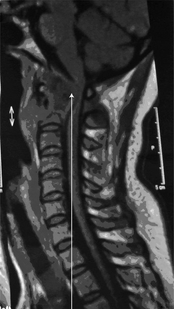
Magnetic resonance imaging (MRI) revealed atlanto-axial subluxation, probably secondary to an inflammatory process involving the odontoid process and the C2 vertebral body, and causing severe spinal cord stenosis and compression at the level of C1 and C2. The patient was therefore diagnosed with tuberculous spondylitis, with C1–C2 destruction and atlanto-axial subluxation with myelopathy (Figure 1).
Patient one: sagittal magnetic resonance imaging scan showing atlanto-axial subluxation (arrow) involving the odontoid process and C2 vertebral body, causing severe spinal stenosis and cord compression at C1–C2. P = posterior

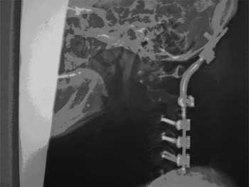
The patient underwent posterior cervical spine instrumentation with occipital cervical fusion, as well as anterior decompression and drainage of a abscess via an endoscopic, endonasal approach (Figure 2). Histopathological examination and culture confirmed tuberculous spondylitis with abscess.
Patient one: lateral X-ray showing posterior instrumentation with occipito-cervical fusion.

By one week post-operatively, the patient's upper and lower limb motor function had improved significantly (from 3/5 to 5/5), and she had regained her sensory function. She was recommenced on antituberculosis medication and continued to recover. At the time of writing, she was still under observation.
Case two
A 57-year-old Malay man presented to the orthopaedics service with a one-month history of localised neck pain and paraesthesia in both arms. These symptoms had been followed by two weeks of progressive weakness of all limbs, to the point that the patient had become bed-bound.
Physical examination revealed obvious quadriparesis (grade 3/5 and 4/5 in the upper and lower limbs, respectively). Sensory function was noted to be grossly intact, and anal tone was normal. All cranial nerves were intact. However, the patient's head was tilted to the left side, and he was unable to sit up or ambulate independently.
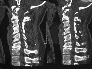
Computed tomography scanning revealed a defect at the base of the odontoid process, which was tilted posteriorly. Lytic destruction of the right lateral mass of C1 was also noted. An MRI scan revealed oedema at the level of C1 to C2, with an antero-posterior diameter of 8 mm at the respective spinal canal (Figure 3). Chest and systemic TB investigations gave negative results.
Patient two: sagittal computed tomography scans of the neck, demonstrating a lytic defect at the base of the odontoid process (arrow), which is tilted posteriorly. L = left

The patient was treated empirically for spinal TB, based on the clinical information and the risk of cord compression, as well as the benefit of antituberculosis chemotherapy in preventing further dissemination. In addition, spinal cord decompression was performed via an endoscopic, endonasal approach. A posterior spinal fusion stabilised the cranio-vertebral junction.
Post-operatively, the patient made a remarkable recovery. His upper limb paraesthesia resolved over the first 48 hours. On the fifth day following decompression, the patient began to ambulate with the aid of a physiotherapist. Within a week, he regained 5/5 muscle strength in all four limbs. The patient was discharged on the eighth day post-decompression. At the time of writing, he remained under observation.
Surgical technique
Surgery was performed in collaboration with the orthopaedic team. A tracheostomy was performed initially to secure the airway during the peri-operative period. Stabilisation of the atlanto-axial joint was needed prior to decompression.
A posterior nasal septectomy and wide bilateral sphenoidotomies (with removal of the sphenoid sinus floor) were completed, using a rigid, 0°, 4 mm, 18 cm rod lens endoscope (Karl Storz, Munich, Germany) and an osteotome. This enabled wide exposure of the nasopharynx, and allowed unimpeded, bimanual use of instruments through both nostrils. Once a wide surgical corridor had been created, the nasopharyngeal mucosa and pharyngobasilar fascia were stripped from the remaining roof of the nasopharynx and lower clivus. Dissection continued in a caudal direction, limited to the area in between the tori tubarius.
The atlanto-occipital membrane and longus capitii muscles were then excised, enabling visualisation of the anterior arch of the atlas. The cervico-vertebral junction was then identified, and the arch of C1 was drilled out using a Midas Rex high speed drill with 3 mm diamond burr (Medtronic, Minneopolis, USA) to gain access to the dens of C2. Debris and granulation tissue within the lytic cavity at the base of the odontoid process were removed and any pus was drained. Biopsies were obtained for histopathological assessment and microbiological culture. The stripped nasopharyngeal mucosa, fascia and muscles were re-affixed with fibrin glue.
Discussion
A literature review revealed various reported methods of ventral surgical access to the atlanto-axial joint.
Previously, the transoral-transpharyngeal approach to the odontoid process was deemed the ‘gold standard’.Reference Kassam, Snyderman, Gardner, Carrau and Spiro2 Its major drawback was a limited surgical field, which compromised access to the upper cervical spine region. Possible complications included: potential contamination by oral flora; dehiscence of the pharyngeal wall; tongue oedema and ischaemic necrosis due to prolonged compression of the tongue, with subsequent upper airway obstruction; velopharyngeal insufficiency; dental damage from surgical retractors; dysphonia; and dysphagia.Reference Sakou, Morizono and Morimoto3, Reference Alfieri, Jho and Tschabitscher4
The transmandibular-circumglossal approach provided a wider surgical exposure, but its popularity was limited by the need for a wide surgical incision, and by complications such as lingual nerve damage and malocclusion.Reference Kassam, Snyderman, Gardner, Carrau and Spiro2, Reference Kingdom, Nockels and Kaplan5
Wolinsky et al. have described an endoscopic, transcervical odontoidectomy, with the reported advantages of obviating the inherent risk of traversing the oropharynx and the need for prolonged intubation and enteral tube feeding.Reference Wolinsky, Sciubba, Suk and Gokaslan6 Nonetheless, these authors' case series was limited.
Kassam et al. have described an endoscopic, endonasal odontoidectomy.Reference Kassam, Snyderman, Gardner, Carrau and Spiro2 Possible drawbacks of this technique include the need to traverse the nasal and nasopharyngeal cavities, and contamination with these cavities' respective normal flora. Such contamination may increase the risk of post-operative meningitis, especially in the event of cerebrospinal fluid leakage; however, this risk is low. The vidian nerves and eustachian tubes are also at risk if exposure is too wide.
• Tuberculosis can cause extensive osseo-ligamentous destruction at the cranio-vertebral junction, leading to atlanto-axial instability and compression of vital cervico-medullary centres
• This may manifest as quadriparesis, bulbar dysfunction and respiratory insufficiency
• This paper describes two patients presenting with spinal stenosis and cord compression secondary to cranio-vertebral tuberculosis, who were successfully decompressed via an endoscopic, endonasal approach
In our limited experience, the endoscopic, endonasal approach provides excellent access to the cranio-vertebral junction as well as to the atlanto-axial regions ventrally. Moreover, in experienced hands and using correct surgical technique, no surgical complications were noted. Our patients were able to commence oral intake immediately after surgery. The endoscopic, endonasal approach is less traumatic anatomically and thus appears to reduce patient morbidity substantially. We found the endoscopic, endonasal approach to spinal cord decompression to be rewarding: complete resolution of compressive myelopathy was obtained in both our patients.
Conclusion
From our limited experience of using an endoscopic, endonasal approach for the surgical management of cranio-vertebral junction stenosis, we conclude that its feasibility and lack of anatomical trauma to critical structures result in reduced post-operative morbidity and hospital stay. This technique appears to be superior to other, contemporary techniques and routes of access. However, a larger case series and long-term follow up are needed to determine the reproducibility and validity of this technique's potential benefits.