INTRODUCTION
In recent decades, scientists have been showing an increasing interest in invasive species and their modes of entry (Chandra & Gerhardt, Reference Chandra and Gerhardt2008; Hulme et al., Reference Hulme, Bacher, Kenis, Klotz, Kühn, Minchin, Nentwig, Olenin, Panov, Pergl, Pyšek, Roques, Sol, Solarz and Vilà2008). Estuaries have been subjected to multiple invasions by planktonic organisms (Cordell et al., Reference Cordell, Rassmussen and Bollens2007). The most common planktonic invasive forms are crustaceans, such as copepods, which have been introduced into estuaries and continental freshwaters throughout the world (Reid & Pinto-Coelho, Reference Reid and Pinto-Coelho1994; Bollens et al., Reference Bollens, Cordell, Avent and Hooff2002, Reference Bollens, Breckenridge, Cordell, Bollens and Kalata2012). The successful invasion and propagation of species in a particular habitat depends upon their environmental tolerance to changing ambient physical and chemical features (Riccardi & Rossetti, Reference Riccardi and Rossetti2007; Bozkurt & Akin, Reference Bozkurt and Akin2012). Invasions by non-native harpacticoid copepods have been reported in some countries. For example, the harpacticoid copepod Harpacticella paradoxa is regarded as having been introduced to the Pacific North-west, USA (Cordell et al., Reference Cordell, Rassmussen and Bollens2007), several sand-dwelling harpacticoids were recorded in the Laurentian Great Lakes (Horvath et al., Reference Horvath, Whitman and Last2001) and the meiofaunal harpacticoid Enhydrosoma lacunae Jakubisiak, 1933 was introduced to the eastern tropical Pacific from the Caribbean sea (Gómez, Reference Gómez2003). Information regarding harpacticoids is generally limited for Indian waters (Krishnaswamy, Reference Krishnaswamy1956; Wells & Rao, Reference Wells and Rao1987; Mantha et al., Reference Mantha, Moorthy, Altaff, Dahms, Sivakumar and Hwang2012) and particularly for the Vellar estuary (Krishnamurthy, Reference Krishnamurthy1967; Santhanam & Perumal, Reference Santhanam and Perumal2003; Perumal et al., Reference Perumal, Santhanam and Rajkumar2008; Santhanam et al., Reference Santhanam, Perumal, Ananth and Shenbaga2012). In Indian waters, the copepods of the family Ameiridae have been recorded from the Chennai coast (Mantha et al., Reference Mantha, Moorthy, Altaff, Dahms, Sivakumar and Hwang2012) alongside the south-east continental shelf of India (Ansari, et al., Reference Ansari, Lyla, Ajmal Khan, Manokaran and Raja2013), however, as yet, a detailed taxonomic description for the Ameiridae is lacking. In spite of non-indigenous species being recorded from marine habitats (Bollens et al., Reference Bollens, Breckenridge, Cordell, Bollens and Kalata2012), only a few have been studied in detail.
So far, no confirmed records of non-native harpacticoid copepods have been reported from the Vellar estuary. Ameiridae is the third largest harpacticoid family, next to Canthocamptidae Sars, 1906 and Miraciidae Dana, 1846 and is currently classified into 46 valid genera (Walter & Boxshall, Reference Walter and Boxshall2011). The genus, Nitokra, belongs to the family Ameiridae and is known to contain 45 valid species (Fuentes-Reines & Suarez-Morales, Reference Fuentes-Reines and Suarez-Morales2014). The primary taxonomic subdivisions in this family have been based on the segmentation in the swimming legs (Karanovic, Reference Karanovic2006; Karanovic & Krajicek, Reference Karanovic and Krajicek2012).
Nitokra affinis is a widespread species, recorded from the following different geographical regions: The Suez Canal Zone (Gurney, Reference Gurney1927), Mangrove Bay (Willey, Reference Willey1930), Ireland (Roe, Reference Roe1958), Southern Italy (Chappuis, Reference Chappuis1938) and the northern Adriatic of Yugoslavia, near Rijeka (Petkovski, Reference Petkovski1954). Recently, Nitokra affinis californica was recorded in the South China Sea (Chertoprud et al., Reference Chertoprud, Gheerardyn and Gomez2011) and Nitokra affinis colombiensis, from the Caribbean coast of Colombia (Fuentes-Reines & Suarez-Morales, Reference Fuentes-Reines and Suarez-Morales2014). The harpacticoid copepod, Nitokra affinis is a common inhabitant of coastal areas, living in interstitial water (Matias-Peralta et al., Reference Matias-Peralta, Yusoff, Shariff and Mohamed2012). Because of its coastal habitat, Nitokra affinis adapt to eurythermic and euryhaline conditions and to various environmental stresses (Matias-Peralta et al., Reference Matias-Peralta, Yusoff, Shariff and Arshad2005). In the genus Nitokra, there is great interspecific variability, as well as relatively small, differential characteristics among species, resulting in numerous synonyms during identification (Wells & Rao, Reference Wells and Rao1987; Apostolov & Marinov, Reference Apostolov and Marinov1988; Karanovic & Pesce, Reference Karanovic and Pesce2002). To overcome this problem, the DNA barcoding approach was established as a reliable tool for the identification of inter- or intra-species and early life history stages (Bucklin et al., Reference Bucklin, Steinke and Blanco-Bercial2011; Laakmann et al., Reference Laakmann, Gerdts, Erler, Knebelsberger, Martínez Arbizu and Raupach2013). This paper deals with the first distributional record of the non-indigenous species (NIS), Nitokra affinis from the Vellar estuary.
MATERIALS AND METHODS
Description of the study area
The Vellar estuary is located in Parangipettai, where it opens into the Bay of Bengal in the south-east of India (Latitude 11°29′N Longitude 79°46′E). This estuary is separated into a marine, brackish and tidal, and freshwater zones based on salinity characteristics (Ramamoorthy, 1954). Heavy floods that occur during the north-east monsoon season (October–December) and the consequent mixing of water, draining from adjacent mangroves, backwater and irrigation channels, influence the physico-chemical properties of the Vellar estuary to a large extent (Figure 1).
Map of the Vellar estuary showing the collection site.

Plankton sampling and identification
Zooplankton samples were collected by the horizontal towing of a plankton net (0.35 m mouth diameter), made up of bolting silk cloth (No. 10: Mesh size 158 μm) for 20 min in the Vellar estuary during our plankton sampling in 2010. The collected samples were immediately transported to the laboratory, vigorously aerated using an aeration pump and thoroughly rinsed to reduce contamination by other zooplankter. Then the zooplankton was screened to isolate those size fractions containing predominantly adult copepods and later-stage copepodites. This was achieved by a first screening through a 500-μm mesh to eliminate fish and prawn larvae. Then, the samples were rinsed for 2 h in a zooplankton washer fitted with a 190 μm mesh sieve to remove rotifers, nauplii of copepods and barnacles. After rinsing, the remaining adult and larger copepodites were immediately preserved in 5% formalin. Specimens of the new harpacticoid described here were isolated, separated and their morphological features were evaluated to detect the family, genus and species of the copepod based on standard keys (Vervoort, Reference Vervoort1964; Karanovic & Cho, Reference Karanovic and Cho2012). Copepods were observed under a dissecting binocular microscope (Olympus CH20i) and their appendages were photographed with a Sony Corp Digital Still Camera. Drawings were made at 40× magnification with a drawing tube mounted on a binocular microscope (Olympus CH20i). For Field Emission Scanning Electron Microscope (FE SEM) analysis, two females were dehydrated using higher concentrations of ethanol solution. Abbreviations used in the text include: R, rostrum; A1, antennule; A2, antenna; Md, mandible; Mx1, maxillule; Mx2, maxilla; Mxp, maxilliped; P1–P4, 1st–4th swimming legs; P5, leg 5; enp, endopod; exp, exopod; and ae, aesthetasc.
Genomic DNA isolation, PCR and DNA sequencing
Genomic DNA was extracted from the tentatively identified copepod, i.e. ‘Nitokra affinis’, using a Qiagen DNeasy tissue kit, following the manufacturer's protocol. The Polymerase Chain Reaction (PCR) was performed using 1 μl (50 ng) of the template DNA, 2 μl (10pmol) of each 18S rDNA targeting primers (IF–5′-TACCTGGTTGATCCTGCCAGTAG-3′ and 5R–5′-CTTGGCAAATGCTTTCGC-3), 10 μl of PCR Master mix (Amplicon) and 5 μl of double-distilled water to, making the final volume 20 μl. The amplification of 18S rDNA began with an initial denaturation at 94°C for 5 min, followed by 30 cycles at 94°C (60 s), 52°C (60 s) and 72°C (60 s), with a final elongation at 72°C for 3 min, before being transferred to 4°C until further analysis. PCR products were confirmed on an agarose gel (1.5%). DNA sequencing of the amplified product was performed at Bioserve Biotechnologies Pvt. Ltd. (Hyderabad, India). The sequences were initially edited in Gene tool and Bio-edit software packages and the edited sequences were submitted to GenBank.
RESULTS
Nitokra affinis (Gurney, Reference Gurney1927).
Based on the morphological features (Vervoort, Reference Vervoort1964), the collected copepod was tentatively identified as Nitokra affinis. The specific identification required the examination of a fourth pair of legs.
DESCRIPTION
FEMALE
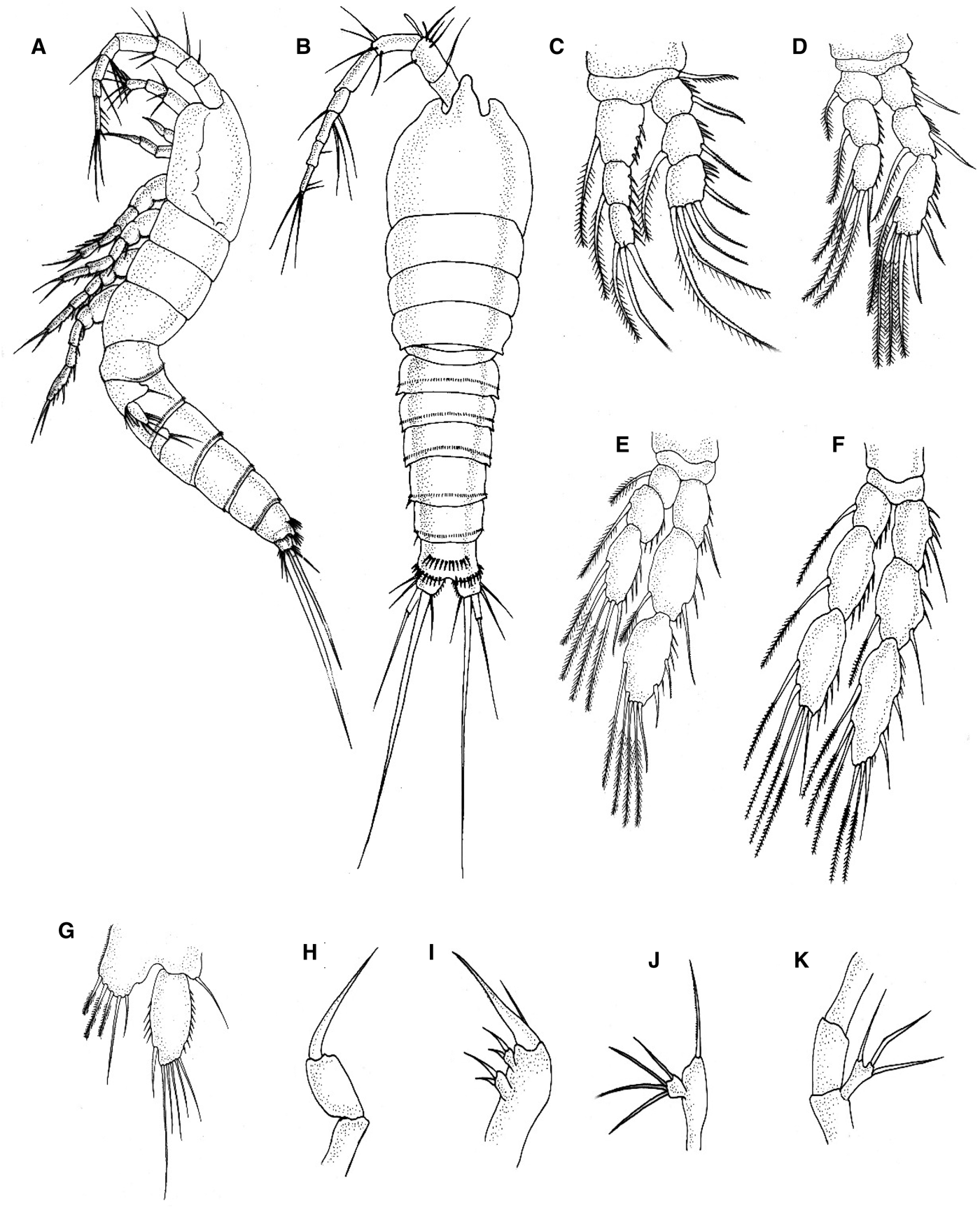
Body length (from tip of the rostrum to caudal rami) 571 μm. Body slender, cephalosome rounded anteriorly and tapering behind from posterior border of cephalothorax (Figures 2A & 5B). Prosome comprised of cephalothorax and three free pedigerous somites. Prosome and abdomen were of nearly the same length and separated by ring-shaped constrictions. Urosome comprised fifth pedigerous somite, genital double somite and three free abdominal somites. Genital double somite short with a row of small cuticular spines dorsolaterally (Figure 4F). The posterior margin of each abdominal somite had stronger spinules dorsally. Somites 1, 2 and 4 were equal in length but somite 3 was slightly longer (Figure 5B). Antennule (A1) slender, longer than cephalic somites, 8-segmented (segment 1 and 2 slightly swollen, 4th segment as long as wide and aesthestasc is present on segment 4), number of appendages: 1(0), 2(6), 3(6), 4(2 + 1 aesthestasc), 5(2), 6(2), 7(2), 8(5) (Figures 2C & 4C, D).
Nitokra affinis female. (A) Dorsal view (10× magnification); (B) Lateral view (10× magnification); (C) Antennule (A1) (40× magnification); (D) Rostrum (R) (40× magnification); (E) First swimming leg. (P1) Number indicates setae on the third segment of exopod (40× magnification); (F) Third swimming leg showing exopod and endopod (P3) (40× magnification).
Antenna (A2) in which X-segmented exopod 1 was armed with three finely plumose setae. Endopod two X-segmented and armed with seven setae. Rostrum (R) small, conical with rounded apex, placed between two depressions at insertion of antennules and half the length of first antennule segment (Figures 2D & 5B). Maxilla (Mx2) with small praecoxal and coxal endite, each carrying two setae. Basal endite stronger, with strong curved spine. In addition, there is a fine seta on the basal endite (Figure 5I). Mandible (Md) fairly big basipodite with single strong seta. No exopodite; endopodite unsegmented and small, carries four marginal setae (Figure 5J). Anal somite short with row of spinules, dorsally and laterally close to the joint with caudal rami. Rounded anal operculum with 10–13 strong spinules close to the posterior margin (Figures 4G & 5B). Caudal rami distinctly broader than long. Five marginal setae and one appendicular seta (two lateral, one outer apical seta, one middle apical seta and one dorsal seta). Two lateral setae and one dorsal setae fine and short. Apical setae lengthened, thickened and well developed. Middle apical seta strongest one and about twice as long as the outer apical setae. Reaches to two-thirds of the body length. Outer apical seta somewhat longer than three free abdominal somites together with caudal rami (Figure 5B).
First swimming leg (P1), in which exopod and endopod three, segmented. EXP1 without inner setae. EXP two with plumose inner setae. EXP three, armed with three naked spines and two geniculate setae. Endopod three segmented. ENP one, not elongated, slightly shorter than the sum of EXP one and EXP two. ENP one with inner setae on distal third of inner margin. ENP two with inner setae. ENP three with one outer spine, one geniculate seta and one plumose inner seta apically (Figures 2E & 5C). Swimming legs P2–P4 with three-segmented endopodites and exopodites. Endopod segment short which extended only to the middle of the third exopod segments (Figures 2F, 3A, C, 4B & 5D–F).
(A) Fourth swimming leg (P4). Numbers 1–8 indicate the setae and spines on the third segment of the exopod (40× magnification); (B) Female. (P5) Exopodite of leg 5. Numbers 1–6 indicate the marginal setae (40× magnification). (C) Female. (P2) Second swimming leg. Numbers 1–7 indicate setae and spine on the third segment of the exopod (40× magnification).
Nitokra affinis, FE-SEM photographs. (A) Habitus, lateral view; (B) Third exopod segment of P2 with seven setae, dorsal view; (C) Cephalothorax and antennule; (D) Antennule; (E) Fifth pedigerous, genital and abdominal somite, lateral view; (F) Dorsal spinules at the urosome; (G) Caudal ramus and setae, lateral view; (H) egg sac, lateral view.
(A) Habitus, male, Lateral view. (B–K) Female: (B) Dorsal view; (C) P1 (first swimming leg); (D) P2 (second swimming leg); (E) P3 (third swimming leg); (F) P4 (fourth swimming leg); (G) P5 (fifth swimming leg); (H) Maxilliped; (I) Maxilla; (J) Mandibular palp; (K) Antenna with exopodite.
ARMATURE FORMULA FOR SWIMMING LEGS
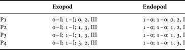
P1–P4, with detailed spine/setae formula (spines in Roman numerals, setae in Arabic numerals) are shown in Table 1.
Armature formula for swimming legs.

Swimming leg P5 with exopod twice as long as broad, ovate and armed with six marginal setae. Baso-endopodite short, reaching half the length of exopodite, with five setae (Figures 3B & 5G).
MALE
Almost similar to female but slender. In male, prosome and abdominal segments are separated less clearly because circular or ring constrictions are not present as in female. Rostrum smaller than in female. First and second abdominal somites separate and of equal length. Abdominal somites 1–4 with completely encircling distal row of spinules. Anal somites slightly shorter than fourth abdominal somite fringed with spinules as in female. Caudal rami and setae almost similar. Segmentation and setae/spine arrangement of P1–P4 similar to female (Figure 5A).
SWIMMING LEG P5
Exopod twice as long as broad and ovate, armed with six marginal setae. Baso-endopodite short, armed with four setae.
BLAST
The 18S rDNA sequence of Nitokra affinis is 769 bp long (GenBank Accession number JX438707.1). Blast similarity in GenBank showed the closest match for Nitokra affinis (94%) with Nitocra hibernica (GenBank accession number EU380305.1), followed by 93% similarity with Cancrincola plumipes (L81938.1), 91% similarity with Argestigens sp. (EU380306.1), Harpacticus sp. (EU380285.1) and 90% similarity followed by Eurycletodes laticauda (EU380285.1), Paramphiascella fulvofasciata (EU380285.1), Bradya sp. (EU380285.1), Amphiascoides atopus (EU380285.1) and Itunella muelleri (EU380285.1).
DISCUSSION
The Vellar estuary is considered to be a most productive habitat with a rich diversity of copepods that enhance the fish productivity (Santhanam et al., Reference Santhanam, Perumal, Ananth and Shenbaga2012). Harpacticoid copepods constitute the second-most abundant meiofauna taxon next to nematodes (Mantha et al., Reference Mantha, Moorthy, Altaff, Dahms, Sivakumar and Hwang2012). In spite of their relative importance in estuarine ecosystems, the taxonomic studies of copepods have been hampered due to the shortage of taxonomists. The present report pertains to the first distributional record of the invasive species, Nitokra affinis, which co-occurred with Euterpina acutifrons from the Vellar brackish water environment. Species of the genus Nitokra generally occur in fresh, brackish and high-saline environments and some species are commensals (Karanovic & Pesce, Reference Karanovic and Pesce2002). The detailed morphological analyses of the presently collected copepod showed that there were eight elements in the third exopodal segment of the fourth leg (Figures 3A & 5F), in contrast to the seven elements found in other species of the genus Nitokra (Vervoort, Reference Vervoort1964; Karanovic & Cho, Reference Karanovic and Cho2012), which confirms that the presently collected copepod is Nitokra affinis and it is the first distributional record from the Vellar estuary. Generally, some of species of the genus Nitokra exhibit a three-segmented exopod on the first leg and the last segment of the exopod bears five spines/setae, respectively. Presumably, the presently collected Nitokra affinis belongs to the third group of the genus Nitokra because of the arrangement in spines/setae of the P1EXP2–3, as reported earlier by Gómez et al. (Reference Gómez, Carrasco and Morales-Serna2012). In the specimens collected here, the first endopodal segment of leg 1 was slightly shorter than the exopodal segment (Figures 2E & 5C), which is different from Vervoort's (Reference Vervoort1964) description where it was stated that the first endopodal segment was styliform and longer than the whole endopodite. This difference makes our species a distinctly independent subspecies of Nitokra affinis. Available data for the subspecies confirmation of Nitokra affinis are insufficient and we are, therefore, unable to end with a conclusive statement based only on morphological traits. Thus, concentrating on the subspecies level through morphological traits could hinder this task. DNA sequencing may solve the confusion pertaining to the subspecies allocations within Nitokra affinis (Fuentes-Reines & Suarez-Morales, Reference Fuentes-Reines and Suarez-Morales2014).
Harpacticoid copepods are the second most abundant group of meiofauna – they have several synonyms and there are still several undiscovered species (Karanovic & Pesce, Reference Karanovic and Pesce2002; Mantha et al., Reference Mantha, Moorthy, Altaff, Dahms, Sivakumar and Hwang2012). In India, barcoding studies to discriminate cryptic or subspecies in copepods are scarce. Presently, we have sequenced 18SrDNA using the reported copepod so as to compare its molecular features with the already available information for different species of the genus, Nitokra. The first sequence of 18S rDNA of the presently analysed species, i.e. Nitokra affinis, has been submitted to GenBank (JX438707.1). It showed the closest similarity (94%) with the copepod Nitokra hibernica. The 6% of blast difference between our species with that of Nitokra hibernica could be related to their morphological variations in terms of segmentation, structure and arrangement of spines/setae in the swimming legs (P1–P4). In Nitokra hibernica, the thrid exopodal segments of legs (P2–P4) have five, five and seven spines/setae (Lesko et al., Reference Lesko, Hudson, Reid and Chriscinske2003) but in our specimen there are seven, seven and eight spines/setae, respectively. Therefore, the barcode characterization provided meaningful evidence in support of the genus Nitokra.
The genus Nitokra is reported for the first time from the Vellar estuary. Although we are uncertain of the exact date of its introduction here, it must have been a recent event, as we only obtained it during our sampling efforts in 2010. Taxonomists pertaining to the morphological and molecular identification of copepods are woefully inadequate and there is an absence of any authenticated data or information about the invasive species/description of a new species for the past few years. Several earlier researchers have focused mainly on the ecological diversity of copepods from the Vellar estuary (Santhanam & Perumal, Reference Santhanam and Perumal2003; Perumal et al., Reference Perumal, Santhanam and Rajkumar2008; Santhanam et al., Reference Santhanam, Perumal, Ananth and Shenbaga2012). The cosmopolitan species, particularly harpacticoid copepods viz. E. acutifrons, Macrosetella gracilis and Microsetella norvegica, are commonly found in Indian waters (Chertoprud et al., Reference Chertoprud, Gheerardyn and Gomez2011). However, in view of the scattered nature of data about harpacticoid copepods in Indian waters, further progress in studying the invasive species of the Vellar estuary is very difficult; under these circumstances the present first distributional record is a significant development here.
The species composition and distributional ranges of harpacticoid copepods in tropical and subtropical waters of the South China Sea, the Philippines islands, the Internal Malayan Archipelago, New Guinea, the Yellow sea and the Andaman and Nicobar islands have been recently reported and the copepod Nitokra affinis was reported to have occurred in the tropical waters of Nha-Trang Bay, Vietnam (Chertoprud et al., Reference Chertoprud, Gheerardyn and Gomez2011) and this is the only report from the Vellar estuary that provides evidence of its occurrence in our nearest site of tropical waters.
The invasive copepod Nitokra affinis is broadly consistent with the expectations based on life history and is generally characterized as a good invader with a high dispersal capacity and physiological adaptation to tolerate and reproduce at a wide range of environmental conditions (Ehrlich, Reference Ehrlich, Mooney and Drake1986; McMahon, Reference McMahon2002; Bozkurt & Akin, Reference Bozkurt and Akin2012). Many species of Nitokra, due to their diverse distributional habitats such as rocks, pools, lagoons and sandy beaches are periodically exposed to fluctuating salinity regimes (Matias-Peralata et al., Reference Matias-Peralta, Yusoff, Shariff and Arshad2005). Hence, the presently recorded non-indigenous invasive Nitokra affinis could thrive successfully in the dynamic brackish water environs of the Vellar estuary, in spite of its interstitial nature.
The recent entry of Nitokra affinis into the Vellar estuary could be possible through one of the following three modes: Firstly, the introduction pathway of Nitokra affinis into the Vellar estuary could be related to Ship-born invasion. It has been reported that the mode of transportation of alien species to new habitats through ballast water discharges has dramatically increased over the past century (Ruiz, et al., Reference Ruiz, Rawlings, Dobbs, Drake, Mullady, Huq and Colwell2000; Bollens et al., Reference Bollens, Cordell, Avent and Hooff2002; Goulletquer et al., Reference Goulletquer, Bachelet, Sauriau, Noel, Leppäkoski, Gollasch and Olenin2002; Brylinski et al., Reference Brylinski, Antajan, Raud and Vincent2012). Copepod invasion due to ship-born ballast-water discharge has recently been reported in many countries (Gómez & Arbizu, Reference Gómez and Arbizu2004; Seuront, Reference Seuront2005; Cordell et al., Reference Cordell, Rassmussen and Bollens2007; Mohamed, Reference Mohamed2011; Bollens et al., Reference Bollens, Breckenridge, Cordell, Bollens and Kalata2012; Brylinski et al., Reference Brylinski, Antajan, Raud and Vincent2012; Temnykh & Nishida, Reference Temnykh and Nishida2012). A number of major ports are located nearer to our present study area: Karaikal, Chennai and Tuticorin. Of these, the Chennai port is well connected to Port Blair (Andaman and Nicobar Islands), Malaysia and Yangoon through the sea-route. It handles a variety of cargos and has regular services to Singapore, Malaysia, Thailand, Myanmar, Sri Lanka, Korea, China, Mediterranean, Europe, Australia and the United States of America during the last few decades. As a result, a billion tonnes of ballast water has been filled from one port of the ocean and discharged into the other port. During this process, the introduction of this species might have occurred. There is no evidence of an earlier occurrence of this species from the Vellar estuary through plankton survey records pertaining to earlier decades.
The second possibility of introduction of this new species could have been through hull fouling. Copepods, especially representatives belonging to Nitokra, are often found as commensals of other marine invertebrates (Karanovic & Pesce, Reference Karanovic and Pesce2002). They may have been attached to the hulls of ships during fouling and during this process became transported to the Vellar estuary (Cordell et al., Reference Cordell, Rassmussen and Bollens2007). It was reported that several harpacticoid copepods have the capability for migration capacity to move for a distance of hundreds of kilometres, with bottom-water drifts or on fragments of macrophytes (Hauspie & Polk, Reference Hauspie and Polk1973). Some interstitial forms also showed a lower degree of mobility (Chertoprud et al., Reference Chertoprud, Chertoprud and Kondar2006). In view of the interstitial nature of Nitokra affinis, which has a slow moving ability, its introduction after long-distance migration is quite improbable.
The third possible mechanism of introduction could be through natural physical processes, such as tsunami waves. Some marine invasions are reported to have occurred due to natural dispersal mechanisms (Prakash et al., Reference Prakash, Balamurugan, Ajith Kumar and Balasubramanian2012). Grzelak et al. (Reference Grzelak, Kotwick and Zczuciński2009) has provided data on the long-term impact of a tsunami on the patterns of meiofaunal distribution. On 26 December 2004, a powerful earthquake of approximately 9.3 Mw magnitude nucleated on the north-western coast of Sumatra, resulting in a devastating tsunami for the south-eastern coast of India, between Sri lanka and Indonesia. Chertoprud et al. (Reference Chertoprud, Chertoprud and Kondar2006) stated that harpacticoid copepods could move passively when water currents are rapid. The sudden vertical displacement of columns of the water, followed by earthquakes, created long waves that moved rapidly across the ocean, reaching the coastal areas with a height of up to 10 m (Grzelak et al., Reference Grzelak, Kotwick and Zczuciński2009). The presently recorded non-indigenous species of Nitokra affinis could, therefore, be related to the transportation of tsunami waves. This is the first report on the occurrence of Nitokra affinis in the Vellar estuary, carried by the tsunami waves from the South China Sea. In view of the present report on the occurrence of the non-indigenous Nitokra affinis in the Vellar estuary, further long-term monitoring and research is needed to assess its impact on other cosmopolitan copepod species, such as Macrosetella gracilis, E. acutifrons and Macrosetella norvegica. Therefore, a systematic ecological action plan for invasive species must be implemented to protect and conserve the sensitive ecosystems of estuaries in India.