Flaviviruses are a group of positive-stranded RNA viruses with a global impact on public health as a result of their widespread distribution and their ability to cause significant morbidity and mortality in humans. More than 75 different flaviviruses have been identified, roughly half of which are capable of causing disease in humans. The majority of flaviviruses are transmitted to humans through the bite of a mosquito or tick. Flavivirus infections result in clinical manifestations that range from febrile illnesses to encephalitis and haemorrhagic disease. Several members of this group, such as the four serotypes of dengue virus (DENV), and West Nile virus (WNV), are considered emerging or re-emerging pathogens because in the past decade the incidence of human disease has increased at an alarming rate (Ref. Reference Mackenzie, Gubler and Petersen1). Other flaviviruses of significant clinical importance include yellow fever virus (YFV), tick-borne encephalitis virus (TBEV), and Japanese encephalitis virus (JEV). The development of vaccines for several flaviviruses is being actively pursued with the goal of eliciting protective levels of neutralising antibody.
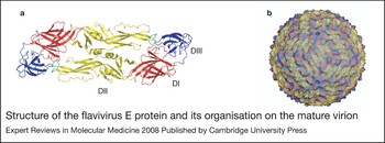
Flaviviruses are small (roughly 50 nm diameter) spherical virions composed of a single copy of an ~11 kb genomic RNA of positive polarity, the capsid protein (C), a lipid envelope derived from the endoplasmic reticulum, and two envelope glycoproteins: envelope (E) and pre-membrane/membrane (prM/M). The atomic structure of the E protein reveals an organisation of three distinct domains separated by short flexible ‘hinges’ (Refs Reference Zhang2, Reference Rey3, Reference Modis4, Reference Modis5). Domain III (DIII) (Fig. 1a; shown in blue) is an immunoglobulin-like domain that is thought to mediate interactions between the virus and structures on the host cell involved in virus attachment (Refs Reference Rey3, Reference Chu6). Domain II (DII) (Fig. 1a; shown in yellow) is an elongated finger-like domain that contains the highly conserved hydrophobic fusion loop that interacts with the membranes of the target cell during fusion (Refs Reference Allison7, Reference Modis8, Reference Bressanelli9). Domain I (DI) (Fig. 1a; shown in red) is a β-barrel structure that connects DII and DIII via flexible hinges that participate in the conformational changes that drive the fusion process (Refs Reference Zhang2, Reference Modis4). An amphipathic stretch of residues referred to as the stem anchor connects the E protein ectodomain to two transmembrane domains that anchor the E protein within the viral membrane. The stem anchor is thought to lie flat against the viral membrane of mature flaviviruses (Ref. Reference Zhang10), be intimately involved in the large rearrangements occurring within and between E proteins during the fusion process, and play a role in interactions with prM (Refs Reference Allison11, Reference Wang, He and Anderson12). The structure of prM is presently unknown.
Structure of the flavivirus E protein and its organisation on the mature virion. (a) Ribbon diagram of a dengue virus E protein dimer with domains II, I and III shown as yellow, red and blue ribbons, respectively. The fusion loop at the tip of DII is shown in green. (b) Cryoelectron reconstruction of the dengue virus mature virion illustrating the arrangement of E proteins on the virion with pseudo-icosahedral symmetry (for a detailed explanation of the icosahedral symmetry patterns of viruses see the weblink for the virus particle explorer: http://viperdb.scripps.edu/) (Ref. Reference Kuhn21). Image kindly provided by Drs Richard Kuhn and Michael Rossmann, Purdue University, IN, USA.

Flaviviruses assemble at the endoplasmic reticulum and bud into the lumen as immature virions (Ref. Reference Mackenzie and Westaway13). Cryoelectron microscopic reconstructions of WNV and DENV immature virions reveal an icosahedral arrangement of 60 trimeric spikes, each composed of prM–E heterodimers in which the prM protein is positioned at the tip of each E protein of the trimer (Refs Reference Zhang14, Reference Zhang15). In this position, prM may prevent low-pH-induced conformational changes that would inactivate the virus particle during egress through mildly acidic compartments of the secretory pathway (Refs Reference Heinz16, Reference Guirakhoo17). During transit through the trans-Golgi network, prM is cleaved by a cellular furin-like protease. This required cleavage step promotes a rearrangement of E protein on the surface of the virion from a heterodimer (prM–E) into an antiparallel homodimer (E–E) and the formation of a mature virus particle (reviewed in Ref. Reference Mukhopadhyay, Kuhn and Rossmann18). Cleavage results in the formation of a small virion-associated M peptide and the release of the N-terminal ‘pr’ portion of the protein (Ref. Reference Stadler19). By contrast to the spikes present on the immature precursor, mature flavivirus virions are relatively smooth and composed of 90 antiparallel dimers arranged with T = 3 pseudo-icosahedral symmetry (Refs Reference Mukhopadhyay, Kuhn and Rossmann18, Reference Mukhopadhyay20, Reference Kuhn21) (Fig. 1b).
Following clathrin-mediated internalisation of a flavivirus into the cell, fusion is orchestrated by a series of conformational changes within and between E proteins arrayed on the surface of the virion in response to exposure to the mildly acidic environment of the endosome (Refs Reference Kimura, Gollins and Porterfield22, Reference Gollins and Porterfield23, Reference Holzmann24). These involve the dissociation of the E protein homodimers present on the mature virion (a reversible step) and the formation of E protein trimers (an irreversible step) (Refs Reference Stiasny25, Reference Stiasny26, Reference Allison27). This process involves rotation between the three domains of the E protein and results in the projection of DII away from the surface of the virion while positioning the fusion loop to interact with the target cell membrane. Following insertion of the fusion loop into the target cell membrane, viral and cellular membranes are brought into close apposition as the E protein folds back upon itself with the stem anchor region fitting into grooves on the exterior of the trimer (Refs Reference Modis8, Reference Bressanelli9). While structurally distinct, these rearrangements represent a functionally analogous process to the well-characterised fusion process of class I fusion glycoproteins such as influenza haemagglutinin and human immunodeficiency virus (HIV)-1 gp120 (reviewed in Refs Reference Eckert and Kim28, Reference Colman and Lawrence29). Efforts are under way to develop small-molecule inhibitors of virus entry orchestrated by the class II fusion proteins of alpha- and flaviviruses (Refs Reference Liao and Kielian30, Reference Bai31, Reference Hrobowski, Garry and Michael32), based on the demonstrated success of fusion and entry inhibitors that target the HIV-1 envelope proteins (Ref. Reference Reeves and Piefer33).
Humoral immunity against flavivirus infection
Humoral immunity is an essential aspect of immune-mediated protection from flavivirus infection (Refs Reference Diamond34, Reference Diamond35, Reference Oliphant36, Reference Tesh37, Reference Wang38, Reference Ben-Nathan39, Reference Camenga, Nathanson and Cole40, Reference Roehrig41, Reference Mathews and Roehrig42, Reference Kreil43, Reference Kreil44, Reference Beasley45). The primary target for neutralising antibodies is the E protein, although antibodies specific for prM and nonstructural (NS) proteins have been observed (Refs Reference Vazquez46, Reference Pincus47, Reference Falconar48, Reference Colombage49, Reference Chung50, Reference Chung51, Reference Shu52, Reference Churdboonchart53, Reference Gibson, Schlesinger and Barrett54). Antibodies specific for NS proteins (proteins not incorporated into virions) do not directly neutralise virus infectivity, but may protect via other effector mechanisms of antibodies as discussed below. More than 12 distinct epitopes have been identified on the surface of the E protein that elicit antibodies characterised by varying degrees of neutralisation potency in vitro and efficacy in vivo (Refs Reference Oliphant36, Reference Beasley and Barrett55, Reference Oliphant56, Reference Crill and Chang57, Reference Roehrig, Bolin and Kelly58, Reference Roehrig, Mathews and Trent59, Reference Heinz60, Reference Mandl61). The antigenic domains of E proteins were initially characterised by mapping and competition experiments with monoclonal antibodies (mAbs); three antigenic domains (C, A and B) were identified, which were later correlated with the structural domains DI, DII and DIII on the E protein (Fig. 1) (Ref. Reference Mandl61). Many of the most potent neutralising antibodies characterised to date recognise the upper lateral surface of DIII that protrudes off the surface of the virion (DIII-lr) (Refs Reference Oliphant36, Reference Beasley and Barrett55, Reference Sukupolvi-Petty62). While humans produce antibodies of this specificity in response to natural infection, recent studies indicate that the human humoral immune response to flavivirus infection is narrower than anticipated, with antibody specificity focused on determinants around the fusion loop at the tip of DII. B-cell repertoire analysis of three WNV-infected humans revealed that only 8% of WNV-specific B-cell clones produced antibodies specific to DIII, whereas almost half produced antibody that bound determinants in DII, particularly the fusion loop (Ref. Reference Throsby63). Functional studies of the polyclonal response of WNV-infected mice, horses and humans indicate that the neutralisation activity of sera is not dependent upon antibodies directed against the DIII-lr epitope (Refs Reference Oliphant64, Reference Sanchez65).
Factors that determine the neutralisation potency of antibodies
Flavivirus neutralisation is a ‘multiple’ hit phenomenon requiring engagement by more than a single antibody (reviewed in Refs Reference Della-Porta and Westaway66, Reference Burton, Saphire and Parren67). Neutralisation occurs when the number of antibodies bound to an individual virion exceeds a required threshold (Ref. Reference Pierson68). In this regard, two biochemical factors play a significant role in determining when an antibody exceeds the stoichiometric requirements for neutralisation: antibody affinity and the accessibility of epitopes on the virus particle.
The strength of binding between antibody and viral antigen (affinity) determines the fraction of epitopes on the virus particle occupied by antibody at any given concentration (referred to as antibody occupancy) and is a primary determinant of neutralisation potency (Ref. Reference Klasse and Sattentau69). Thus, it is no surprise that differences in neutralisation potency between antibodies often can be accounted for by differences in the strength of antibody–antigen interactions. Integrating data from measurements of the avidity of antibody–virion interactions and the concentration of antibody required to inactivate 50% of the virus allows an estimate of antibody occupancy when the virus is neutralised. For the most potent neutralising antibodies against flaviviruses, neutralisation appears to occur at a relatively low occupancy (Refs Reference Pierson68, Reference Gromowski and Barrett70). However, some high-affinity antibodies exhibit rather limited neutralisation potency and inhibit infection only at very high concentrations relative to their affinity for viral antigens. In fact, for some antibodies, even complete occupancy of epitopes on the virion is not sufficient to exceed the threshold for neutralisation (Ref. Reference Pierson68).
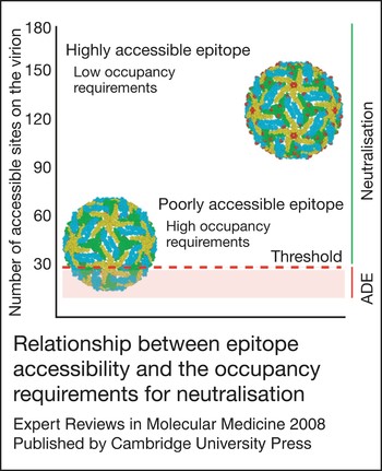
The pseudo-icosahedral arrangement of E proteins on the virion displays the E protein in three distinct chemical environments defined by proximity to the two-, three- or fivefold axes of symmetry (Refs Reference Mukhopadhyay20, Reference Kuhn21). From the perspective of the antibody, epitopes in each of these environments may be differentially accessible for antibody binding because of steric constraints imposed by adjacent E proteins on the virus particle. As a result, the number of sites available for binding may differ among structurally distinct epitopes on the virion. Accessibility is a significant factor that modulates antibody potency and shapes the occupancy requirements for neutralisation. Antibodies that bind highly exposed determinants may exceed the stoichiometric threshold for neutralisation by binding the virion at relatively low occupancy (Ref. Reference Pierson68). By contrast, epitopes that are predicted to be poorly exposed may require nearly complete occupancy to achieve threshold requirements for neutralisation (Refs Reference Pierson68, Reference Stiasny71) (Fig. 2).
Relationship between epitope accessibility and the occupancy requirements for neutralisation. The accessibility of epitopes recognised by two different antibodies on the mature West Nile virion is illustrated using molecular modelling: residues that form each determinant are illustrated as solid spheres. E proteins are coloured according to their proximity to the twofold, threefold or fivefold symmetry axes (blue, green and yellow, respectively). The number of accessible binding sites for each antibody is indicated on the left, whereas the ‘threshold’ for neutralisation is indicated as a red line [modelled in this instance as 30 monoclonal antibodies (mAbs) based on studies using the mAb E16] (Ref. Reference Pierson68). To exceed the threshold requirements for neutralisation, only a fraction of highly accessible determinants must be simultaneously occupied by antibody (a low occupancy requirement). By contrast, a significantly greater percentage of poorly accessible epitopes must be bound to achieve the same number of antibodies docked on the average virion (a high occupancy requirement). Not all epitopes appear to exist on the average virion at levels that exceed this threshold.

The stoichiometry of antibody binding that defines the neutralisation threshold for flaviviruses has been estimated for only one antibody and epitope (Ref. Reference Pierson68). The WNV-specific mAb E16, which binds an epitope on the lateral surface of DIII, potently neutralises virus infection in vitro (Ref. Reference Oliphant36) and is protective in vivo even when administered several days after virus infection (Refs Reference Morrey72, Reference Morrey73). Structural studies of E16 in complex with DIII on the intact virion demonstrate that this antibody cannot bind E proteins around the fivefold axis of symmetry. Thus, this potently neutralising antibody can recognise only 120 of the 180 E proteins on WNV (Refs Reference Nybakken74, Reference Kaufmann75). As this antibody neutralises at an occupancy of roughly 25%, it is predicted that approximately 30 mAbs are required for neutralisation (Ref. Reference Pierson68). By comparison, molecular modelling studies suggest that many of the other epitopes recognised by neutralising antibodies are less accessible on the mature virion (Refs Reference Oliphant56, Reference Pierson68, Reference Stiasny71). How antibodies bind ‘cryptic’ epitopes yet neutralise virus infectivity is difficult to explain using existing static models of virion structure and is an area of active investigation.
Mechanisms of neutralisation
At present, the cell biology of the flavivirus entry pathway is not well understood. The first step in the process likely involves the interaction of virions on the surface of target cells with one or more cellular factors. Presumably, these interactions trigger access for the virus to the endocytic pathway via clathrin- and Rab5-mediated endocytosis and transport processes (Refs Reference Krishnan76, Reference van der Schaar77, Reference Gollins and Porterfield78, Reference Chu and Ng79) where fusion occurs in a pH-dependent fashion. Cellular factors that play a required role as a flavivirus ‘receptor’ have not yet been rigorously defined despite considerable effort. While many candidate receptors have been proposed (reviewed in Ref. Reference Anderson80), due in part to the extremely broad tropism in vitro of many flaviviruses, it remains difficult to distinguish molecules that play an essential role in the virus entry pathway from those that promote more efficient and durable attachment of virions to the cell surface. Furthermore, the requirements and characteristics of a cellular receptor for flaviviruses have not been formally established as several lines of evidence suggest exposure to acidic pH is the sole requirement for the conformational changes in the E protein that drive membrane fusion (Refs Reference Gollins and Porterfield23, Reference Stiasny, Koessl and Heinz81, Reference Stiasny82).
Blocking virus attachment
Antibodies have the potential to neutralise the infectivity of flaviviruses by interfering with several steps of the virus entry pathway including attachment, internalisation and fusion. Antibody-mediated neutralisation of several viruses has been reported to occur by blocking the attachment of viruses to the cell (reviewed in Ref. Reference Klasse and Sattentau69). Perhaps the most well-characterised example are antibodies that block the binding of the HIV-1 envelope protein to the CD4 receptor or CCR5 coreceptor on T cells (reviewed in Ref. Reference Pantophlet and Burton83). While the cellular factors involved remain unclear, antibodies may block flavivirus infection by inhibiting the interactions between virions and the cell surface during the attachment step. mAbs specific for DIII have been shown in some studies to block infection at this stage (Ref. Reference He84). Several lines of indirect evidence suggest DIII plays an important role in virus attachment, including: (1) DIII protrudes the farthest from the surface of the virion; (2) many of the mutations that impact tropism or virulence map to DIII (Refs Reference Jennings85, Reference Mandl86, Reference Holzmann87, Reference Jiang88); and (3) soluble forms of DIII can block infection (Ref. Reference Chu6). Thus, blockade of the binding step is an attractive model for the neutralising mechanism of some DIII-specific mAbs.
The calcium-dependent (C-type) lectin DC-SIGN (CD209) is a well-characterised ‘attachment factor’ for flaviviruses. CD209 plays an important role in the infection of immature monocyte-derived dendritic cells by DENV in vitro (Refs Reference Tassaneetrithep89, Reference Navarro-Sanchez90), and polymorphisms in the CD209 promoter are associated with protection against dengue fever (Ref. Reference Sakuntabhai91). Furthermore, transfection of a variety of cells lines with plasmids encoding either CD209 or the related molecule CD209L increases their capacity to support DENV infection (Refs Reference Tassaneetrithep89, Reference Navarro-Sanchez90, Reference Lozach92). However, CD209-augmented infection does not require internalisation of the attachment factor, suggesting other processes and molecules at the cell surface are involved (Ref. Reference Lozach92). Like DENV, infection of cells in vitro by WNV can be enhanced by interactions with CD209L and to a lesser extent CD209, but the role (if any) of these lectins during WNV infection in vivo has not been examined (Refs Reference Davis93, Reference Davis94). As the spatial arrangement of the carbohydrate-recognition domains at the end of CD209 and CD209L tetramers is an important aspect of efficient ligand binding, it is conceivable that antibodies docked on virions may inhibit flavivirus infection by disrupting required multivalent binding of the sugars arrayed on the E protein (Refs Reference Pokidysheva95, Reference Mitchell, Fadden and Drickamer96). Thus, DIII-independent inhibition of virion attachment may also be a mechanism of neutralisation and may contribute to cell-type-dependent differences in neutralisation potency observed for some mAbs.
Inhibiting viral membrane fusion
mAbs also have the potential to neutralise infectivity at steps downstream of binding, perhaps by inhibiting the conformational changes in the E protein associated with membrane fusion. Pioneering electron microscopy studies by Gollins and Porterfield suggest that West Nile virions complexed with neutralising quantities of antibody can be internalised by target cells, suggesting a post-attachment mechanism of neutralisation (Ref. Reference Gollins and Porterfield97). Neutralisation by the WNV DIII-specific mAb E16 also occurs at a post-attachment step of the viral entry pathway (Ref. Reference Nybakken74). Structural and cryoelectron reconstruction analysis suggests docking of this antibody to the virion imposes steric constraints on the low-pH-mediated rearrangements of the E proteins that drive fusion (Refs Reference Kaufmann75, Reference Nybakken98). Of significant interest, the ability of mAbs to directly block flavivirus fusion has been recently demonstrated using TBEV. Using a cell-free system in which labelled flavivirus virions are induced to fuse with synthetic lipid membranes, mAbs to some but not all epitopes on the TBEV E protein directly blocked fusion (Ref. Reference Stiasny99). Using similar approaches, the WNV DIII-specific mAb E16 has been shown to also directly and completely block membrane fusion (B. Thompson, J. Smit, M. Diamond and D. Fremont, unpublished).
While specific mechanisms of neutralisation can readily be demonstrated using individual in vitro approaches, neutralisation by a single antibody may occur via multiple mechanisms that operate simultaneously, depending on how many antibodies bind to the virion. For example, the WNV-specific mAb E16 neutralises infection at a relatively low occupancy of epitopes on the virion. Several lines of evidence suggest this inhibition occurs primarily by blocking the conformational changes in E protein required for fusion. However, when individual virions are coated with saturating quantities of E16, blockade of attachment may also occur (Ref. Reference Nybakken74). Furthermore, antibodies that neutralise infection only at full occupancy may block fusion in some experimental contexts, but may not neutralise infection in the endosome via this mechanism where the concentration of antibody falls below that required for saturation and inhibitory function.
Viral clearance via Fc-dependent effector functions of antibodies
Antibodies may also inhibit flavivirus infection by activating Fc-dependent effector functions including complement activation. Studies using animal models of flavivirus infection highlight the importance of an intact complement system for humoral immunity (Refs Reference Mehlhop100, Reference Mehlhop and Diamond101). Virus opsonisation with the classical pathway complement components C1q, C4b and C3b may promote the formation of C5b–C9 membrane attack components that result in direct lysis of the virion. The efficiency of this process for flaviviruses may be limited by the small surface area of viral membrane exposed in the context of the mature virion. Complement may also augment the neutralisation potency of antibodies directly by modulating the occupancy requirements for neutralisation: increasing antibody avidity or increasing the steric effects of bound antibody may more efficiently result in a blockade of virus attachment or fusion (Refs Reference Della-Porta and Westaway66, Reference Meyer102, Reference Feng, Mozdzanowska and Gerhard103). Finally, neutralisation potency may also reflect complement-independent Fcγ-receptor-dependent clearance pathways. The differential capacity of antibodies of IgG subclasses to interact with complement and/or Fcγ receptors identifies a layer of complexity beyond epitope specificity for determining the potency of neutralising antibodies (Refs Reference Schlesinger and Chapman104, Reference Schlesinger, Foltzer and Chapman105).
Antibody-dependent enhancement of infection
Antibody-dependent enhancement of infection (ADE) describes the dramatic increase in infection of cells bearing Fcγ receptors or complement receptors in the presence of subneutralising concentrations of antibody or immune sera. In this context, antibodies are believed to play a role in exacerbating disease following DENV infection (Ref. Reference Halstead106). Clinical manifestations of DENV range from a self-limiting acute, febrile illness (dengue fever) to a potentially fatal syndrome characterised by plasma leakage and shock (dengue haemorrhagic fever; DHF) (Ref. Reference Rothman107). Four related serotypes of DENV circulate in nature, each capable of causing the full spectrum of DENV-related disease. Prospective clinical studies clearly demonstrate that sequential infection with two DENV serotypes is associated with a more severe disease course (Ref. Reference Vaughn108). The most direct link between ADE and the clinical outcome of DENV infection comes from investigations of the unusually large number of DHF cases following primary infection observed in children of DENV-immune mothers during the first year of life (Ref. Reference Halstead, Nimmannitya and Cohen109). At birth, DENV-specific passively acquired antibodies are present at a relatively high concentration and exhibit neutralising activity in vitro. However, as the infant ages, maternally acquired antibody wanes to levels that no longer neutralise virus, and allows for enhancement of infection in vitro (Ref. Reference Kliks110). The waning antibody titres of infants to levels that support ADE in vitro parallels the risk of DHF following primary DENV infection during the first year of life. In a broader context, antibodies elicited by primary infection with one serotype of DENV may bind related viruses introduced during secondary infection with reduced avidity, resulting in engagement of the virion with a stoichiometry that does not permit virus neutralisation yet can support ADE.
The phenomenon of ADE has been established using several viral systems in vitro (Refs Reference Klimstra111, Reference Iankov112, Reference Huang113, Reference Wallace114, Reference Takada115, Reference Girn, Kavoosi and Chantler116, Reference Littaua, Kurane and Ennis117, Reference Takeda, Tuazon and Ennis118, Reference Tamura, Webster and Ennis119, Reference Morens and Halstead120, Reference Morens, Halstead and Marchette121, Reference Gimenez122, Reference Gimenez, Keir and Cash123, Reference Osiowy, Horne and Anderson124, Reference Mady125, Reference Gould and Buckley126, Reference Barrett and Gould127, Reference Schlesinger and Brandriss128, Reference Robinson, Montefiori and Mitchell129, Reference Halstead and O'Rourke130). Furthermore, in some circumstances, passive transfer of antibody has been shown to result in significant increases in viral load in animal models of DENV and WNV infection (Refs Reference Goncalvez131, Reference Halstead132, Reference Mehlhop133), as well as a more rapid progression to disease in heterologous challenge experiments performed with the related JEV and Murray Valley encephalitis viruses (Refs Reference Wallace114, Reference Broom134). Gollins and Porterfield suggest that enhancement of flavivirus infection can be explained in part by more-efficient virus binding and internalisation in the presence of antibody (Refs Reference Gollins and Porterfield78, Reference Gollins and Porterfield135). These studies suggest that enhancement is an opsonic phenomenon in which antibodies increase the efficiency of virus attachment to the cell surface. However, other mechanisms are possible, including increasing the efficiency of post-attachment steps in the replication cycle following Fcγ-receptor-mediated signalling, delivery of antibody-bound virions to more favourable locations in the endocytic compartment, direct alterations in the fusion process, and the antibody-dependent release of autocrine or paracrine factors (cytokines, interferons, chemokines and nitric oxide) that modulate virus replication (Refs Reference Chareonsirisuthigul, Kalayanarooj and Ubol136, Reference Suhrbier and La Linn137, Reference Hober138, Reference Lidbury and Mahalingam139, Reference Mahalingam and Lidbury140).
Clinical implications/applications
The most potent inhibitory mAbs against flaviviruses neutralise infection by engagement of a relatively small fraction of the available epitopes on the average virion (Refs Reference Pierson68, Reference Gromowski and Barrett70). As a practical consequence, neutralisation at low occupancy requires lower concentrations of antibody and can occur even with lower-affinity antibodies. By contrast, neutralisation by antibodies that recognise epitopes on poorly accessible structures requires engagement of a larger fraction of epitopes on the virion to reach the threshold required for neutralisation (Fig. 2). Thus, neutralisation is achieved only at relatively high concentrations of antibody. Of interest, some epitopes on the virion are accessible at a frequency very close to the threshold for neutralisation and thus may not elicit antibodies with significant neutralisation potential even at full occupancy.
Integrating new information that defines in biochemical terms the potency of neutralising antibodies into the design of next-generation flavivirus immunogens and antibody therapeutics may increase efficacy and reduce the potential for ADE. As the phenomena of neutralisation and enhancement appear related simply by the stoichiometry of antibody binding to individual virions, most epitopes have the capacity to elicit antibody capable of promoting ADE (Ref. Reference Pierson68). However, antibodies specific for determinants that are poorly accessible are not only less potent due to the large occupancy requirements for neutralisation, but also more likely to promote enhancement on Fcγ-receptor-bearing cells over a wide range of concentrations. Of note, many of the epitopes raised by natural infection exhibit these properties using in vitro tests, and are poorly protective in vivo (Refs Reference Oliphant56, Reference Throsby63). Thus an important goal for vaccine development against flaviviruses in general, and the four serotypes of DENV in particular, will be to redirect the humoral immune response away from poorly accessible structures to target more accessible determinants that elicit highly potent neutralising antibodies.
In addition to their role as effector molecules in response to vaccination, antibodies may be effective therapeutics as suggested by clinical improvement in WNV-infected patients treated with immune γ-globulin (Refs Reference Hamdan141, Reference Shimoni142). Antibodies that neutralise infection at low occupancy (i.e. low plasma concentrations) have greater therapeutic potential and a decreased risk for ADE in vivo. In light of its potency in vitro and in vivo (Ref. Reference Oliphant36), including the ability to protect rodent models of WNV infection even when administered several days post-infection (Refs Reference Morrey72, Reference Morrey73), human clinical trials with the humanised version of the WNV-specific mAb E16 are planned for the treatment of severe WNV disease.
Research in progress and outstanding questions
Based on recent studies, a composite picture has emerged as to the location of epitopes that are recognised by the most strongly neutralising antibodies. Although preliminary experiments suggest the most potently inhibitory mAbs against WNV block the pH-dependent fusion step, these results need to be confirmed with a larger panel of antibodies, including those that recognise related flaviviruses. The ongoing identification of attachment and entry receptors for flaviviruses will undoubtedly impact our understanding of antibody neutralisation. Cell-specific differences in receptor usage may affect the mechanism and potency of antibody inhibition. Moreover, further study is warranted to explain why crossreactive mAbs that recognise the fusion loop in DII, a cryptic epitope on the mature virion, differentially neutralise flaviviruses. The current static model of the arrangement of E proteins on the mature virion might require revision.
A fundamental understanding of the mechanisms of antibody-mediated neutralisation may have significant implications for the generation of novel antibody-based therapeutics, epitope-targeted vaccines, or peptide inhibitors of WNV infection. WNV and other flaviviruses may be well suited to ‘reverse vaccinology’ – the identification and targeting of specific structural protein epitopes that elicit protective antibodies. In this strategy, epitopes that are poorly protective would be eliminated or masked in favour of epitopes that elicit strongly protective antibodies. This could be achieved through selective epitope mutation or deletion, epitope masking with N-linked carbohydrates, subunit (i.e. DIII alone) vaccines, or through generation of novel variants that display desired epitopes. Vaccines that elicit potently neutralising antibodies that block fusion could be safer and more effective against a range of flavivirus infections.
Acknowledgements and funding
The authors thank members of their laboratories, and also Daved Fremont, Richard Kuhn and John Roehrig, for helpful discussions. The authors also thank the reviewers of the manuscript for their comments and suggestions. The work was supported by the Pediatric Dengue Vaccine Initiative (M.S.D. and T.C.P.), NIH [AI061373 (M.S.D.) and U54 AI057160 (Midwest Regional Center of Excellence for Biodefense and Emerging Infectious Diseases Research)], and the Intramural Research Program of NIAID at NIH.