Hypoplastic left heart syndrome is a congenital heart disease with the most improved outcomes over the last 30 years.Reference Feinstein, Benson and Dubin 1 Although the debate over the optimal treatment approach continues, the three-stage surgical management is the primary option for most families.Reference Feinstein, Benson and Dubin 1 Available surgical procedures for first-stage palliation in hypoplastic left heart syndrome include the Blalock–Taussig shunt, Sano shunt, and hybrid procedure.Reference Feinstein, Benson and Dubin 1 Neonates undergoing first-stage palliation are at a high risk for cardiopulmonary dysfunction with subsequent extubation failure and prolonged mechanical ventilation leading to poor outcomes.Reference Shakti, McElhinney and Gauvreau 2 , Reference Devor, Kang and Wellnitz 3
Pulmonary dead space is a fraction of inspired air that does not participate in gas exchange.Reference West and Coryell 4 Elevated dead space fractions have been associated with increased risk of morbidity and mortality in children undergoing congenital heart surgery.Reference Ong, Stuart-Killion and Daniel 5 Furthermore, dead space fractions seem to help identify patients at risk of extubation failure and prolonged mechanical ventilation after congenital heart surgery.Reference Shakti, McElhinney and Gauvreau 2 , Reference Devor, Kang and Wellnitz 3 Several risk factors related to congenital heart surgery influence dead space, such as cardiopulmonary bypass duration, residual shunting, post-operative cardiac output, pulmonary microvascular thrombosis, pulmonary vasculopathy, pulmonary hypertension, as well as the degree of pulmonary blood flow especially in single-ventricle patients.Reference Shakti, McElhinney and Gauvreau 2 , Reference Devor, Kang and Wellnitz 3 , Reference Székely, Sápi and Király 6 – Reference Taylor, Smetana and Frantz 9
In patients undergoing first-stage palliation, the type of shunt (Blalock–Taussig versus Sano) utilised has been associated with changes in dead space.Reference Shakti, McElhinney and Gauvreau 2 However, an evaluation of dead space fractions in patients undergoing the hybrid procedure has not been performed to date. Given this gap in knowledge, the goals of the present study were to characterise changes in dead space fraction during the first 120 post-operative hours in neonates undergoing stage 1 palliation, including the hybrid procedure; document whether dead space fraction varied between shunt type (Blalock–Taussig and Sano) and hybrid procedure; and determine the association between dead space fraction and outcomes.
Materials and methods
Study setting
The study was conducted in a multidisciplinary pediatric cardiac intensive care unit (ICU) in a quaternary, free-standing children’s hospital. It was designed as a retrospective chart review. Approval from the institutional review board was obtained, with waiver of informed consent and waiver of Health Insurance Portability and Accountability Act authorisation.
Study population
From all children who underwent cardiac surgery between June, 2013 and December, 2016 in our institution, patients with hypoplastic left heart syndrome were identified. This study period was selected due to a routine use of the Philips Respironics NM3 Respiratory Profile Monitor (Philips Respironics, Wallingford, CT) in the post-operative care of patients in our cardiac ICU. We included hypoplastic left heart syndrome patients undergoing first-stage surgical palliation utilising the Norwood procedure, with either Blalock–Taussig or Sano shunts, or the hybrid procedure. To homogenise the population of patients, only hypoplastic left heart syndrome and its variants were enrolled, including mitral atresia/aortic atresia, mitral stenosis/aortic stenosis, mitral stenosis/aortic atresia, mitral atresia/aortic stenosis, and variants (defined as atresia or stenosis of one of the valves [mitral or aortic] and a small annular size of the other). Exclusion criteria included patients who died or were placed on extracorporeal membrane oxygenation in the operating room or within 120 hours of cardiac ICU admission; patients with unbalanced or maligned atrioventricular canal and Shone’s complex; patients with known pulmonary abnormalities or diseases (such as hypoplastic lungs or airway malformations), endotracheal tube leak >20%, tracheostomy, or genetic syndromes; as well as patients with incomplete NM3 respiratory data or lack of paired arterial blood gas and end-tidal measurements. Patients who were extubated within the 120-hour period of cardiac ICU admission were not excluded, and their data were collected while they remained on invasive mechanical ventilation until the first extubation.
Data collection and definitions
Eligible patients were identified through our institutional cardiac ICU database. Medical records were reviewed retrospectively to obtain patient characteristics, diagnostic information, and surgical data. Information regarding hemodynamic and respiratory data as well as outcomes was also collected. Respiratory and hemodynamic data were recorded at 0, 6, 12, 18, 24, 36, 48, 72, 96, and 120 hours post-operatively or until extubation. Day 1 was defined as the initial post-operative 24 hours; day 2 was defined as 24–48 hours after surgery; and days 3–5 were defined as post-operative hours 72–120. Patient characteristics included weight and age at the time of operation, gender, gestational age, cardiac anatomy category, non-cardiac anomalies, presence of genetic syndrome, type of surgical procedure, shunt size and bypass, cross clamp and circulatory arrest times, as well as maximum post-operative lactate. Given the importance of the cardiopulmonary interactions in this fragile population, dead space fractions along with other respiratory dynamics data, hemodynamic parameters, and fluid balance were collected at each of the post-operative time points. The respiratory information recorded by the NM3 monitor included fraction of dead space ventilation to tidal ventilation (Vd/Vt), mean airway pressure, arterial to end-tidal carbon dioxide (PaCO2–EtCO2) gradient, and dynamic compliance. The NM3 device monitors volumetric capnography using a non-dispersive infrared absorption technology. The breath-by-breath curve analysis from volumetric capnography provides the data needed to estimate dead space and other respiratory dynamics. The arterial to end-tidal carbon dioxide gradient was monitored using paired arterial blood gas and end-tidal measurements using time-based capnography. The hemodynamic information collected at each post-operative interval included heart rate, mean arterial pressure, and cerebral somatic oximetry. Given that fluid overload can impair lung compliance, information regarding net fluid balance at each of the post-operative intervals was also collected. Ventilator data including respiratory rate, mean airway pressure, positive end expiratory pressure, peak inspiratory pressure, and tidal volume per patient weight were also collected. At all of the above time periods, a data point for each of these respiratory and hemodynamic parameters was collected from every patient. Post-operative outcomes were duration of mechanical ventilation, hospital length of stay, and hospital mortality. Duration of mechanical ventilation was defined as time in days from surgical palliation until extubation. Stage 1 palliation with either Blalock–Taussig shunt, Sano shunt, or hybrid procedure was performed according to the decision of each patient’s surgeon and cardiologist. At our institution, hybrid procedure was usually performed in patients deemed to be poor candidates for Blalock–Taussig and Sano shunts. Post-operative hemodynamic and respiratory support for stage 1 palliation for hypoplastic left heart syndrome is generally standardised in our cardiac ICU but did vary according to the assigned intensivist’s direction. Patients undergoing palliation for hypoplastic left heart syndrome are usually ventilated in pressure-regulated volume control, with a positive end expiratory pressure 5–10 cmH2O, tidal volume 6–8 ml/kg, and respiratory rate for adequate carbon dioxide clearance. FiO2 is minimised to maintain adequate pulmonary to systemic flow (Qp/Qs) ratio using arterial oxygen saturation of 75–85% as surrogate. Early extubation to non-invasive positive pressure ventilation or nasal cannula is usually the norm but depends on intensivist preference and patient’s clinical status. Epinephrine, vasopressin, and milrinone are the standard of care for inotropic, vasopressor, and lusotropic support. Continuous muscle relaxation is not routinely provided.
Statistical analysis
Demographic, anatomic, and intraoperative characteristics, as well as outcomes for the overall cohort of patients and across the three categories, were described using the sample median and interquartile range (IQR) for continuous variables, and frequency and percentage for categorical variables. To address whether there were differences between the three groups, the Kruskal–Wallis test was utilised to compare continuous variables, while chi-squared and Fisher’s exact tests were used to compare categorical variables. The linear mixed model ascertained differences in mean dead space and other respiratory and hemodynamic parameters over time between surgical approaches with the Blalock–Taussig shunt as the reference group. We also utilised a linear mixed model to determine the differences in heart rate, mean arterial pressure, cerebral oximetry, net fluid balance, arterial to end-tidal carbon dioxide gradient, fraction of dead space ventilation to tidal ventilation, and dynamic compliance, including subgroup analyses to examine the trends over the time period for each group. Kruskal–Wallis analysis was used to determine the differences in ventilator parameters, maximum post-operative lactate level, duration of mechanical ventilation, and hospital length of stay. All demographic and clinical characteristics were assessed for possible confounding bias via linear regression. All variables that changed the original point estimate associating surgical procedures and mean dead space by >10% were added to the final model for adjustment. Therefore, age, gender, weight, cardiac anatomy category, and maximum post-operative lactate were added to the final model. Finally, multivariable linear regression calculated the mean differences of mechanical ventilation duration and length of stay relative to surgical procedures, respectively. All p-values were two-sided and p < 0.05 was considered statistically significant. All analyses were performed using Stata version 14 (StataCorp., College Station, Texas, USA).
Results
Patient and surgical characteristics
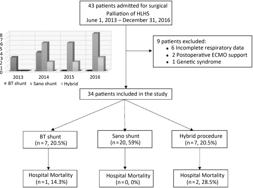
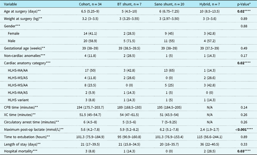
Forty-three patients were admitted for stage 1 palliation of hypoplastic left heart syndrome from June, 2013 to December, 2016. Nine patients were excluded from the study due to incomplete respiratory data (n = 6), need for post-operative extracorporeal membrane oxygenation support (n = 2), and presence of genetic syndrome (n = 1). A total of 34 patients were included in the study (Fig 1). Of these patients, 7 (20.5%) underwent Blalock–Taussig shunt, 20 (59%) underwent Sano shunt, and 7 (20.5%) underwent hybrid procedure. All Sano patients had a 5 mm shunt, while 42.8% and 57.2% of Blalock–Taussig patients had a 3 and 3.5 mm shunt, respectively. The median time to successful extubation was 101.3 hours (IQR = 75.9–184.9). Twenty patients (59%) were extubated before the end of the 120-hour period. The type of surgical approach varied over time (Fig 1). No significant differences were observed in demographic, anatomic, and intraoperative variables across the three surgical approach categories, except for significantly increased age at surgery in the hybrid group and increased number of patients with mitral atresia/aortic atresia anatomy in the Sano group (Table 1). Maximum post-operative lactate was significantly lower in patients undergoing the hybrid procedure compared to Blalock–Taussig and Sano shunt patients (Table 1). Post-operative length of mechanical ventilation and hospital length of stay were similar between groups, while mortality was significantly higher for the hybrid group (Table 1). No statistical differences were observed in ventilator management across the three surgical groups (Supplementary Table S1).
Patient flow chart and surgical distribution over time.

Patient demographic, anatomic, intraoperative, and outcome variables

AA = aortic atresia; AS = aortic stenosis; CPB = cardiopulmonary bypass; HLHS = hypoplastic left heart syndrome; MA = mitral atresia; MS = mitral stenosis; N/A = not applicable; XC = cross clamp.
* Kruskal–Wallis test to compare continuous measures across groups. Fisher’s exact test to compare categorical variables across groups.
** Median (Q1–Q3).
*** n (%).
**** (p <0.05).
Post-operative dead space
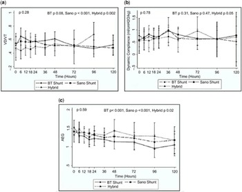
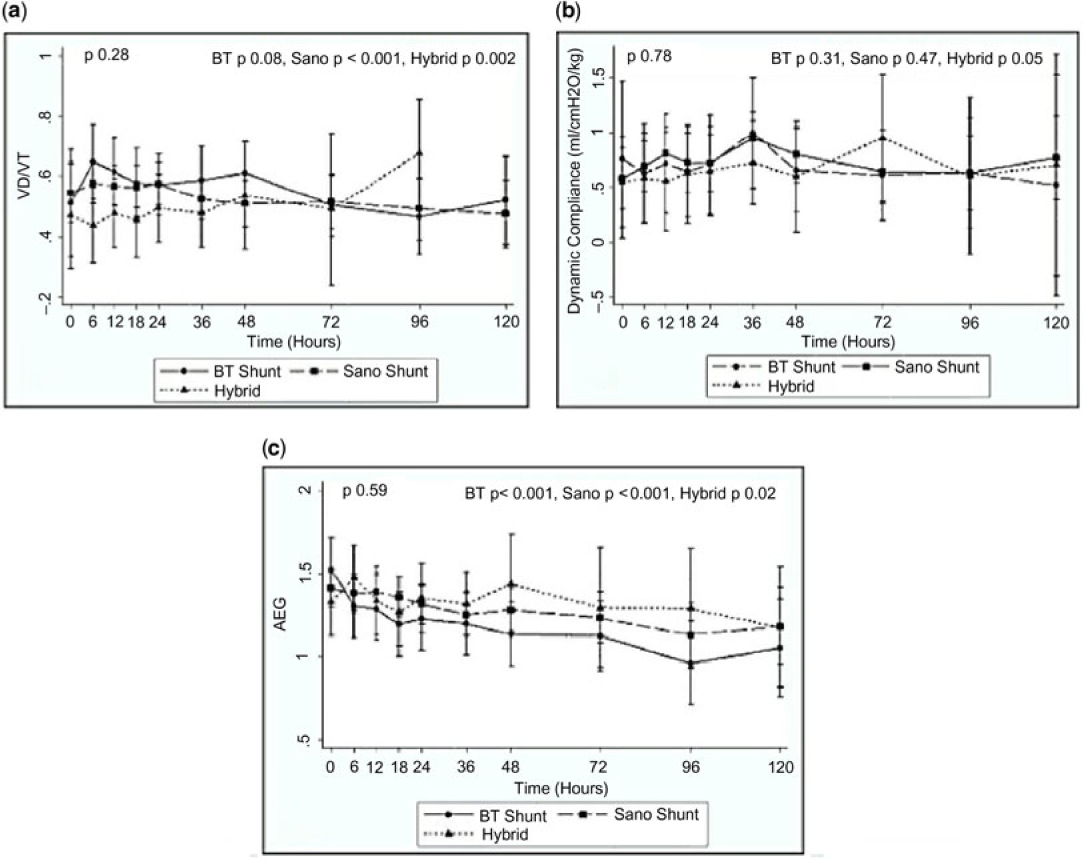
Changes in dead space during the first 120 post-operative hours across the three surgical groups are represented in Figure 2a. The range of dead space of each patient at all time points was 0.23–0.87. Dead space significantly decreased over time in Sano patients, while there was a trend towards decreased dead space in the Blalock–Taussig shunt group (Fig 2a). Patients undergoing the hybrid procedure had a statistically significant increase in dead space fraction over time (Fig 2a). Table 2 represents the maximum dead space values during the study period and the mean dead space values on days 1, 2, and 3–5. Mean dead space on day 1 was significantly lower for the hybrid group (Table 2).
Respiratory parameters during the first 120 post-operative hours after stage 1 surgical palliation for hypoplastic left heart syndrome. ( a ) Dead space ratio (Vd/Vt); ( b ) dynamic compliance; and ( c ) arterial to end-tidal carbon dioxide gradient (AEG). Data were stratified by surgical approach. Linear mixed model with the individual as the cluster for all specified outcomes was utilised to determine the differences between groups. The p-value comparing overall differences between the groups is shown on the upper left. The p-value for each surgical group, representing the trend over time, is shown on the upper right. BT, Blalock–Taussig.
Maximum dead space and mean dead space ratios on day 1, day 2, and days 3–5 stratified by surgical approach

*Kruskal–Wallis test to compare Vd/Vt measures across groups. **(p <0.05).
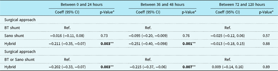
Dead space was then evaluated using multivariable linear regression at different time points after adjusting for variables that were significant in bivariate analysis, including age at surgery, gender, weight, cardiac anatomy category, and maximum post-operative lactate. Analysis was stratified by surgical approach and by comparing hybrid patients with patients with both types of shunt as a combined group. Mean dead space was found to be significantly lower for the hybrid group on days 1 and 2, but not on days 3–5 (Table 3). A multivariable linear mixed model, after adjusting for the same variables and time, demonstrated that mean dead space was significantly lower over time for the hybrid group on days 1 and 2, but not on days 3–5 (Table 3).
Linear mixed model of the mean dead space ratios over different time periods stratified by surgical approach and comparing hybrid with BT and Sano shunts as a combined group

* Linear mixed model to ascertain the estimated mean differences in Vd/Vt over the first 24, 36–48, and 72–120 hours, respectively adjusting for age at surgery, gender, weight, time, cardiac anatomy category, and maximum post-operative lactate with patient as the random effect. **(p <0.05).
Other post-operative respiratory, hemodynamic, and fluid balance parameters
Changes in other respiratory parameters during the first 120 post-operative hours across the three surgical groups are represented in Figure 2(b)–(d). Dynamic compliance and arterial to end-tidal carbon dioxide gradient were not overall different across the post-operative intervals stratified by surgical procedure (Fig 2b and c). No significant differences were observed in dynamic compliance between the groups over time (Fig 2b) or when mean dynamic compliance was evaluated on days 1, 2, and 3–5. Over time, we also observed that the arterial to end-tidal carbon dioxide gradient significantly decreased for all surgical groups (Fig 2c) but was not different between groups. Dead space fraction correlated with arterial to end-tidal carbon dioxide gradient across all time points (p < 0.001). Changes in hemodynamic parameters and fluid balance during the first 120 post-operative hours across the three surgical groups are represented in Supplementary Figure S1.
Outcome measures
Associations between dead space and outcomes (duration of mechanical ventilation and hospital length of stay) were assessed using multivariable models. Models were adjusted for confounders significant in bivariate analysis, including age at surgery, gender, weight, cardiac anatomy category, and maximum post-operative lactate. After adjusting for these confounders, maximum dead space and mean dead space on days 1 and 2 did not correlate with log duration of mechanical ventilation and log hospital length of stay. We used mean daily dead space values to categorise the cohort into two groups (≤0.6 and >0.6). We used this cut-off value because of previous literature demonstrating no difference in the duration of mechanical ventilation with a 0.5 cut-off in single-ventricle patients and suggesting that a higher dead space cut-off might be useful.Reference Devor, Kang and Wellnitz 3 Patients with a mean dead space >0.6 on days 1 and 2 had similar mechanical ventilation duration and hospital length of stay than those with mean dead space ≤0.6 on each day. However, patients with a mean dead space >0.6 on days 3–5 had a significant decrease in mechanical ventilation duration compared to those with mean dead space ≤0.6 on days 3–5 (p < 0.001). This finding persisted when the cohort was analysed excluding the hybrid group (p = 0.003). We then categorised patients based on the duration of mechanical ventilation as ≤120 and >120 hours. We used this cut-off because 120 hours was our post-operative study time period. Patients with mean dead space >0.6 on days 3–5 had a significantly decreased likelihood of remaining on mechanical ventilation for >120 hours (p = 0.02). Supplementary Table S2 illustrates these associations. Other dead space cut-off values were used to evaluate this association, but none significantly correlated with outcomes. We were not able to investigate the associations of dead space with extubation failure in our study.
Discussion
This study is the first, to date, to examine dead space fractions in patients undergoing the three stage 1 palliative surgeries for hypoplastic left heart syndrome, including the hybrid procedure. The primary finding is that dead space was different for patients with Blalock–Taussig and Sano shunts compared to the hybrid procedure. Over time, dead space decreased in patients undergoing Blalock–Taussig and Sano shunts, while it increased in those undergoing the hybrid procedure. This finding may reflect the waning impact of bypass in Blalock–Taussig and Sano patients. No differences were observed in dead space and pulmonary compliance between the groups across each time point or when analysed as maximum values. However, multivariable analysis demonstrated lower mean dead space in the hybrid group on days 1 and 2 and over time on each day. This finding may be indicative of the absence of bypass effect on the pulmonary vasculature in these patients. No differences in dead space were observed between Blalock–Taussig and Sano shunts at any time points. This finding conflicts with previous data from Shakti et al who demonstrated higher maximum dead space in Sano shunt patients compared to Blalock–Taussig shunt patients within the 48-hour post-operative period.Reference Shakti, McElhinney and Gauvreau 2 The differences between our results and the study by Shakti et al may be attributable to several factors. Study design disparity may explain this difference as the study by Shakti et al did not include patients with a hybrid procedure and only assessed infants undergoing Blalock–Taussig and Sano shunts. Differences in patient populations may also have influenced the study results, as there are discrepancies in gestational age, shunt size in patients undergoing Blalock–Taussig, and unfavourable cardiac anatomy between both studies. Post-operative physiologic changes related to diastolic runoff in Blalock–Taussig patients may also justify this difference.
Interestingly, dead space significantly increased over time in hybrid patients. This may be attributable to several factors, including post-operative worsening of right ventricular function, decreased myocardial contractility, and possibly increased right ventricular afterload with decreased pulmonary blood flow due to pulmonary artery banding.Reference Bugnitz, Miao and Berger 10 , Reference Kobayashi, Natarajan and Turner 11 A combination of these cardiovascular factors with failure of the lungs to adjust to the decrease in pulmonary blood flow may explain our findings.
Similar to previous studies, we also found that dead space may be a useful predictor of length of mechanical ventilation in patients undergoing stage 1 palliation for hypoplastic left heart syndrome, regardless of surgical approach.Reference Shakti, McElhinney and Gauvreau 2 Our study is the first, to date, to demonstrate that mean dead space >0.6 in post-operative days 3–5 is associated with a significant decrease in mechanical ventilation in patients undergoing stage 1 palliation for hypoplastic left heart syndrome. One might expect increased dead space to be associated with prolonged mechanical ventilation; however, cardiopulmonary interactions in patients with univentricular physiology may be very different from patients with biventricular physiology, as noted in previous studies.Reference Devor, Kang and Wellnitz 3 We believe that increased dead space in the late post-operative phase may indicate better balanced pulmonary to systemic flow, with decreased pulmonary blood flow leading to increased systemic perfusion. We speculate that improved haemodynamics could then possibly facilitate earlier withdrawal of mechanical ventilation in this patient population. Improvement over time in other post-operative non-cardiac risk factors for prolonged invasive ventilation, such as lung disease, airway and/or vocal cord edema, fluid overload, phrenic nerve inflammation, or renal dysfunction, could have also influenced the association.Reference Gupta, McDonald and Goyal 12 , Reference Wernovsky, Kuijpers and Van Rossem 13 This intriguing finding suggests that clinicians should consider very differently dead space in neonates undergoing stage 1 palliation for hypoplastic left heart syndrome compared to other critically ill infants requiring mechanical ventilation.
Limitations
Our findings should be interpreted with caution due to the limitations of the methods. First, our experience comes from a single-centre retrospective database review with a small sample size. Second, we were not able to adjust for confounding factors, not identified in our dataset, that may be associated with differences in lung compliance and duration of mechanical ventilation (such as lung disease, airway and/or vocal cord edema, fluid overload, phrenic nerve inflammation, or renal dysfunction). Third, the absence of guidelines for post-operative management and variability of surgical approach over time, especially for patients undergoing hybrid procedure, could reflect differences in practitioner preferences. Fourth, the lack of adjustment for inotropic score may have masked the significant difference in right ventricular function and subsequently pulmonary blood flow in our cohort. Fifth, the finding of dead space >0.6 at post-operative days 3–5, associated with a decreased duration of mechanical ventilation, being statistically significant may not have clinical relevancy. Finally, despite our efforts to streamline monitoring of pulmonary function, our results could have been affected by the learning curve in the use of NM3 Respironics monitor as a standard of care for intubated patients in our institution.
Conclusions
Dead space was different for patients with Blalock–Taussig and Sano shunts compared to the hybrid procedure. In patients who underwent the hybrid procedure, dead space was significantly lower on days 1 and 2. Mean dead space >0.6 in post-operative days 3–5 was associated with a decreased duration of mechanical ventilation for all surgical groups. The measurement of dead space fraction in this vulnerable group may be of value in their clinical care. Extended prospective studies would be useful to further define the role of dead space fraction and precise pulmonary mechanical data in improving outcomes.
Author ORCIDs
Pilar Anton-Martin 0000-0003-1215-3285
Acknowledgements
The authors acknowledge the Divisions of Cardiac Critical Care Medicine, Cardiology and Cardiothoracic Surgery for the excellent patient care provided.
Supplementary Material
To view supplementary material for this article, please visit https://doi.org/10.1017/S1047951119000076.
Financial Support
This research received no specific grant from any funding agency, commercial, or not-for-profit sectors.
Conflict of Interest
None.
Ethical Standards
The authors assert that all procedures contributing to this work comply with the ethical standards of the relevant national guidelines on human experimentation, Department of Health and Human Services Code of Federal Regulations (45 CFR 46) Policy for Protection of Human Research Subjects, and with the Helsinki Declaration of 1975, as revised in 2008, and has been approved by the institutional review board of Phoenix Children’s Hospital.