





Introduction
The family Parmeliaceae has over 2700 species worldwide (Lücking et al. Reference Lücking, Hodkinson and Leavitt2017). Due to its diverse habitats, more than 16% of these known species are found in Mexico, with growth forms including crustose, foliose and fruticose lichens (Herrera-Campos et al. Reference Herrera-Campos, Lücking, Pérez-Pérez, Miranda González, Sánchez, Barcenas-Peña, Carrizosa, Zambrano, Ryan and Nash2014, Reference Herrera-Campos, Pérez-Pérez and Nash2016). Among the genera of Parmeliaceae is the foliose genus Bulbothrix Hale (Nash Reference Nash2016). As currently defined, this genus is not a monophyletic lineage, falling into two separate clades, one including the genus Parmelinella Elix & Hale (Divakar et al. Reference Divakar, Crespo, Blanco and Lumbsch2006; Crespo et al. Reference Crespo, Kauff, Divakar, del Prado, Pérez-Ortega, de Paz, Ferencova, Blanco, Roca-Valiente and Núñez-Zapata2010, Reference Crespo, Divakar, Leavitt and Lumbsch2016).
The genus Bulbothrix s. lat. has a wide distribution, occurring from tropical-subtropical to temperate regions (Michlig & Benatti Reference Michlig and Benatti2015), and is most common in semi-arid and secondary forests (Hale Reference Hale1976). Currently, there are 66 accepted species in the genus (Benatti Reference Benatti2014; Masson et al. Reference Masson, Benatti and Sérusiaux2015; Zhang et al. Reference Zhang, Wang, Liu, Li, Shi, Ye, Yang and Wang2016; Kirika et al. Reference Kirika, Divakar, Buaruang, Leavitt, Crespo, Gatheri, Mugambi, Benatti and Lumbsch2017), c. 70% of which are found in South America, its main centre of distribution, followed by South Africa (Hale Reference Hale1976). In Mexico, nine species have been recognized: Bulbothrix atrichella (Nyl.) Hale, B. confoederata (W. L. Culb.) Hale, B. coronata (Fée) Hale, B. fungicola (Lynge) Hale, B. isidiza (Nyl.) Hale, B. laevigatula (Nyl.) Hale, B. subdissecta (Nyl.) Hale, B. tabacina (Mont. & Bosch) Hale and B. ventricosa (Hale & Kurok.) Hale (Nash Reference Nash2016).
Species of the genus Bulbothrix s. lat. have small, adnate thalli (generally ≤ 10 cm in diameter), are irregular to dichotomously lobate, the lobes sublinear, imbricate, elongate and plane, with rounded, subrounded or crenate apices; isidia are the most common propagation structure, and soredia or pustules are found only on a small number of species; the apothecia are smooth or coronate (crowned with ciliate bulbs); the marginal cilia have a bulbate base and each has simple or variably branched apices (Hale Reference Hale1975; Elix Reference Elix1993; Nash Reference Nash2016). The bulbs are enlarged or swollen structures composed of rounded cells and containing a hollow cavity filled with an unidentified oily substance (Hale Reference Hale1975; Benatti Reference Benatti2011). These structures are considered taxonomically significant for species identification (Benatti Reference Benatti2011, Reference Benatti2014).
The presence of these structures allowed the identification of a species new to science and a new record for Mexico. These findings emphasize the importance of continued exploration of diverse ecosystems and the examination of herbarium specimens, thereby contributing to the knowledge of lichen biota in Mexico, one of the megadiverse countries.
Materials and Methods
Morphological examination of the specimens was conducted using a Zeiss Stemi 305 stereoscopic microscope, and microscopic preparations were studied in distilled water with a Zeiss Primo Star optical microscope. Taxonomic keys by Benatti (Reference Benatti2012a, Reference Benatti2013a, Reference Benatti2014), Bungartz et al. (Reference Bungartz, Benatti and Spielmann2013), Michlig & Benatti (Reference Michlig and Benatti2015) and Nash (Reference Nash2016) were followed.
To accurately distinguish true bulbs and pycnidia (Fig. 1), the clearing procedure proposed by Benatti (Reference Benatti2011) was employed. Small portions of the thallus containing structures resembling bulbs or pycnidia were removed from the upper cortex and placed on a microscope slide with a few drops of commercial sodium hypochlorite. The procedure was repeated for bulbs on the thallus margin and apothecia, as well as for rhizines. The characteristic oily substance of the bulbs turns amber over time (Fig. 1B), becoming reddish amber when the sample is nearly dry, a change not observed in pycnidia.
Differences between bulbs and pycnidia of Bulbothrix bulbillosa (8755) and Bulbothrix buapana sp. nov. (9870). A, ciliate bulbs on the lobe margin of B. bulbillosa after the clearing process. B, bulbs on the thallus of B. buapana , showing the amber colour obtained after the clearing process. C, oily substance of a bulb of B. buapana. D, pycnidium of B. buapana. Scales = 50 μm. In colour online.

Figure 1 Long description
A four-panel micrograph showing lichen structures. Each panel includes a white scale bar representing 50 micrometers in the bottom right corner.
* Panel A: Micrograph of Bulbothrix bulbillosa showing ciliate bulbs on the lobe margin. Two dark, spherical bulbs are embedded within a translucent, fibrous tissue layer. A black pointer indicates the center of the left bulb.
* Panel B: Micrograph of Bulbothrix buapana showing bulbs on the thallus. Two distinct amber-colored spherical bulbs are visible against a pale, textured background. A black pointer indicates the lower-right bulb.
* Panel C: Micrograph of Bulbothrix buapana showing an oily substance. A central bulb is ruptured or open, revealing a translucent interior indicated by a black pointer. Two smaller, clear oily droplets are visible on the upper-left margin of the structure.
* Panel D: Micrograph of Bulbothrix buapana showing pycnidia. Three circular, radiating structures are visible against a green and yellow textured thallus. A black pointer indicates the center of the bottom-most pycnidium, which shows a dense, fibrous radial pattern.
The new species was investigated by also taking measurements of all relevant structures, including the lobes, rhizines, cilia, apothecia and spores (Kirika et al. Reference Kirika, Divakar, Buaruang, Leavitt, Crespo, Gatheri, Mugambi, Benatti and Lumbsch2017). For ascospore measurements, 11 specimens with well-developed apothecia were selected. From each specimen, three apothecia were chosen and 30 spores were measured from each apothecium, resulting in a total of 990 spores. Spore measurements are given as arithmetic mean value (x̄) ± 1.96 × standard deviation; values in parentheses represent minimum and maximum values, and Q represents length/width ratio of the spores (Masson Reference Masson2012; Masson et al. Reference Masson, Benatti and Sérusiaux2015). The thickness of the upper cortex, lower cortex, algal layer, medulla and apothecial layers was also measured. The distinction of the apothecial layers followed the method proposed by Ferencova (Reference Ferencova2012), which divides the proper exciple into three parts: the hyaline layer beneath the subhymenium, the intermediate layer, and the basal layer immediately above the algal layer. The thickness of each layer is then measured.
Secondary metabolites were analyzed using the standardized thin-layer chromatography (TLC) method, with solvent C (toluene:acetic acid, 200:30, 230 ml) (Elix Reference Elix2014). To confirm the identification of gyrophoric acid and distinguish it from lecanoric acid, the microcrystallization technique proposed by Orange et al. (Reference Orange, James and White2001) was employed, using GAW solvent (glycerol:ethanol:water, 1:1:1) and Parmotrema tinctorum (Despr. ex Nyl.) Hale as a control species for the presence of lecanoric acid (Fig. 2). For microchemical spot tests, 10% potassium hydroxide (K), sodium hypochlorite (C), and p‑phenylenediamine (P) were applied to the cortex and medulla (Orange et al. Reference Orange, James and White2001).
Differences between gyrophoric acid and lecanoric acid. A, C+ pink reaction (gyrophoric acid) in Bulbothrix buapana sp. nov. B, C+ red reaction (lecanoric acid) in Parmotrema tinctorum. C, gyrophoric acid crystals in GAW. D, lecanoric acid crystals in GAW. Scales: C & D = 50 μm. In colour online.
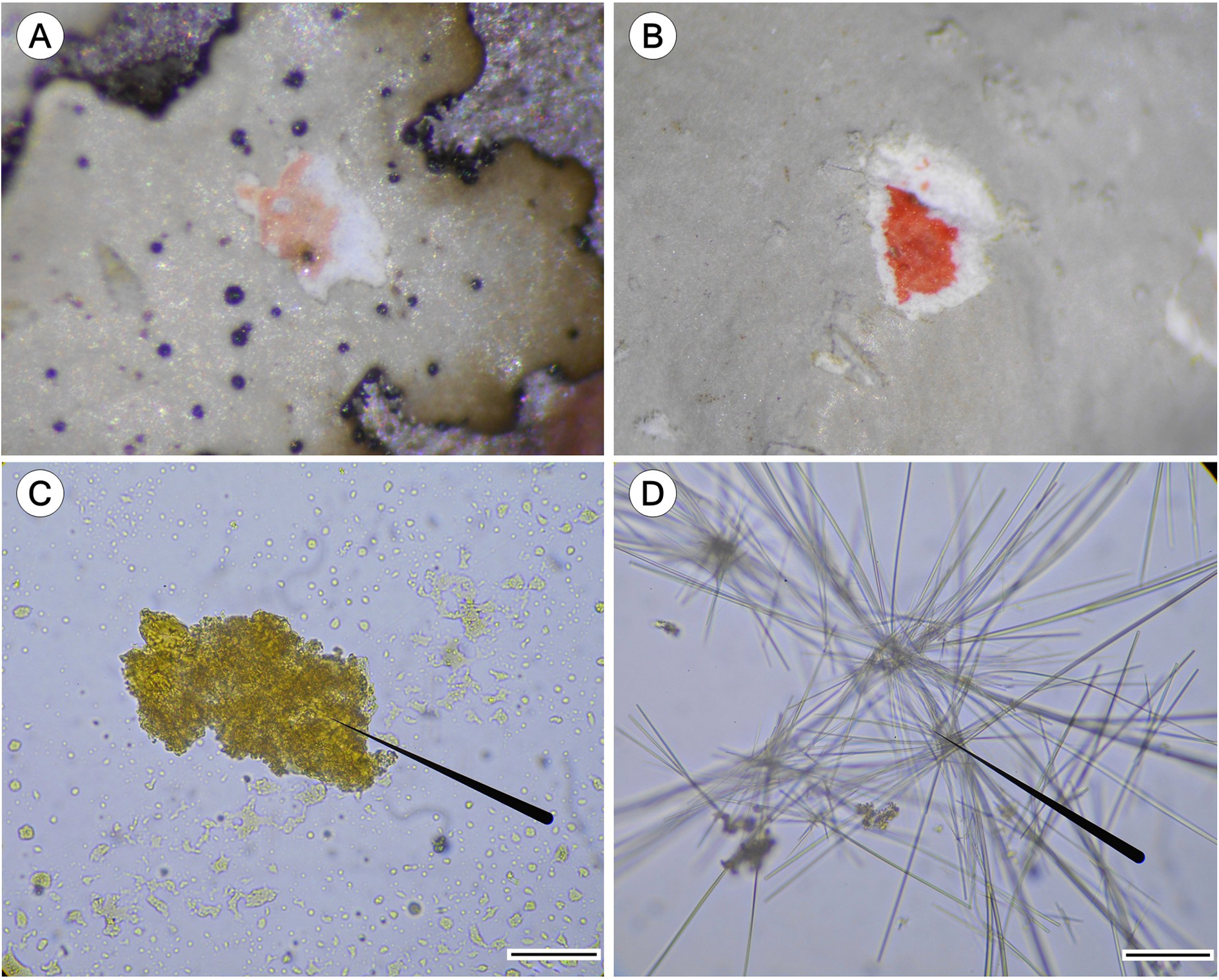
Figure 2 Long description
A four-panel photo arranged in a two-by-two grid.
* Panel A (top-left): A macro photo of Bulbothrix buapana showing a pale pink C plus reaction on the lichen surface, indicating the presence of gyrophoric acid. The lichen thallus is light gray with small dark spots.
* Panel B (top-right): A macro photo of Parmotrema tinctorum showing a vibrant red C plus reaction on the lichen surface, indicating the presence of lecanoric acid.
* Panel C (bottom-left): A micrograph of gyrophoric acid crystals in G A W. The crystals appear as a dense, yellowish-brown amorphous cluster. A black pointer line indicates the central mass. A scale bar in the bottom right represents 50 mu m.
* Panel D (bottom-right): A micrograph of lecanoric acid crystals in G A W. The crystals form long, thin, colorless needles radiating in star-like clusters. A black pointer line indicates a central intersection of the needles. A scale bar in the bottom right represents 50 mu m.
To generate a distribution map of Bulbothrix species in Mexico, data from Nash (Reference Nash2016) and the Consortium of Lichen Herbaria (Consortium of Lichen Herbaria 2024) were used. The map was produced in ArcMap (Esri 2016) using political boundary layers from CONABIO (2023).
The specimens examined are deposited in the herbarium of the Facultad de Ciencias Biológicas of the Benemérita Universidad Autónoma de Puebla (HUAPLI), the herbarium of the Universidad Autónoma de Aguascalientes (HUAA), the herbarium of the Jardín Botánico Nicolaita ‘Melchor Ocampo’ of the Universidad Michoacana de San Nicolás de Hidalgo (EBUM), and the herbarium of the Instituto de Ecología (XAL).
Taxonomy
Bulbothrix buapana Hernández Alberdin, Pérez-Pérez & Benatti sp. nov
MycoBank No.: MB 861518
Bulbothrix buapana sp. nov. is similar to B. coronata , but differs in having ciliate bulbs on the upper cortex, with bulbate marginal cilia either lacking apices or bearing simple apices. It differs from B. laeviuscula (Räsänen) Benatti, as B. buapana has rhizines with dark bulbate bases and gyrophoric acid in the medulla, and from Bulbothrix bulbillosa Benatti et al. in having broader lobules (> 1.5 mm) and lacking isidia.
Type: Mexico, Oaxaca, locality El Huamúchil, Santiago Huajolotitlán; 17°47′36.8″N, 97°42′42.5″W, 1685 m, on the bark of Vachellia farnesiana, in a transition zone between tropical dry forest and xerophilous scrub, 30 November 2021, Hernández Alberdin L. E. & Pérez-Pérez R. E. HUAPLI 8745 (HUAPLI—holotype).
Bulbothrix buapana sp. nov. (holotype). A, type specimen. B, thallus and lobes. C, detail of ciliate bulbs on the upper cortex and margin. D, lower cortex. E, coronate apothecia. Scales: A = 1 cm; B–E = 1 mm. In colour online.

Figure 3 Long description
A multi-panel photo series showing the holotype of Bulbothrix buapana.
Panel A at the top shows the full type specimen, a foliose lichen with a light greyish-tan thallus and rounded lobes, featuring a scale bar of 1 centimeter in the bottom right.
Panel B at the middle-left shows a close-up of the thallus and lobes with small black dots across the surface.
Panel C at the middle-right provides a high-magnification view of ciliate bulbs, appearing as dark purple or black spherical protrusions along the upper cortex and margins.
Panel D at the bottom-left displays the lower cortex, which is dark brown to black with a textured, rhizinate surface.
Panel E at the bottom-right shows several coronate apothecia, which are cup-shaped reproductive structures with brown centers and white, beaded margins.
Scale bars for panels B through E represent 1 millimeter.
Anatomy of Bulbothrix buapana sp. nov. (holotype). A, cross-section of an apothecium showing asci. B, lactophenol blue staining to identify the layers of the proper exciple (Ferencova Reference Ferencova2012); type II apothecium, SH = subhymenium, HL = hyaline layer, IL = intermediate layer, BL = basal layer. C, ascospores. D, conidia. E, detail of the coronate apothecium. Scales: A & B = 50 μm; C & D = 10 μm; E = 0.1 mm. In colour online.

Figure 4 Long description
A multi-panel figure containing five sections labeled A through E.
Panel A. A cross-section micrograph of an apothecium. A black pointer indicates the curved inner layer. The structure shows a dense layer of elongated asci arranged vertically beneath a dark outer crust. Scale bar equals 50 micrometers.
Panel B. A high-magnification micrograph with lactophenol blue staining showing layered tissues. From the inner tissue moving outward, four layers are identified by arrows and initialisms. S H points to the subhymenium. H L points to the hyaline layer. I L points to the intermediate layer. B L points to the basal layer. Scale bar equals 50 micrometers.
Panel C. A micrograph showing five oval-shaped ascospores with a yellowish-green center and a clear outer wall. Scale bar equals 10 micrometers.
Panel D. A micrograph showing numerous small, needle-like conidia scattered across the frame. Scale bar equals 10 micrometers.
Panel E. A top-down macro photograph of a coronate apothecium. It features a circular, reddish-brown central disc surrounded by a thick, white, scalloped margin. Small dark purple or black spots are distributed along the outer edge of the white margin. Scale bar equals 0.1 millimeters.
Thallus foliose, grey to dark grey, becoming brownish in the herbarium; with olivaceous margins, subcoriaceous, corticolous, moderate to strongly adnate to the substratum, 2–6 cm diam., sublinear-laciniate; upper cortex 15–35(–40) μm thick, algal layer 35–90 μm thick, medulla white, 45–90 μm thick, lower cortex 15–60 μm thick. Lobes 0.5–3.0(–3.5) mm wide, sublinear to subirregular, contiguous to slightly imbricate, anisotomically, dichotomously or occasionally irregularly branched; apices plane to subconvex, truncated to rounded; margins plane, sinuous to crenate, irregular or entire, the axils oval to irregular, with bulbate cilia; cilia black, contiguous and abundant along the margins, with semi-immersed to immersed bulbate bases c. 0.02–0.12(–0.15) mm wide, the bulbs usually without an apex at early stages; apices simple or furcate, 0.03–0.10(–0.20) × c. 0.02 mm. Medulla white. Upper cortex smooth and continuous, occasionally with fissures in the older parts of the thallus, emaculate, without pruina, with abundant, scattered or grouped ciliate bulbs 0.03–0.13(–0.15) mm wide. Soredia, pustules and isidia absent. Lacinules absent to scarce, adventitious, occasionally growing in the centre of the thallus, at the margins of the oldest laciniae, simple, flat, 0.2–0.5 × 0.1–0.3(–0.4) mm, truncate or rounded, concolorous with the upper cortex. Lower cortex dark or pale brown at the margins, eventually turning black in the central parts, shiny, slightly papillate, densely rhizinate in the centre, and scarce at the margins. Rhizines brown to pale brown at the thallus margins, turning black at the centre but occasionally still with brown apices, simple to dichotomously or irregularly branched, 0.05–0.40(–1.00) mm long, partially with darker bulbate bases, abundant and evenly distributed.
Apothecia plane to slightly concave, adnate to sessile, 0.3–4.3(–5.0) mm diam., larger at centre than at the younger parts of the thallus, laminal to submarginal, margins subcrenate to crenate, coronate, with usually contiguous bulbs 0.04–0.10 mm away from each other, wide; amphithecium smooth, with ciliate bulbs. Disc brown, epruinose, imperforate; epithecium 5.0–13.5(–15) μm; hymenium 30–50 μm; subhymenium 10–13 μm, hyaline layer 10.5–19.0(–26.0) μm, intermediate layer 12–20 μm, basal layer 20–36 μm. Ascospores colourless, simple, ellipsoid to oval, 6.5–10.0(–11.5) × (4.0–)4.5–5.5(–6.5) μm (x̄ = 8.26 × 5.09; x̄ 1 = 8.27 × 5.06, x̄ 2 = 7.97 × 5.05, x̄ 3 = 8.63 × 5.08, x̄ 4 = 8.01 × 5.09, x̄ 5 = 8.01 × 5.08, x̄ 6 = 8.47 × 5.16, x̄ 7 = 8.21 × 5.12, x̄ 8 = 8.40 × 5.15, x̄ 9 = 7.97 × 5.12, x̄ 10 = 8.80 × 5.08, x̄ 11 = 8.06 × 5.04), Q = (1.20–)1.62(–2.0); epispore 0.7–1.0 μm.
Pycnidia submarginal to laminal, immersed, with black ostioles. Conidia fusiform or weakly bifusiform, 6.5–8.5(–9.0) × 1.0–1.7(–2.0) μm.
Chemistry
Upper cortex K+ yellow, UV−, with atranorin; medulla K−, C+ rose, KC+ rose, P−, UV−, with gyrophoric acid.
Etymology
The specific epithet ‘ buapana ’ is dedicated to the Benemérita Universidad Autónoma de Puebla (BUAP), as a tribute by the first author to honour his alma mater.
Ecology and distribution
Bulbothrix buapana was frequently found in the tropical dry forest areas of the municipality of Santiago Huajolotitlán, Oaxaca State. In addition to Oaxaca, it was also collected from areas within the states of Michoacán and Morelos, also in tropical dry forest. It was collected on the phorophytes Bursera spp., Escontria chiotilla , Fraxinus uhdei , Lysiloma, Malpighia mexicana, Plumeria rubra , Prosopis sp., Schinus molle , Vachellia farnesiana and others still not identified, both in the understorey and in the lower branches of the canopy.
Notes
Bulbothrix buapana sp. nov. is very similar to B. coronata , as both species have coronate apothecia and gyrophoric acid as the main medullary component. The new species differs in having ciliate bulbs on the upper cortex, with bulbate marginal cilia either lacking apices or bearing simple apices, whereas B. coronata has cilia with initially simple or forked apices that soon become dichotomously or irregularly branched (Benatti Reference Benatti2013b). Bulbothrix buapana also resembles B. laeviuscula , as both have coronate apothecia, ciliate bulbs on the upper cortex, and marginal cilia without apices or with simple apices. However, B. buapana has rhizines with dark bulbate bases, whereas B. laeviuscula has rhizines without bulbate bases and lacks medullary substances (Benatti Reference Benatti2012c). Bulbothrix bulbillosa may be interpreted as the isidiate counterpart of B. buapana , as both species are characterized by coronate apothecia, ciliate bulbs on the upper cortex, and gyrophoric acid in the medulla. Bulbothrix bulbillosa differs in having narrower lobules (< 1.5 mm) and in producing simple to branched isidia with ciliate bulbs (Bungartz et al. Reference Bungartz, Benatti and Spielmann2013).
Additional specimens examined
Mexico: Michoacán: Lago de Camécuaro National Park, Tangancícuaro, in secondary vegetation of tropical dry forest, on trees of Fraxinus uhdei and Schinus molle , 19°53′56.2″N, 102°12′34.7″W, 1732 m, 2024, Rangel Osorni 3353, 3362, 3376, 3377 (EBUM). Morelos: El Zapote, Puente de Ixtla, in a transition zone between tropical dry forest and Quercus forest, on trees of Bursera sp. and Lysiloma, 18°27′59.8″N, 99°19′21.1″W, 18°28′01.9″N, 99°19′16.5″W, 18°28′03.2″N, 99°19′14.5″W, 1897–1971 m, 2024, Pérez-Pérez & Vidal Figueroa 9870–80 (HUAPLI). Oaxaca: El Huamúchil, Santiago Huajolotitlán, in a transition zone between tropical dry forest and xerophilous scrub, on trees of Bursera spp., Escontria chiotilla , Malpighia mexicana, Plumeria rubra , Prosopis sp. and Vachellia farnesiana, 17°47′36.8″N, 97°42′42.5″W, 17°47′35.5″N, 97°42′36.3″W, 17°47′37.5″N, 97°42′23.4″W, 17°47′38.8″N, 97°42′29.8″W, 1685–1758 m, 2021, Hernández Alberdin & Pérez-Pérez 8743, 8744, 8746–49, 8753, 8754, 8757–64, 9014–18, 9858–68 (HUAPLI); San Francisco Sola, Sola de Vega, in secondary vegetation of tropical dry forest, on trees of Vachellia sp., 16°29′57.6″N, 96°57′31.2″W, 1425 m, 2021, Hernández Alberdin & Pérez-Pérez 8499 (HUAPLI).
Bulbothrix bulbillosa Benatti, A. A. Spielm. & Bungartz
Bulbothrix bulbillosa , new record for Mexico. A, thallus. B, detail of ciliate bulbs on the upper cortex and margin. C, lower cortex. D, isidia with bulbs. Scales: A = 1 cm; B = 1 mm; C & D = 0.5 mm. In colour online.

Figure 5 Long description
A multi-panel photograph containing four labeled sections.
* Panel A, top-left: A macro view of the lichen thallus, appearing as a flat, irregular, grayish-green leafy structure against a white background. A black scale bar in the bottom right represents 1 centimeter.
* Panel B, top-right: A close-up view of the upper cortex and margin. The surface is pale gray and covered with numerous small, dark purple or black spherical bulbs. A black scale bar in the bottom right represents 1 millimeter.
* Panel C, bottom-left: A view of the lower cortex, showing a tan to light brown underside with a distinct dark purple or black scalloped margin. A black scale bar in the bottom right represents 0.5 millimeters.
* Panel D, bottom-right: A high-magnification view of isidia with bulbs. The surface is densely packed with small, cylindrical, or branched outgrowths tipped with dark spherical bulbs. A black scale bar in the bottom right represents 0.5 millimeters.
Thallus foliose, greenish grey, with olivaceous margins, becoming dusky grey in the herbarium, subcoriaceous, corticolous, strongly adnate to the substratum, 0.5–6.0 cm diam., sublinear-laciniate. Lobes 0.3–1.5(–2.2) mm wide, dichotomously anisotomously or trichotomously branched, contiguous to slightly imbricate; apices plane, truncate to subtruncate; margins plane, smooth to sinuous or subirregular, entire, ciliate, axils oval; cilia black, abundant along the margins, absent only at the apices of the laciniae, with semi- immersed to immersed basal bulbs c. 0.05(–0.10) mm wide, sometimes the bulbs without an apex; apices initially simple, eventually becoming furcate and then turning slightly dichotomously or irregularly branched, 0.02–0.15(–0.20) × c. 0.02 mm. Upper cortex continuous to occasionally cracked on older parts, smooth, with numerous laminal ciliate bulbs 0.03–0.08(–0.10) mm wide; maculae absent, sometimes with visible scars left by fallen isidia. Lacinules generally rare and sparse, adventitious, very short, randomly distributed along the margins or on upper cortex, simple, flat, 0.1–0.7 × 0.1–0.3(–0.5) mm, truncate or acute, underside concolorous with the lower margin. Soredia and pustulae absent. Isidia scarce to common, laminal, initially granular, soon becoming smooth cylindrical, straight to slightly tortuous, 0.05–0.35(–0.55) × 0.04–0.7(–0.11) mm, simple to rarely little-branched, erect, firmly attached or occasionally broken off, concolorous with the thallus, typically densely ‘peppered’ with black ciliate bulbs, apically darkening (brownish). Medulla white. Lower cortex black to dark brown, ±shiny, smooth to subrugose, moderately to densely rhizinate; marginal zone shiny, attenuate, light brown turning darker at the transition to the centre, smooth, papillate. Rhizines light to dark brown or blackish, initially simple, soon becoming dichotomously or irregularly branched, partially with subtle basal bulbs, 0.10–0.50 × c. 0.03 mm, frequent to abundant, equally distributed.
Apothecia not found.
Pycnidia very scarce, laminal, with black ostioles. Conidia not found.
Chemistry
Upper cortex K+ yellow, UV− with atranorin; medulla K−, C+ rose, KC+ rose, P−, UV−, with gyrophoric acid.
Specimens examined
Mexico: Aguascalientes: Los Adobes, Calvillo, in tropical dry forest, on trees of Manihot caudata , 21°48′28.43″N, 102°41′15.55″W, 1858 m, 2021, Simijaca 3475 (UAA). Oaxaca: El Huamúchil, Santiago Huajolotitlán, in a transition zone between tropical dry forest and xerophilous scrub, on trees of Bursera spp. and Escontria chiotilla , 17°47′36.8″N, 97°42′42.5″W, 17°47′38.8″N, 97°42′29.8″W, 1685–1758 m, 2021, Hernández Alberdin & Pérez-Pérez 8750–52, 8755, 8756, 9869 (HUAPLI); San Francisco Sola, Sola de Vega, in secondary vegetation of tropical dry forest, on trees of Bursera sp., Opuntia sp. and Vachellia sp., 16°29′57.6″N, 96°57′31.2″W, 1425 m, 2021, Hernández Alberdin & Pérez-Pérez 8496–98 (HUAPLI).
The genus Bulbothrix Hale s. lat. in Mexico
Based on previously reported records by Nash (Reference Nash2016) and the review of available herbarium material, 11 species are recorded across 14 states of Mexico (Table 1, Figs 6 & 7). Bulbothrix bulbillosa is reported for the first time in Mexico from the states of Aguascalientes and Oaxaca; B. fungicola and B. tabacina are newly recorded for the state of Oaxaca, B. isidiza for the state of Veracruz, B. subdissecta for the state of Puebla, and B. ventricosa (previously reported from Puebla (Pérez-Pérez et al. Reference Pérez-Pérez, Silva-Espejo, Figueroa-Castro and Castañeda-Posadas2024)) for the states of Morelos and Oaxaca.
Description of the main characteristics that define the species of Bulbothrix s. lat. in Mexico. The species data correspond to the descriptions provided by Benatti (Reference Benatti2012a, Reference Benatti b, Reference Benatti2013a, Reference Benatti b, Reference Benatti2014), Michlig & Benatti (Reference Michlig and Benatti2015) and Nash (Reference Nash2016).

Table 1. Long description
The table contains 10 columns: Species, Lobes (m m wide), Upper cortex, Lower cortex, Apothecia, Isidia, Spores (mu m), Spot tests in medulla, Medullary chemistry, and Distribution.
* B. atrichella: 0.5–2.5 m m; without laminal ciliate bulbs; dark to brown margins; ecoronate; absent isidia; spores 6–8 x 5–6; K minus, C plus rose, K C plus rose, P minus; gyrophoric acid; SON.
* B. buapana: 0.5–3.0 m m; with laminal ciliate bulbs; dark to pale brown margins; coronate; absent isidia; spores 6.5–10 x 4.5–5.5; K minus, C plus rose, K C plus rose, P minus; gyrophoric acid; MICH, MOR, OAX.
* B. bulbillosa: 0.3–1.5 m m; with laminal ciliate bulbs; black to dark brown; coronate; present with ciliate bulbs; spores 5.5–6.0 x 5.0–5.5; K minus, C plus rose, K C plus rose, P minus; gyrophoric acid; AGU, OAX.
* B. confoederata: 0.5–1.2 m m; without laminal ciliate bulbs; black to brown; ecoronate; absent isidia; spores 5–7 x 3–5; K minus, C plus bright red, K C plus bright red, P minus; lecanoric acid; CHIH.
* B. coronata: 0.5–1 m m; without laminal ciliate bulbs; black; coronate; absent isidia; spores 5–10 x 4–5; K minus, C plus rose, K C plus rose, P minus; gyrophoric acid; BCS, CHIA, CHIH, GUA, MICH, NULE, OAX, SON.
* B. fungicola: 0.3–0.7 m m; without laminal ciliate bulbs; black; coronate; present with ciliate bulbs; spores 8–10 x 4–6; K minus, C plus rose, K C plus rose, P minus; gyrophoric acid; CHIA, OAX.
* B. isidiza: 1.5–4 m m; without laminal ciliate bulbs; uniformly brown; ecoronate; present isidia; spores 7–14 x 5–8; K plus yellow to deep red, C minus, K C minus, P plus orange; salazinic acid; CHIA, JAL, VER.
* B. laevigatula: 0.5–2 m m; without laminal ciliate bulbs; dark to brown margins; ecoronate; present isidia; spores 5–7 x 3–4; K minus, C plus bright red, K C plus bright red, P minus; lecanoric acid; CHIA, MICH, TAM, VER.
* B. subdissecta: 0.5–1.2 m m; without laminal ciliate bulbs; dark to pale brown margins; ecoronate; present isidia; spores 8–10 x 4–6; K minus, C plus rose, K C plus rose, P minus; gyrophoric acid; CHIA, PUE, SON, VER.
* B. tabacina: 1.5–5 m m; without laminal ciliate bulbs; black to dark brown margins; ecoronate; present isidia; spores 9–15 x 5–8; K plus yellow to deep red, C minus, K C minus, P plus orange; salazinic acid; CHIA, JAL, OAX, VER.
* B. ventricosa: 1.4–4.5 m m; with laminal ciliate bulbs; black with small dark brown spots; ecoronate; present isidia; spores 13–17 x 7–8; K plus yellow to orange, C minus, K C minus, P plus yellow; norstictic acid; CHIA, MOR, OAX, PUE, VER.
*AGU = Aguascalientes, BCS = Baja California Sur, CHIA = Chiapas, CHIH = Chihuahua, GUA = Guanajuato, JAL = Jalisco, MICH = Michoacán, MOR = Morelos, NULE = Nuevo León, OAX = Oaxaca, PUE = Puebla, SON = Sonora, TAM = Tamaulipas, VER = Veracruz.
Map of the distribution of Bulbothrix species in Mexico. In colour online.

Figure 6 Long description
A map of Mexico with state borders outlined in gray. A legend in the bottom-left lists eleven species with corresponding symbols. A compass rose is in the Northeast and a scale bar from 0 to 750 K m is in the Southeast.
Species distribution by region:
* Northwest: Bulbothrix confoederata (dark blue pentagon), Bulbothrix coronata (green hexagon), and Bulbothrix atrichella (yellow circle with dot) are clustered in the northern states of Chihuahua and Sonora. A single Bulbothrix coronata point is located at the southern tip of Baja California Sur.
* Central and Northeast: Bulbothrix coronata is scattered through the Sierra Madre Oriental. Bulbothrix bulbillosa (magenta circle) and Bulbothrix laevigatula (red triangle) appear in the central highlands.
* West Coast: Bulbothrix tabacina (brown circle) and Bulbothrix buapana sp. nov. (green circle with cross) are found near the Pacific coast in Jalisco and Colima.
* South and Southeast: This region has the highest density and diversity. Bulbothrix ventricosa (light blue triangle) and Bulbothrix subdissecta (purple asterisk) are concentrated in the Veracruz and Oaxaca regions. Bulbothrix fungicola (orange star) and Bulbothrix isidiza (teal square) are located further East in the Chiapas region near the Guatemalan border.
Some Bulbothrix species that were identified in this study. A, B. buapana on Vachellia farnesiana. B, B. buapana. C, B. bulbillosa. D, B. fungicola. E, isidia of B. fungicola. F, B. isidiza. G, isidia of B. isidiza. H, B. tabacina. I, isidia of B. tabacina. J & K, B. ventricos a. L, isidia of B. ventricosa. Scales = 0.5 mm. In colour online.

Figure 7 Long description
A multi-panel photo collection organized in a grid of four rows and three columns. Each panel includes a white scale bar representing 0.5 mm in the bottom right corner.
* Panel A. A wide shot of Bulbothrix buapana growing on the textured bark of a Vachellia farnesiana branch.
* Panel B. A close-up of B. buapana showing a pale grey thallus with dark bulbate cilia along the margins.
* Panel C. A close-up of B. bulbillosa featuring a lobed thallus with numerous small dark spots across the surface.
* Panel D. B. fungicola showing a light-colored thallus with distinct dark, irregular margins.
* Panel E. A high-magnification view of the isidia of B. fungicola, appearing as dense, granular outgrowths.
* Panel F. B. isidiza showing smooth, rounded lobes with dark edges.
* Panel G. A high-magnification view of the isidia of B. isidiza, showing a textured, bumpy surface.
* Panel H. B. tabacina featuring a broad, pale thallus with slightly undulating margins.
* Panel I. A high-magnification view of the isidia of B. tabacina, showing fine, hair-like or granular projections.
* Panel J. A wide shot of B. ventricosa, appearing as a vibrant light green lichen spreading across a tree branch.
* Panel K. A close-up of B. ventricosa showing the rounded lobe structure and dark marginal cilia.
* Panel L. A high-magnification view of the isidia of B. ventricosa, showing a dense cluster of pale, globular structures.
Bulbothrix bulbillosa was initially considered endemic to the Galapagos Islands (Bungartz et al. Reference Bungartz, Benatti and Spielmann2013); however, van den Boom & Sipman (Reference van den Boom and Sipman2014) reported it from the Dominican Republic, and Benatti et al. (Reference Benatti, Käffer, Martins and Lemos2015) reported it for the first time from the state of Rio Grande do Sul, Brazil. Additionally, it has also been recorded in El Salvador (Botanic Garden and Botanical Museum Berlin, 2016; GBIF record 1212037355). Its occurrence in Mexico represents the first record of the species in North America.
Furthermore, the state of Chiapas in Mexico exhibits the highest species richness, with a total of seven species, followed by Oaxaca with six species and Veracruz with five species. Bulbothrix coronata is the most frequently recorded species, reported from eight states, followed by B. ventricosa , currently known from five states, and B. laevigatula , B. subdissecta and B. tabacina each recorded from four states (Table 1).
As mentioned above, ciliate bulbs represent an important taxonomic character that must be considered when examining specimens. The use of complementary techniques, such as microcrystallization, is strongly recommended, particularly given the inherent challenges in identifying certain secondary metabolites via TLC. Specifically, the differentiation between gyrophoric and lecanoric acids is notably difficult due to their near-identical R f classes in standard solvent systems (such as systems A or C). Furthermore, both compounds exhibit nearly indistinguishable spot colours after charring, both under visible and ultraviolet light. Such chromatographic overlap can lead to misinterpretation of results; therefore, caution and the use of authentic standards are required when attempting to differentiate these depsides solely through TLC (Orange et al. Reference Orange, James and White2001; Norouzi et al. Reference Norouzi, Sohrabi, Yousefi and Boustie2023).
This became evident when some of the specimens examined were initially identified as B. tabacina ; however, they actually corresponded to B. ventricosa , as the thalli exhibited bulbs on the upper cortex and contained norstictic acid (Benatti Reference Benatti2012b). Other specimens previously identified as B. fungicola were, after morphological review and microcrystallization tests, reassigned to B. isidiza , B. tabacina and B. ventricosa. Some specimens identified as B. laevigatula were in fact B. bulbillosa , since they displayed bulbs on the upper cortex and gyrophoric acid. Similarly, specimens previously identified as B. coronata showed bulbs on the upper cortex and gyrophoric acid in the medulla, and were therefore reassigned to B. buapana.
It is therefore recommended to conduct a new review of specimens deposited in other herbaria, particularly those previously identified as B. coronata , due to its close resemblance to B. buapana. Additionally, a taxonomic key to the Bulbothrix species distributed in Mexico is presented.
Key to the Bulbothrix s. lat. species from México

Long description
A three-column taxonomic key table. The first column lists the couplet number, the second column provides morphological or chemical descriptions, and the third column indicates the next couplet or the identified species name.
* Couplet 1: Thallus without propagules and apothecia usually present leads to 2. Thallus with isidia and apothecia lacking or sometimes present leads to 5.
* Couplet 2 (from 1): Apothecia coronate and amphithecia with ciliate bulbs leads to 3. Apothecia ecoronate and amphithecia lacking ciliate bulbs leads to 4.
* Couplet 3 (from 2): Upper cortex densely covered with ciliate bulbs leads to B. buapana sp. nov. Upper cortex lacking ciliate bulbs leads to B. coronata.
* Couplet 4 (from 2): Medulla C plus and K C plus rose to reddish rose (gyrophoric acid) leads to B. atrichella. Medulla C plus and K C plus distinctly reddish (lecanoric acid) leads to B. confoederata.
* Couplet 5 (from 1): Isidia with ciliate bulbs leads to 6. Isidia lacking ciliate bulbs leads to 7.
* Couplet 6 (from 5): Upper cortex with abundant ciliate bulbs leads to B. bulbillosa. Upper cortex lacking ciliate bulbs leads to B. fungicola.
* Couplet 7 (from 5): Medulla K minus leads to 8. Medulla K plus yellow turning orange or red leads to 9.
* Couplet 8 (from 7): Medulla C plus and K C plus rose to reddish rose (gyrophoric and lobaric acids) leads to B. subdissecta. Medulla C plus and K C plus distinctly reddish (lecanoric acid) leads to B. laevigatula.
* Couplet 9 (from 7): Medulla K plus yellow turning orange or bright red (norstictic acid) leads to B. ventricosa. Medulla K plus yellow turning dark red (salazinic acid) leads to 10.
* Couplet 10 (from 9): Lower cortex usually uniformly brown leads to B. isidiza. Lower cortex black with black or brown margins leads to B. tabacina.
Conclusions
The data obtained in this study indicate that Bulbothrix may have a broad distribution throughout Mexico; it can be found in temperate forests, cloud forests, and even tropical ecosystems, including dry forests. Nevertheless, to date, 18 states of the Mexican Republic lack documented records of this genus. The limited reports may be attributed to the fact that, in some instances, the thalli are small and can easily be overlooked, especially when they are mixed with other species in the field.
Furthermore, future revisions should consider the technique for distinguishing ciliate bulbs from other structures, such as pycnidia. It is suggested to use microcrystallization tests, in addition to thin-layer chromatography with EA solvent, for the separation of lecanoric and gyrophoric acids.
Acknowledgements
We are grateful to the herbaria EBUM of the Jardín Botánico Nicolaita ‘Melchor Ocampo’ at the Universidad Michoacana de San Nicolás de Hidalgo (M.Sc. Violeta Rangel Osornio), XAL of the Instituto de Ecología A.C. (Dr Francisco Gerardo Lorea Hernández), and HUAA of the Universidad Autónoma de Aguascalientes (Dr Diego Simijaca and Dr Gilberto Ocampo) for the loan of specimens. We also thank Dr César Sandoval Ruiz, the biologist Jennifer Gutiérrez Andrew, and Ciro Rodríguez Pérez for their kindness in bringing us twigs with lichens from their respective field trips.
Author Contribution
L. E. Hernández Alberdin: conceptualization, investigation, description and review of species, writing (original draft, review and editing). R. E. Pérez-Pérez: conceptualization, investigation, description and review of species, writing (original draft, review and editing), resources. M. N. Benatti: investigation, species confirmation, writing (review and editing). J. F. Vidal Figueroa and Á. Palillero-Cisneros: chemical tests.
Author ORCIDs
Rosa Emilia Pérez-Pérez, 0000-0001-5874-2849.
Competing Interests
The authors declare none.


 Open access

