Hearts in which the arterial trunks are abnormally related, despite the presence of concordant ventriculo-arterial connections, are extremely rare.Reference Bernasconi, Cavalle-Garrido, Perrin and Anderson1 When found, they are usually seen with normal atrial arrangement. We are aware of only two such cases in which the atrial chambers were mirror imaged.Reference Bernasconi, Cavalle-Garrido, Perrin and Anderson1 Here, we report a unique patient who has parallel arterial trunks, with anterior and left-sided aorta, discordant atrioventricular and concordant ventriculoarterial connections, mirror-imaged atrial arrangement, left-sided heart, ventricular septal defect, and pulmonary stenosis. It is the presence of superior-inferior ventricles, with the left ventricle positioned superiorly, which is the previously undescribed feature.
Case report
A 9-year-old patient was referred for evaluation of cyanosis. Cyanosis had been noted since birth, and after investigations, the presumptive diagnosis was concordant atrioventricular and discordant ventriculo-arterial connections in the setting of mirror-imaged atrial arrangement, with a ventricular septal defect, pulmonary stenosis, and bilateral superior caval veins. A right Blalock-Taussig shunt had been constructed at the age of 2 years, followed by bilateral Glenn anastomosis at the age of 7.
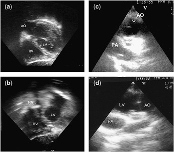
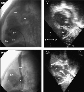
Her recent echocardiogram confirmed the presence of mirror-imaged atrial arrangement and a left-sided heart, but revealed that the atrioventricular connections were discordant. Her morphologically left ventricle was seen to be located leftward, superiorly and anteriorly, with the morphologically right ventricle positioned rightwards and inferiorly, producing the arrangement known as superior-inferior ventricles (Figs. 1 and 2). The aorta was seen to be arising from the anteriorly located morphologically left ventricle, and the pulmonary trunk from the morphologically right ventricle. In other words, the ventriculo-arterial connections were concordant, albeit that the arterial trunks, although arising from their morphologically appropriate ventricles, coursed in parallel fashion as they ascended from the ventricular mass, being abnormally related to each other, with the aorta being in an anterior and left-sided position. The patient also had bilateral infundibulums, a ventricular septal defect, and severe infundibular pulmonary stenosis. The abdominal aorta was right-sided and mildly anterior relative to the left-sided inferior caval vein. Thus, her correct diagnosis was a left-sided heart with mirror- imaged atrial arrangement, discordant atrioventricular and concordant ventriculoarterial connections, parallel arterial trunks with anterior and left-sided aorta, supero-inferior ventricles with superior left ventricle, ventricular septal defect, and pulmonary stenosis. Cardiac catheterization confirmed the diagnosis (Fig. 2)
(a) Subcostal long axis view showing concordant ventriculo-arterial connections with the aorta (AO) arising from the morphologically left ventricle (LV). (b) Modification of the same view shows the pulmonary trunk (PA) arising from the morphologically right ventricle (RV). Note the hypoplastic pulmonary trunk, but the relatively well developed left and right pulmonary arteries. (c) Parasternal short axis view demonstrating anterior and leftward localization of the aorta. (d) Modified short axis view showing anteriorly positioned left ventricle and the aorta arising from it.

(a) Left ventricular angiography demonstrates anterior and superior positioned morphologically left ventricle (LV). The aorta is seen arising from the left ventricle in an anterior position. The pulmonary trunk is posteriorly located. Note the parallel arrangement of the great arteries, and the inferior position of the right ventricle (RV). (b) The subcostal short axis echocardiographic view shows the anterior and superiorly positioned left ventricle and the inferiorly positioned right ventricle. The ventricular septum (ivs) is horizontal, and the ventricles are in superior-inferior position. Angiographic (c) and echocardiographic (d) images show the discordant atrioventricular connections. The superior (svc) and inferior caval vein (ivc) drain to the morphologically right atrium (ra), which is connected to the morphologically left ventricle. Note the similarities between the echocardiographic and angiocardiographic images. Additional abbreviation: MV: mitral valve.

Discussion
Hearts in which the great arteries arise from their appropriate ventricles, but in abnormal relationship to each other, with the aorta positioned anteriorly relative to the pulmonary artery, are extremely rare. Initially, Harris and FarberReference Harris and Farber2 suggested the term “anatomically corrected transposition” to describe this arrangement since, at that time, an anterior position of the aorta was the accepted diagnostic criterion for “transposition”, irrespective of the ventricular origin of the great arteries. Subsequently, Van PraaghReference Van Praagh3 proposed that the lesion should be called “anatomically corrected malposition of the great arteries”.
At that time, bilateral infundibulums were considered to be an essential part of the diagnosis. Since then, comparable cases with absence of one or other infundibulum, or both infundibulums, have been reported.Reference Bernasconi, Cavalle-Garrido, Perrin and Anderson1, Reference Loya, Desai and Sharma4, Reference Cavalle-Garrido, Bernasconi, Perrin and Anderson5 These cases lacking bilateral infundibulums, have previously been described with arcane names, such as “isolated ventricular inversion” or “isolated ventricular noninversion”. Bernasconi and her colleaguesReference Bernasconi, Cavalle-Garrido, Perrin and Anderson1 have now pointed out that the constant feature of all these hearts is the combination of concordant ventriculo-arterial connections and a parallel arrangement of the arterial trunks as they exit from the ventricular mass. It is the infundibular morphology, along with the atrioventricular connections, that differ in these malformations described with the various names. As they emphasized,Reference Bernasconi, Cavalle-Garrido, Perrin and Anderson1 it is inappropriate to describe one variable feature on the basis of another morphological feature that is itself has no constancy. Thus, they proposed that the parallel nature of the concordantly connected arterial trunks as they exit from the ventricular mass should serve as the basis of diagnosis, as opposed to infundibular morphology, albeit that the best descriptors for these entities have yet to be determined.
The arrangement of parallel arterial trunks with concordant ventriculo-arterial connections is seen most frequently in patients with usual atrial arrangement and concordant atrioventricular connections.Reference Bernasconi, Cavalle-Garrido, Perrin and Anderson1 In this setting, the aorta, in the majority of cases, arises in a left-sided and anterior position, as it does in our patient. The abnormality has rarely been described in the setting of mirror-imaged atrial arrangement. We are aware of only two previous cases with concordantly connected but parallel arterial trunks in mirror-imaged atrial arrangement.Reference Anderson, Becker, Losekoot and Gerlis6, Reference Anderson, Arnold and Jones7 Only one of these, as with our patient, had discordant atrioventricular connections.Reference Anderson, Becker, Losekoot and Gerlis6 This previously reported patient also had a ventricular septal defect, but the outflow tracts were unobstructed. Our patient also had supero-inferior ventricles, but with the morphologically left ventricle uppermost. As far as we are aware, all but oneReference Porras, Kratz, Loukas, Van Doesburg, Davignon and Van Praagh8 of previously known cases with supero-inferior ventricles has had the morphologically right ventricle superior to the left ventricle.
The clinical manifestations of patients with this unusual relationship of the arterial trunks depend primarily on the atrioventricular connections and the associated cardiac lesions. In the presence of concordant atrioventricular connections, of course, the circulations potentially will be haemodynamically normal. On the other hand, in patients with discordant atrioventricular connections, as our patient, the systemic and pulmonary circulations are in parallel, and physiologically uncorrected, giving the picture as seen in patients with transposition, or concordant atrioventricular and discordant ventriculo-arterial connections. It was almost certainly because of the parallel nature of the circulations, and the anterior location of the aorta, that our patient was initially misdiagnosed as having “regular” transposition.
Many paediatric cardiologists are unaware of this malformation. Part of the perceived rareness of the lesion, nonetheless, may be due to lack of awareness of its existence during life, since the great majority of the reported cases were diagnosed as pathological curiosities.Reference Bernasconi, Cavalle-Garrido, Perrin and Anderson1, Reference Anderson, Becker, Losekoot and Gerlis6 As shown by us, and others,Reference Shani, Balkin, Zion, Rosenmann and Glaser9 echocardiographic interrogation following the rules of sequential segmental analysis, and giving attention to the ventricular origin and the relationships of the of the great arteries, permits accurate diagnosis of this rare and complex malformation.