Introduction
Over the last 15 years, there has been widespread application of image guidance surgery in rhinology and anterior skull base operations. Image guidance surgery systems have been likened to global positioning systems, allowing intra-operative localisation of anatomical sites based on pre-operatively obtained computed tomography (CT) scans.Reference Roth, Lanza, Zinreich, Yousem, Scanlan and Kennedy1–Reference Metson, Gliklich and Cosenza3 Image guidance surgery depends on external reference frames, digitally stored CT images, patient–computer interface systems and a calibration registration scheme to locate and display the position of a surgical instrument.Reference Metson, Cosenza, Gliklich and Montgomery4, Reference Metson, Cosenza, Cunningham and Randolph5
Image guidance surgery was first introduced in the field of neurosurgery and was used to alleviate pain and reduce tremor by enabling the accurate ablation of specific parts of the cerebrum. Image guidance surgery has progressed slowly in the field of ENT in the UK. This is perhaps because of initial software problems and a hardware that required regular updating. These initial challenges have successfully been overcome and today the available technology is robust and reliable. In their review article on the workings of image guidance surgery, Labadie et al. confirmed our findings that sub-millimetric accuracy makes image guidance surgery highly reliable.Reference Labadie, Davis and Fitzpatrick6
In the USA, image guidance surgery is used in approximately 45 per cent of an estimated 400 000 endoscopic sinus surgery procedures performed per year.Reference Hepworth, Bucknor, Patel and Vaughan7 Due to this high usage, the American Academy of Otolaryngology–Head and Neck Surgery (AAO–HNS) has published guidelines on the use of image guidance surgery in rhinology. Approved indications include: revision endoscopic sinus surgery; extensive sinonasal polyposis; distorted anatomy; excision of benign and malignant sinonasal tumours; cerebrospinal fluid (CSF) rhinorrhoea; and surgery involving the frontal, posterior ethmoid and sphenoid sinuses.
Although image guidance surgery has become an integral part of advanced rhinology and anterior skull base surgery, it does not replace sound anatomical knowledge of these complex regions.
Glasgow Royal Infirmary (north Glasgow) was fortunate enough to acquire the expensive hardware system required for image guidance surgery early on, and as a result we have carried out possibly the largest and longest series of image-guided surgical procedures in rhinology and anterior skull base surgery in the UK.
Aims
This study aimed to: examine and categorise the application of image guidance surgery within our rhinology and anterior skull base practice; compare image guidance surgery cases with non-image guidance surgery cases (performed by the senior author (G W McG)); and audit our practice against the AAO–HNS guidelines on image guidance surgery use in rhinology.
Materials and methods
This study comprised all patients who underwent image guidance surgery and non-image guidance surgery between October 2005 and September 2011. Data were prospectively collected using our image guidance surgery BrainLab VectorVision® computer database. All case records and operation notes were reviewed in terms of patient profile, diagnosis, nature of surgery, pathology and complications.
BrainLab is a passive marking system that uses optical tracking technology for anatomical localisation. The hardware is an integrated system consisting of a monitor and an infrared camera array on an adjustable arm. BrainLab enables the wireless tracking of surgical instruments, which gives the operator control via a computer interface and touch screen monitor.
High resolution CT imaging of the nose and paranasal sinuses is undertaken pre-operatively. This is followed by the three-dimensional reconstruction of images in the axial, coronal and sagittal planes. Reconstructed images are stored on an optical disc and transferred to the computer in the operating theatre. Patient registration is performed intra-operatively using a wireless handheld laser device that projects numerous virtual points onto the patient's surface anatomy. These virtual markers are detected by the navigation system and a precise topographic registration is calculated. Accurate registration is confirmed by performing an anatomical check on known structures (fiducials) prior to commencing surgery.
Results
The total number of procedures undertaken during the study period was 308. There were 174 image guidance surgery cases (56 per cent), comprising 106 males and 68 females, with an age range of 16 to 83 years (mean age was 55.4 years). There were 134 non-image guidance surgery cases (44 per cent), comprising 75 males and 59 females, with an age range of 19 to 81 years (mean age was 53.4 years). There was no significant difference between the image guidance surgery and non-image guidance surgery groups in terms of age and sex (independent sample t-test, p > 0.05).
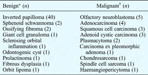
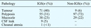
The image guidance surgery procedures consisted of 71 operations for neoplastic disorders (40 per cent) and 103 for non-neoplastic disorders (60 per cent). Details of all neoplastic cases operated on using image guidance surgery are shown in Table I. The non-image guidance surgery group included 11 neoplastic disorders (8 per cent) and 123 non-neoplastic disorders (92 per cent). Table II shows the different clinical diagnoses for the image guidance surgery and non-image guidance surgery groups.
Neoplastic cases managed with image-guided surgery

*A total of 50 benign cases. †A total of 21 malignant cases.
Clinical diagnosis for both groups*

* For image guidance surgery and non-image guidance surgery groups. IGS = image guidance surgery
Image guidance surgery usage
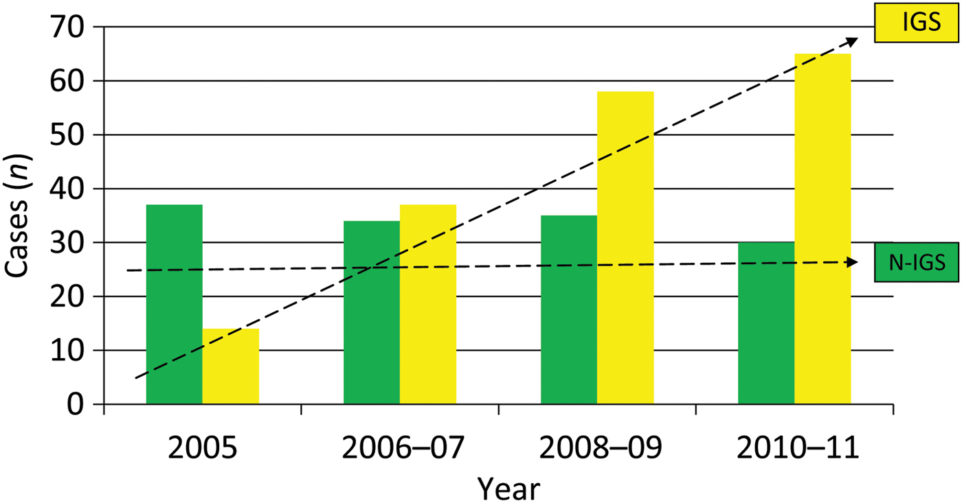
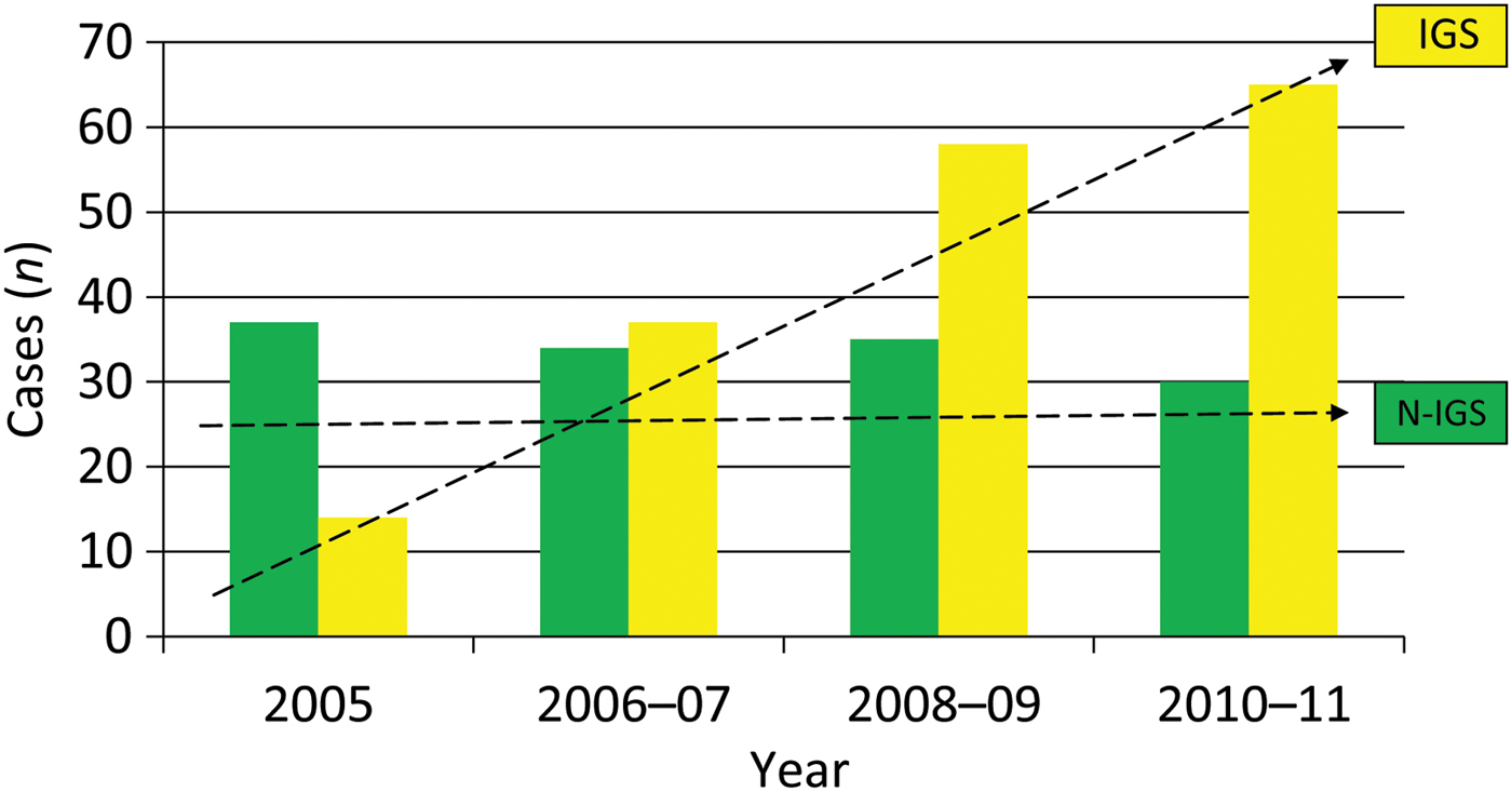
A review of the use of image guidance surgery between 2005 and 2011 revealed a steady year on year increase in its application. Figure 1 demonstrates the number of cases in the image guidance surgery and non-image guidance surgery groups over a six-year period. Although there was an increase in image guidance surgery use, it is interesting to note that the number of non-image guidance surgery procedures were similar each year.
Image guidance surgery and non-image guidance surgery cases undertaken between 2005 and 2011 (arrows indicate the trends over time). IGS = image guidance surgery; N-IGS = non-image guidance surgery
Categorisation of surgery
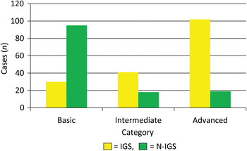
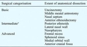
To enable a detailed analysis, we categorised endoscopic endonasal procedures as basic, intermediate or advanced based on the extent of anatomical dissection (as detailed in Table III). Figure 2 demonstrates that image guidance surgery was used more in intermediate (n = 41) than in basic cases (n = 30), and was used more in advanced (n = 103) than in intermediate cases. However, in the non-image guidance surgery group, the majority of operations were in the basic category (n = 95), with smaller numbers in the intermediate (n = 20) and advanced categories (n = 19).
Categorisation of image guidance surgery and non-image guidance surgery procedures. IGS = image guidance surgery; N-IGS = non-image guidance surgery

Categorisation of endoscopic endonasal procedures

*Intermediate surgical categorisation refers to the basic dissections plus those listed as intermediate.
Audit of image guidance surgery use
An audit of our image guidance surgery use against the AAO–HNS guidelines revealed 94 per cent compliance in the management of benign and malignant sinonasal tumours. (The remaining 6 per cent of tumour operations were undertaken using non-image guidance surgery for examination under anaesthetic, or to obtain tissue samples or for tumour debulking.) There was 89 per cent compliance for surgery involving the frontal, posterior ethmoid or sphenoid sinuses (11 per cent of the operations performed in this region were for benign diseases; non-image guidance surgery was used in these cases to establish physiological drainage pathways). For revision endoscopic sinus surgery, 87 per cent compliance was noted. (The 13 per cent of non-image guidance revision functional endoscopic sinus surgical procedures were basic procedures involving nasal polypectomy and re-establishment of middle meatal antrostomies.)
Cost implications and setting up
There are a number of systems available in the market and initial investment continues to be a major stumbling block in acquiring image guidance surgery equipment. Depending on system specifications, the cost of image guidance surgery equipment ranges from £40 000 to £100 000. In addition, there is an initial learning curve associated with setting up an image guidance surgery system. This issue, along with cost analysis per patient, has been discussed previously by Metson et al. Reference Metson, Cosenza, Gliklich and Montgomery4 In our experience, the use of image guidance surgery adds an extra 5 to 10 minutes to the overall operating time.
Complications
Complications of surgery were defined as major (e.g. death, catastrophic bleeding, blindness, CSF leak) or minor (e.g. bleeding, infection, headache). There were no major complications in either group. All complications recorded were in the minor category. In the image guidance surgery group, five complications (3 per cent) were reported: bleeding, infection, lip numbness, post-operative headache and temporary diplopia. The former two occurred in patients whose procedures were in the intermediate category and the latter three were reported by patients who underwent advanced surgery. In the non-image guidance surgery group, one patient (less than 1 per cent), whose procedure was in the intermediate category, suffered post-operative bleeding.
Discussion
Image guidance surgery is an expanding and evolving technology which has demonstrated steady growth since the mid-1990s. Prior to 2005, the north Glasgow institute was using an earlier version of image guidance technology. Only in 2005 did we feel that our technology was sufficiently stable to allow us to evaluate its clinical application.
Early published literature, which focused on the technical aspects of image guidance surgery, was soon followed by reports confirming its intra-operative accuracy. Most of the current literature published on image guidance surgery is based on data series from the USA.Reference Metson8–Reference Citardi and Batra11 The AAO–HNS, in their official statement, summarise that there is sufficient evidence to support the use of image guidance surgery in clarifying complex anatomy during endoscopic sinus and skull base surgery, and also state that image guidance surgery is not an experimental or investigational tool. In our series, the largest single group of image guidance surgery procedures were for tumour resection, followed by sinonasal polyposis. Although the majority of polyp surgery in our unit has not been image-guided, the proportion of cases for which image guidance surgery has been utilised is significant. These are represented by our revision endoscopic sinus operations and cases with distorted anatomy.
The introduction of any new technology tends to generate an initial enthusiasm which often fails to be sustained. This was not the case in our practice, as seen in Figure 1. We found image guidance surgery particularly useful for patients requiring advanced endoscopic sinus surgery and tumour surgery.
• Image guidance surgery is an evolving and expanding technology in rhinology and anterior skull base surgery
• This paper reports the largest series of image-guided ENT surgical procedures in the UK
• Image guidance surgery should be the standard practice for advanced endoscopic sinus surgery and tumour surgery
• We argue the need for a policy statement for image guidance surgery use in the UK
Although we demonstrated a high compliance rate with the AAO–HNS guidelines, there is potential for improvement. We would argue the need for a policy statement on the use of image guidance surgery in rhinology and anterior skull base surgery in the UK.
The complication rate was higher in the image guidance surgery group than the non-image guidance surgery group. However, all complications occurred in patients undergoing tumour resection categorised as an intermediate or advanced surgical procedure. As 94 per cent of tumour operations were performed using image guidance surgery, the higher complication rate may be due to a confounding variable associated with case mix. In order to definitively address the safety profile and efficacy of image guidance surgery, a randomised study would be required wherein patients with similar pathology were randomly assigned into image guidance surgery and non-image guidance surgery groups, and their outcomes monitored. Such a study is unlikely to be possible and may be ethically unfeasible; it may therefore prove impossible to acquire level I evidence on image guidance surgery use.