Introduction
Although the centre of diversity of Hypogymnia in Asia appears to be in south-west China (McCune & Wang Reference McCune and Wang2014), a number of endemic species have been found in central China (Wei & Wei Reference Wei and Wei2005, Reference Wei and Wei2012) and Far East Russia (Tchabanenko & McCune Reference Tchabanenko and McCune2001; McCune & Tchabanenko Reference McCune and Tchabanenko2002). The present paper continues that exploration by describing a new, relatively rare, esorediate species from Far East Russia and central China.
Materials and Methods
We applied standard microscopy and chemical spot test methods. Many specimens were subjected to thin-layer chromatography (TLC), using the standard methods of Culberson (Reference Culberson1972) and reference tables of Chicita Culberson (1996, unpublished). Fragments of specimens were extracted in acetone at room temperature, spotted on aluminum-backed silica gel plates (Merck 5554/7 Silica gel 60 F254), run in solvent systems A and C of Culberson (Reference Culberson1972), lightly brushed or sprayed with 10% H2SO4, and charred in an oven at 100°C. No attempt was made to distinguish chloroatranorin from atranorin; nor was water applied to detect fatty acids.
Apothecial anatomy and measurements were studied with bright-field microscopy on hand-cut sections mounted in water with no staining. Ascospore and spermatia sizes were measured in sets of nine and expressed as the median and range. Spores were photographed, cut and pasted onto a single canvas, then measured with the line tool in PhotoShop with View|Info displayed. These raw values were converted to micrometres using a conversion factor calibrated from a stage micrometer.
The unusual morphology of lobes of the genus Hypogymnia has necessitated some terminology seldom used in other lichens. See McCune & Wang (Reference McCune and Wang2014) for further explanation of terminology for lobe cavities and perforations, and for further details on the methods.
Hypogymnia papilliformis McCune, Tchabanenko & X. L.Wei sp. nov.
MycoBank No.: MB 809097
Similar to H. delavayi but differing in the more often isotomic dichotomous branching, upper surface becoming strongly verrucose or papillose, and absence of 3-hydroxyphysodic acid.
Type: Russia, Primorsky Krai, Uglovaya Pad, Lazovsky Reserve, 43°0′N, 134°4′E, 600 m, mixed conifer–broadleaved forest, on Pinus koraiensis, 12 November 1981, S. Tchabanenko 13 (OSC —holotype; HMAS-L— isotype).
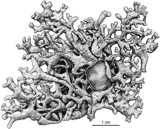
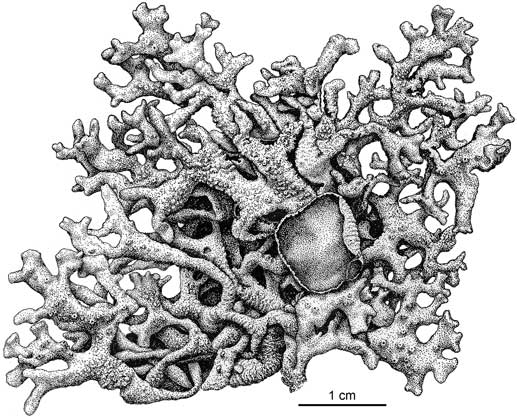
Hypogymnia papilliformis, habit (part of holotype). Drawing by Alexander Mikulin.
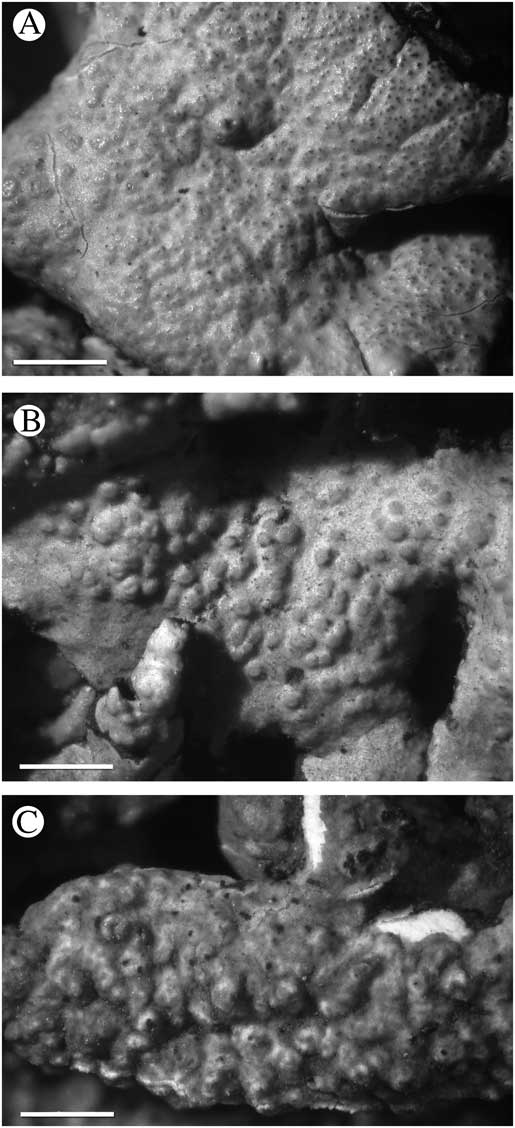
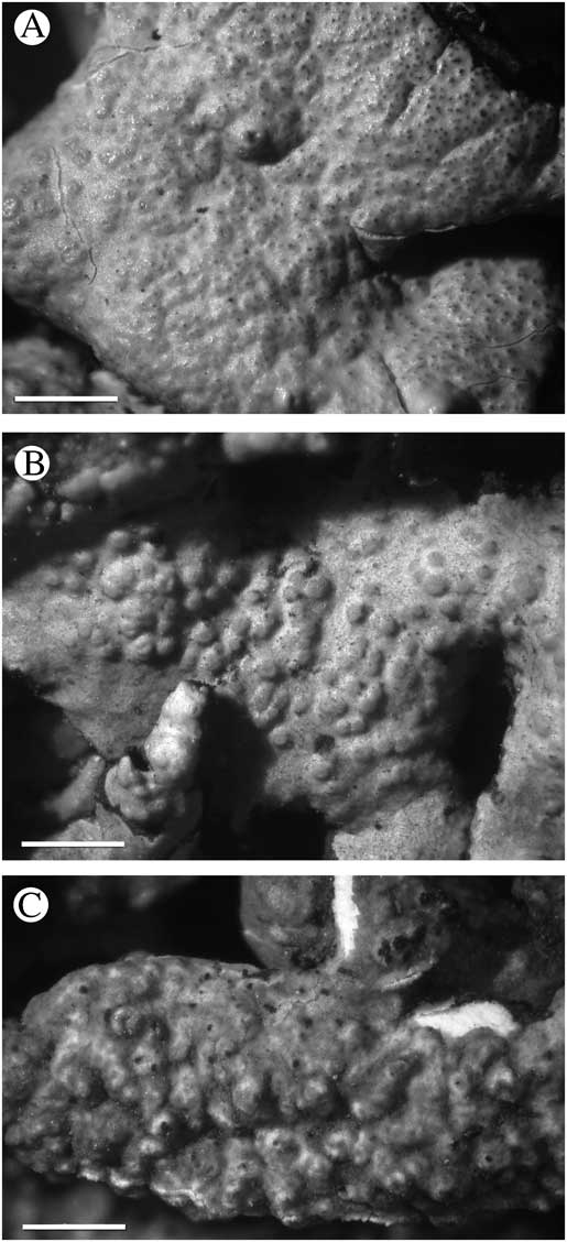
Hypogymnia papilliformis, variation in upper surface. A, initial stages of development of papillae (Skirina 22711); B, well-developed papillae (Skirina 22711); C, mixed papillae and rugosity (Xu & Yang 1511). Scales: A–C = 1 mm.
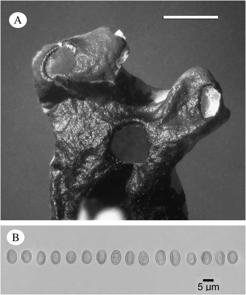
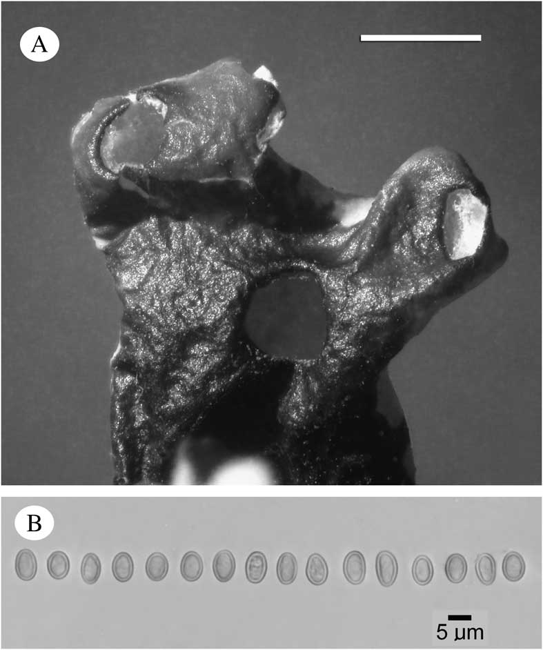
Hypogymnia papilliformis details. A, perforations in lobes viewed from below (Skirina 22711); B, ascospores (holotype). Scale: A = 1 mm.
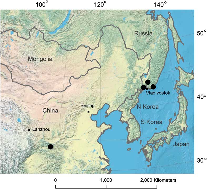
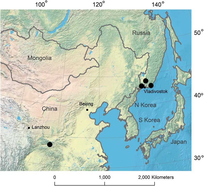
Known world distribution of Hypogymnia papilliformis (solid dots). In colour online.
Thallus appressed to suberect, to 8 cm diam.; texture cartilaginous; branching variable, isotomic dichotomous or sometimes with adventitious side branches; lobes separate to centrally subcontiguous, commonly 1·5–4·0(–5·0) mm broad, short or elongate, hollow, even to somewhat nodulose in outline, lateral budding occasional; upper surface becoming distinctly rugose to verrucose or papillose, epruinose, pale greenish grey, dark mottling absent or very sparse, black border not visible; lower surface near lobe tips brown to dark brown, less often black, matt or semi-glossy; lobe tips and axils usually perforate, the holes lacking a differentiated rim; ceiling of lobe cavity off-white to dark (check 1 cm back from lobe tip); floor of lobe cavity dark brown; soredia, isidia, and lobules lacking.
Apothecia occasional, sessile to substipitate, the receptacle funnel-shaped; disc brown, to 8 mm diam.; epihymenium pale brown, POL−; hymenium 45–50 μm tall; subhymenium c. 15 μm tall, POL−; hypothecium POL−, 22–32 μm tall; ascospores 6·0–7·0 × 4·1–4·9 μm (median 6·5 × 4·5 μm).
Pycnidia (spermatogonia) common; spermatia rod-shaped to weakly bifusiform, 4·8–6·4 × 0·9–1·1 μm (median 5·6 × 0·9 μm).
Secondary chemistry. Atranorin, physodic acid (major), 2′-O-methylphysodic acid (minor), and vittatolic acid (minor accessory); medulla K−, C−, KC+ orange-red, P−.
Etymology. The epithet ‘papilliformis’ refers to the papillose texture that develops on the upper surface.
Ecology and substratum. Hypogymnia papilliformis is so far known from bark and wood of conifers in mixed conifer-broadleaved forests at elevations of 600–1500 m. The species occurs at temperate latitudes in the mountains of Shaanxi Province, China, and Far East Russia (southern Sikhote-Alin Range, Primorsky Krai).
Specimens examined. China: Shaanxi Province: Ningshan County, Taibai Mountain, 33°26′N, 108°27′E, 1500 m, on bark, 2005, L. Xu & J. Yang 1511(HMAS-L 85008).—Russia: Primorsky Krai: Hasansky District, Kedrovaya pad Reserve, Kedrovaya River, conifer-broadleaved forest, 43°5′41·15"N, 131°33′40·71"E, 600 m, on Pinus koraiensis, 1994, Tchabanenko 184 (SAKH); Anuchinsky District, Tigrovaya River, near village of Tigrovyi, on twigs of Abies nephrolepis, 2008, Skirina 22711 (SAKH); Lazovsky District, Lazovsky Pass, 43°30′7·3"N, 133°35′19·1"E, c. 900 m, 22 ix 2009, Tchabanenko s.n. (SAKH).
Discussion
Hypogymnia papilliformis appears to be a rare species. At this point, we have so few records and know so little about its habitat that it is difficult to describe with any precision the ecological conditions necessary for its survival.
The only known reference to the species in the literature is Tchabanenko (Reference Tchabanenko2000) as H. yunnanensis Y. M. Jiang & J. C. Wei (synonym of H. delavayi; McCune Reference McCune2012), before the distinctions between H. delavayi and the new species were understood. Although Rassadina (Reference Rassadina1971 and other papers) reported H. delavayi from the Russian Far East, the name was applied to various esorediate imperforate species, including what we now know as H. arcuata Tchabanenko & McCune and H. sachalinensis Tchabanenko & McCune.
The species most similar to H. papilliformis is the true H. delavayi from south-west China. Hypogymnia papilliformis is predominantly isotomically branched while H. delavayi usually develops subpinnate branching with numerous side lobes that are±perpendicular to the main lobes. The ceiling of the lobe cavity is mid brown to dark brown in H. papilliformis, while the ceiling is often pale brownish or white in H. delavayi. Also, H. papilliformis lacks 3-hydroxyphysodic acid, and thus has a K– medulla, while H. delavayi always contains 3-hydroxyphysodic acid as a major substance and is thus K+ slowly reddish brown.
Hypogymnia sachalinensis is perhaps the species in the Russian Far East most likely to be confused with H. papilliformis. They are, however, readily distinguished by the narrower (mostly 0·5–1·5 mm broad), relatively smooth lobes of H. sachalinensis, in contrast to the broader (mostly 2–4 mm broad), verrucose or papillose lobes of H. papilliformis. Furthermore, H. sachalinensis always lacks 2′-O-methylphysodic acid, while all specimens of H. papilliformis tested so far contain this substance.
The phylogenetic affinities of H. papilliformis are obscure, although we presume a close relationship with H. delavayi on morphological grounds. Unfortunately, we lack specimens collected recently enough to be useful in DNA sequencing, based on our current poor results with specimens even a few years old. Both species share a tendency to open branching, a verrucose or less commonly rugose upper surface, perforations in the lobe tips and axils, and ascospores broadly overlapping in size and shape. Spermatia are also essentially the same size and shape in the two species. No species similar to H. papilliformis are known from other continents.
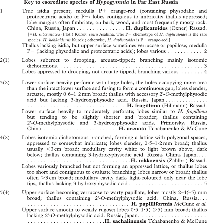
Rassadina (Reference Rassadina1971) included 20 species of Hypogymnia in the Russian Far East, seven of which lack soredia and corticate granules. Most of our species concepts have been refined since then, so we offer below a key to the esorediate species in the Russian Far East. We defer a treatment of the sorediate species because certain problematic species (especially H. pseudophysodes, H. bullata, and H. submundata) require further study.
We thank the curators of CANL, KUN, HMAS, L, OSC, and US who kindly co-operated with loans and visits; Elisa Alphandary for assistance with TLC; Alexander Mikulin for drawing the holotype; and Patricia Muir and anonymous reviewers for checking the manuscript. Financial support was provided by the National Natural Science Foundation of China (31200018).