




No CrossRef data available.
Article contents
- Abstract
- Early steps in cardiac development
- The contribution and influence of cardiac cells derived from the neural crest
- The link between the neural crest and human cardiac malformations
- The contribution and the role of the epicardially derived cells
- Link of the epicardially derived cell to human malformations
- Conclusions
- Footnotes
- References
The quintessence of the making of the heart
Published online by Cambridge University Press: 18 April 2005
Article contents
- Abstract
- Early steps in cardiac development
- The contribution and influence of cardiac cells derived from the neural crest
- The link between the neural crest and human cardiac malformations
- The contribution and the role of the epicardially derived cells
- Link of the epicardially derived cell to human malformations
- Conclusions
- Footnotes
- References
Abstract
In my Mannheimer lecture, designed to meet the needs of a mainly clinical audience, I present aspects of cardiac development that link basic science to clinically relevant problems. During development of the cardiac tube, and its subsequent changes as a dextrally looped structure, which is still connected to the dorsal body wall by a venous and an arterial pole, there are basic requirements. These consist of the development of myocardium, endocardium and the interposed cardiac jelly from the cardiogenic plates. In this primitive heart tube, septation and valvar formation then take place to convert it into a four-chambered heart. I demonstrate that the refining of the above events cannot take place without the addition of extracardiac populations of cells. These are presented as the “quintessence of heart development”, and consist of cells derived from the neural crest, along with epicardially derived cells. Without these contributions, the embryos uniformly die of cardiac insufficiency. Important contributions are made by the cells derived from the neural crest to septation and the formation of the arterial valves, and possibly in differentiation of the central conduction system. The epicardially derived cells are essential for formation of the interstitial fibroblasts and the myocardium, as well as the coronary vascular system. I conclude by discussing specific malformations of the heart that might be linked to these extracardiac contributions.
Keywords
Information
- Type
- Mannheimer Lecture
- Information
- Copyright
- © 2003 Cambridge University Press
Morphogenetic and functional characteristics of cardiac development, combined with the insight into the molecular biological and genetic aspects that drive the formation of the heart, are essential for understanding the aetiology of cardiac malformations in the human population. Study of the pathogenesis of such neonatal cardiac malformations with or without surgical repair, even when extended to include those malformations that can be diagnosed prenatally, has its limitations. This also applies to our understanding of the mechanisms that are disturbed during development. An experimental approach, in which my colleagues and I studied avian and rodent embryos, brought new insights and expanded my horizon in four major areas, specifically:
- Formation and looping of the heart tube.
- Development of the various septal components.
- Differentiation of the atrioventricular and arterial valves.
- The precursors of the embryonic and fetal conduction system.
In recent years, these experimental approaches have been complemented by a fifth dimension, which I have come to regard as the quintessence of cardiac development, hence the title of this paper. Extracardiac derived cells migrate towards, into, and over the primitive heart tube to permit proper formation of the heart. In fact, without these extracardiac contributions, being those derived from the cardiac neural crest along with the cells derived from the pro-epicardial organ, so-called “epicardially derived cells”, the heart does not develop beyond specific stages. Indeed, the embryos uniformly die. In the human, this would be within the first 6–8 weeks of development, in the chicken embryo around the 6th day, at the 32nd stage within the systematisation of Hamburger and Hamilton, and in the mouse around the 15th day of development. Many experimental models, in which essential genes or molecules are disturbed, interfere with these elements derived from extracardiac sources. In this review, based on the Mannheimer Lecture I gave to the Association for European Paediatric Cardiology in 2002, I would like to present a number of examples from the first four dimensions, and add specific data on the relevance of the fifth dimension. The examples provide, where possible, a link between the experimental research and human cardiac malformations.
Early steps in cardiac development
The heart initially develops from the left and right cardiogenic plates that fuse in the midline. The cardiac mesoderm borders on the endoderm of the primitive gut and, at this junction, the first endocardial and endothelial precursors are found.1 From this stage on, the almost straight heart tube is a bilaminar structure, with an outer myocardial and an inner endocardial lining. Extracellular matrix, the cardiac jelly, is found in between (Fig. 1a,b). This tube starts to loop ventrally and to the right, so-called dextral looping. During septation and valvar formation, the outflow tract comes to be positioned in front of the inflow tract concomitant with tightening of the inner curvature and expansion of the right side of the atrioventricular canal, thus permitting formation of the right ventricular inflow (Fig. 2a,b and scanning electron micrograph movie). In this process, the cardiac segments, and the interposed transitional zones, are repositioned by remodelling of the heart tube. The transitional zones are the sinuatrial transition, the atrioventricular canal, the primary fold or primary ring, and the ventriculo-arterial transition (Figs 3a–e, 4a–e and movies). The sinuatrial transition and primary fold are not lined by cushion tissue. They play an important role in the formation of the cardiac conduction system.2 The atrioventricular canal and outflow tract, in contrast, are lined by endocardial cushions that are essential in ventricular septation and valvar formation (see also Ref. 3).

Figure 1. (a) Scanning electron micrograph of ventral view of the heart tube in a chicken embryo at the 10th stage of the classification made by Hamburger and Hamilton. The blood is propelled from the venous pole (V) to the arterial pole (A). (b) Transverse section of the heart tube that is connected to the body wall by the dorsal mesocardium (DM). The myocardium (M) is still thin and separated from the inner endocardial layer (E) by the cardiac jelly (CJ). N: neural tube; DAo: dorsal aortae.

Figure 2. (a) Scanning electron micrograph of a looped heart tube of a chicken embryo at the 17th stage of Hamburger and Hamilton. The dorsal mesocardium has been disrupted in the central part, leaving a connection of the heart tube at the venous pole, and at the arterial pole. In the heart tube there are the first indications of formation of segments: ventricle (V), atrium (A), and transitional zones: atrioventricular canal (AVC) and proximal (POT) and distal (DOT) outflow tract. The inner curvature ([sstarf ]) is remodelled to accommodate transition of the outflow tract over the atrioventricular canal as shown in b, from an embryo at the 22nd stage of Hamburger and Hamilton. An animated view of the process of looping is to be found at: www.medfac.leidenuniv.nl/cask/cadim.htm

Figure 3. Schematic drawings of the ventricle and outflow tract of the heart during looping and septation. Indicated are the transitional zones, specifically the atrioventricular canal (AVC), the primary fold (PF), and the ventriculo-arterial transition (VA). During the process of looping, (b) the arterial segment, or “truncus arteriosus” (TA), will move over the inner curvature to the left (see b, c, and e). The atrioventricular canal becomes partly positioned above the newly formed right ventricular inflow tract (RVI). The primary fold (PF, yellow) forms the embryonic basis of the conduction system, that will differentiate into the definitive atrioventricular conduction system (red in e). Septation of the arterial segment (b, d, e) separates the pulmonary trunk (Pu), connected to the right ventricle (RV), through a long outflow tract from the aorta (Ao) that arises by a short outflow tract from the left ventricle (LV). An animated sequence is available on the website of the Journal at www.greenwich-medical.co.uk AVN: atrioventricular node; MB: moderator band; RAVR: right atrioventricular ring bundle; TO: tricuspid orifice.

Figure 4. Schematic drawings of the sinus venosus (SV) and atrium. The transitional zones are the sino-atrial transition (SA) and the atrioventricular canal (AVC). The sinuatrial transition forms boundaries in both left and right atriums. This transition also forms the basis of the embryonic conduction system, which will differentiate eventually into the sinus node (SAN). Other parts of the transition may play a role in pathology of the conduction system, such as abnormal atrial automaticity. AAVN: embryonic anterior atrioventricular node; CV: caval veins; RVV: right venous valve; LVV: left venous valve; fusion of LVV and RVV in the septum spurium (SP); L: limbus of the oval fossa ovalis; PV: pulmonary veins; CS: coronary sinus; SS: septum secundum; SCV: superior caval vein; ICV: inferior caval vein. An animated sequence is available at: www.medfac.leidenuniv.nl/cask/cadim.htm
Studies from molecular biology have not yet revealed that the genetic make-up of the developing heart yields genes that are specific for given cardiac chambers. Although there are seemingly atrial and ventricular specific genes,4 as well as genes that favour either the left or the right side of the heart, many variations are seen that show only temporary selectivity, or include combinations such as right atrial with left ventricular expression.5 It seems that current data point towards a primitive myocardial heart tube, with subsequent temporo-spatial specifications for the different chambers.4 Recent studies made by our group have shown that the primary myocardial tube forms the wall of the major part of the right atrium, part of the anterior and posterior walls of the left atrium, and various parts of the definitive left and right ventricles. The inflow tract of the right ventricle, and the distal part of the outflow tract, are structures that develop relatively late. After remodelling and differential growth of the compartments, with differentiation of the contributing cellular components, the resultant is a heart with specific morphologic characteristics for each of the four chambers and connecting vessels (Fig. 5a–d). The achievement of the specific characteristics is guided in no small way by the cells derived from extracardiac sources, specifically those from the neural crest and the epicardial organ (Fig. 6).

Figure 5. Overview of the 4 chambers of the heart. (a) View into the right ventricle (RV) shows the characteristic morphological aspects such as a broad muscle band, the supraventricular crest (CS), between the orifices of the tricuspid (TO) and pulmonary valves (Pu). The crest is the proximal margin of the long muscular subpulmonary infundibulum. The continuity of the muscular profiles of the septomarginal trabeculation (TS), the supraventricular crest, and the ventriculo infundibular fold (IF) form the larger part of the boundary between inflow and outflow of the RV. (b) View into the left ventricle (LV) with a smooth septal surface and a fibrous continuity between the leaflets of the mitral (MV) and the aortic valves. The short left ventricular outflow tract ([sstarf ]) forms the connection with the aorta. (c) View into the left atrium (LA) with the attachment points (→) of the valve of the oval foramen clearly visible. The latter partly overlaps, at this stage, with the limbus of the fossa (L) that forms the free rim of the so-called secondary septum in the right atrium. (d) View into the right atrium (RA), into which the systemic veins (SCV: superior caval vein; ICV: inferior caval vein) and coronary sinus (CS) enter. In the upper left hand side the entrance to the right atrial appendage (RAA) is visible. AVN: site of AV node; TO: tricuspid orifice; VFO: valve of the oval foramen.

Figure 6. Schematic picture of neural crest cells derived from the dorsal margin of the neural tube that migrate towards the heart. These cells enter both at the outflow tract (→) as well as at the venous pole (). Also the cells of the epicardial organ, epicardially derived cells, and endothelial cells of the liver migrate towards the heart, specifically spreading from the venous pole ().
The contribution and influence of cardiac cells derived from the neural crest
The cells that enter the heart migrate from the cardiac neural crest, located in the neural plate between the otic placode and the 4th somite, along the pharyngeal arches towards the outflow tract, as well as entering through the inflow tract of the heart. This has been proven by various experimental approaches, the first being the studies of quail-chick chimera made by Kirby,6 followed by the studies using the retroviral transmitted lacZ reporter gene.7
The most recent approach has been provided by transgenic neural crest cell reporter mice.8, 9 We have studied the Wnt1 reporter mouse,10 and were able to find, as in the chicken embryo,11, 12 that there is a contribution of cells from the neural crest to the outflow as well as the inflow tracts (Fig. 7a,b). The latter observation was not described in the initial report on this mouse.10 Both animal models show the role of the cells derived from the neural crest in the formation of the smooth muscle cells of the arteries passing through the pharyngeal arches, as well as the pulmonary trunk. The transgenic mouse, in contrast to the avian studies, also shows the elaborate contribution to the leaflets of the arterial valves. The condensed mesenchyme of the aorto-pulmonary septum, which extends with two prongs into the endocardial ridges of the outflow tract, is also composed of cells derived from the neural crest. In the chicken embryo, these cells are removed by the process of apoptosis. This process correlates closely in time and space with myocardialisation of the embryonic proximal septum of the outflow tract.12 In the mouse embryo, a comparable apoptotic process is seen in this area, but has not been specifically traced to the cells derived from the neural crest. A still unresolved point concerns the contribution of the cells derived from the neural crest to the smooth muscle cells of the proximal coronary arteries. In the chicken, this area is clearly not derived from the neural crest,7 while in the mouse the association remains questionable. The parasympathetic ganglions, and coronary arterial innervation, are clearly derived from the neural crest. Both in chicken11 and mouse (unpublished observations), we have seen cells from the neutral crest becoming associated with the main areas of the conduction system, namely the sinus node, the atrioventricular node, and the penetrating atrioventricular bundle (Figs 3e, 4e). Scattered cells derived from the neural crest were found in the areas occupied by the primitive conduction system, which include the internodal atrial pathways, and the right atrioventricular ring bundle. It has to be stressed, however, that the cells in these areas derived from the neural crest did not differentiate into insulated tracts of conducting cells, nor into Purkinje fibers.

Figure 7. (a) Section of a Wnt1 lacZ reporter mouse at the 15.5 embryonic day, with neural crest cells (blue) in the arterial valves of the aorta (Ao), in the centre of the muscular outflow tract, and surrounding the coronary arteries (CA). (b) Section through the inflow tract, with neural crest cells (→) surrounding the pulmonary veins (PV). CS: coronary sinus; LA: left atrium; LVOT: left ventricular outflow tract; RA: right atrium; Oes: oesophagus; RVOT: right ventricular outflow tract.
The link between the neural crest and human cardiac malformations
The above findings suggest an obvious link between the neural crest and cardiac anomalies involving the aortic arch and the outflow tract. Research on the role of the cells derived from the neural crest in controlling coronary vascular development and tone needs to be expanded. The mouse model clearly supports a link between the neural crest and malformations of the aortic arches along with the bifoliate aortic valve. It is very interesting, in this respect, that studies in mice do not allow differentiation between interruption of the aortic arch at the isthmus as opposed to interruption between the left common carotid and subclavian arteries, as the isthmus is lacking in the mouse as well as in the chicken embryo. This implies that we do not know whether, in aortic coarctation at the site of the isthmus in conjunction with patency of the arterial duct, there is involvement of cells derived from the neural crest. van Mierop13 has already postulated a role for such cells in differentiating between interruption and hypoplasia at these two component parts of the aortic arch. The above argumentation is important for the questions on the role of cells derived from the neural crest in the development of the 22q11 deletion syndrome. Many genes in the deleted region of chromosome 22q11 have been linked causally to the various clinical characteristics of the syndrome. This, indeed, includes many regions that are invaded by cells from the neural crest, such as the facial area, the thymus, the majority of the aortic arch, including the segment between the left common carotid and subclavian arteries, and the cardiac outflow tract. Currently, Tbx1 is the gene that attracts most interest, although it is not expressed in the neural crest.14 Our own studies with retroviral infection of the cells derived from the neural crest with an anti-sense gene known as DGCR6, related to the critical region for Di George syndrome,6 have produced chicks with interruption of the fourth aortic arch and a subarterial ventricular septal defect, lesions typical for the syndrome (unpublished data B. Hierck).
The possible role of cells derived from the neural crest in formation, and thus disturbances, of the definitive conduction systems needs to be established. It is fascinating that all major components of the central conduction system have cells derived from the neural crest adjoining them during a certain embryonic period. The large contribution to the site of pulmonary venous entrance might influence this area, which is well-known as a site of abnormal atrial automaticity. We have shown with immuno-histochemical staining procedures in human embryos,15 as well as recently with a mouse having a reporter gene for the cardiac conduction system,16 that the myocardium surrounding the pulmonary veins has similar characteristics to the embryonic conduction system as seen in other areas.15
The contribution and the role of the epicardially derived cells
Initially, the myocardium of the heart tube is not covered by an outer layer of epicardium. This epicardium derives from the pro-epicardial organ,17, 18 and spreads over the myocardium to provide, through a process of epithelial-mesenchymal transformation, a population of epicardially derived cells that migrate into the various compartments of the heart. These are, in sequence of development, subendocardial, intramyocardial and subendocardial cushion populations,19 followed by smooth muscle cells and adventitial fibroblasts for the coronary vasculature.20 We have not been able to show a coronary endothelial contribution.20, 21 Studies involving ablation of the pro-epicardial organ show that these epicardially-derived cells are essential for the differentiation of the compact myocardium (Fig. 8a,b). In this model, the compact myocardium is thin and lacks organisation. Our hypothesis is that the epicardially-derived cells provide essential signals, either in cell-to-cell contacts, or through interactions with the cellular matrix, that are important for the maturation of the myocardium. Absence of the epicardially-derived cells also leads to disturbances of looping, lack of ventricular septation, and abnormal formation of the endocardial cushions. In case of complete absence of the epicardially-derived cells, the embryo dies with failure of formation of the coronary vasculature. When the outgrowth of epicardium from the venous pole does not take place, or is inhibited, we have observed a collar of cytokeratin-positive mesothelium extending from the arterial pole over the myocardial outflow tract, with a sharp line of demarcation.22 It still has to be resolved whether these mesothelial cells in the outflow tract, which are continuous with the pericardium, grow out actively over the outflow tract, or whether the relative late formation of the outflow tract by the secondary heart field produces this phenomenon. Rescue experiments, in which we inserted a quail pro-epicardial organ in a chicken embryo subsequent to ablation of the host epicardial organ, showed that quail cells eventually again reach the myocardial–arterial border, and thus normalise the situation. In the inhibition studies, we could also show that formation of the mesothelial collar on the outflow tract is accompanied by migration of a population of endothelial cells derived from the splanchnic plexus.22 Normally, such endothelial cells reach the heart from the venous pole.

Figure 8. (a) Section of the heart of a normal chicken embryo at the 29th stage of Hamburger and Hamilton showing the architecture of the ventricular compact myocardium (M) and the trabecular layer, as well as the overlying epicardium (EP). (b) Section through the ventricular region of an embryo at the same stage in which the outgrowth of the epicardial organ has been inhibited. Septation and valvar formation are deficient, and the compact myocardium is markedly thinned, lacking its covering of epicardial cells. END: endocardium, PERI: pericardium. Staining: anti α smooth muscle actin.
Link of the epicardially derived cell to human malformations
The widespread distribution of epicardially derived cells, and the serious impairment of cardiac differentiation when they are absent, offer an important modulating role for these extracardiac cells. Such a role guiding myocardial differentiation might shed a novel light on the origin of certain cardiomyopathies where the primary culprit might be the disturbed interaction between myocytes and fibroblasts. The focus of most studies in this field is still solely on the pathomorphogenesis of the myocardial cell. As epicardially derived cells are also present in the atrioventricular cushions, an influence on valvar differentiation might be expected. The subendocardial position points towards a role in regulating the endocardial–mesenchymal tranformation that, if not properly timed, might result in valvar dysplasia. The epicardially-derived cells have also been postulated by us to play a role in differentiation of the Purkinje fibers.19 The origin of coronary endothelial cells from the arterial pole might explain the abnormal origin of a coronary artery from a pulmonary vessel, or from a subclavian artery. It is well established that, in case of delayed outgrowth of the epicardium in a rescue experiment, in which, through quail epicardial organ grafting, the embryo survives, it is frequent to see abnormalities of the coronary vasculatue, such as single or absent coronary arteries combined with the formation of ventricular–coronary arterial communications.22 These findings might provide an explanation for human cardiac anomalies. In case of pulmonary atresia with intact ventricular septum, we can distinguish cases with and without ventriculo-coronary arterial communications.23 Studies in prenatal life have shown that development of the ventriculo-coronary arterial communications can precede the pulmonary atresia.24 On the basis of our developmental studies, we have speculated on the presence of a primary anomaly in these cases, possibly preceded by epicardial dysfunction.25
Conclusions
In my Mannheimer lecture, reproduced in this review, I hope to have intrigued both basic scientists, paediatric cardiologists, and other workers in the field for the continuous search of developmental mechanisms behind congenital cardiac malformations. The multidisciplinary approach, in which not only genetics but the broad field of cell biology, experimental models and the absolute necessity of combining the findings in the above areas with the clinical spectrum, will enable small steps forward in the immense realm of choices and influences that work upon the developing heart.
Footnotes
The article is the text of the Mannheimer lecture, given on the occasion of the 37th Meeting of the Association of European Pediatric Cardiology, Porto, Portugal, 2002.
References
Drake CJ, Hungerford JE, Little CD. Morphogenesis of the first blood vessels. Ann NY Acad Sci 1998; 857: 155–179.Google Scholar
Blom NA, Gittenberger-de Groot AC, DeRuiter MC, Poelmann RE, Mentink MM, Ottenkamp J. Development of the cardiac conduction tissue in human embryos using HNK-1 antigen expression: possible relevance for understanding of abnormal atrial automaticity. Circulation 1999; 99: 800–806.Google Scholar
Gittenberger-de Groot AC, DeRuiter MC, Bartelings MM, Poelmann RE. Embryology of congenital heart disease. In: Crawford MH, DiMarco JP, (eds). Cardiology, First. Mosby International Limited, London, 2001; pp 2.1–2.10.
Christoffels VM, Habets PEMH, Franco D, et al. Chamber formation and morphogenesis in the developing mammalian heart. Dev Biol 2000; 223: 266–278.Google Scholar
Franco D, Kelly R, Lamers WH, Buckingham M, Moorman AFM. Regionalized transcriptional domains of myosin light chain 3f transgenes in the embryonic mouse heart: morphogenetic implications. Dev Biol 1997; 188: 17–33.Google Scholar
Kirby ML, Gale TF, Stewart DE. Neural crest cells contribute to normal aorticopulmonary septation. Science 1983; 220: 1059–1061.Google Scholar
Bergwerff M, Verberne ME, DeRuiter MC, Poelmann RE, Gittenberger-de Groot AC. Neural crest cell contribution to the developing circulatory system. Implications for vascular morphology? Circ Res 1998; 82: 221–231.Google Scholar
Epstein JA, Li J, Lang D, et al. Migration of cardiac neural crest cells in Splotch embryos. Development 2000; 127: 1869–1878.Google Scholar
Waldo KL, Lo CW, Kirby ML. Connexin 43 expression reflects neural crest patterns during cardiovascular development. Dev Biol 1999; 208: 307–323.Google Scholar
Jiang X, Rowitch DH, Soriano P, McMahon AP, Sucov HM. Fate of the mammalian cardiac neural crest. Development 2000; 127: 1607–1616.Google Scholar
Poelmann RE, Gittenberger-de Groot AC. A subpopulation of apoptosis-prone cardiac neural crest cells targets to the venous pole: multiple functions in heart development? Dev Biol 1999; 207: 271–286.Google Scholar
Poelmann RE, Mikawa T, Gittenberger-de Groot AC. Neural crest cells in outflow tract septation of the embryonic chicken heart: differentiation and apoptosis. Dev Dyn 1998; 212: 373–384.Google Scholar
van Mierop LHS, Kutsche LM. Cardiovascular anomalies in DiGeorge syndrome and importance of neural crest as a possible pathogenetic factor. Am J Cardiol 1986; 58: 133–137.Google Scholar
Lindsay EA, Vitelli F, Su H, et al. Tbx1 haploinsufficieny in the DiGeorge syndrome region causes aortic arch defects in mice. Nature 2001; 410: 97–101.Google Scholar
Blom NR, Gittenberger-de Groot AC, DeRuiter MC, Poelmann RE, Mentink MMT, Ottenkamp J. Development of the cardiac conduction tissue in human embryos using HNK-1 antigen expression. Circulation 1999; 99: 800–806.Google Scholar
Rentschler S, Vaidya DM, Tamaddon H, et al. Visualization and functional characterization of the developing murine cardiac conduction system. Development 2001; 128: 1785–1792.Google Scholar
Vrancken Peeters M-PFM, Mentink MMT, Poelmann RE, Gittenberger-de Groot AC. Cytokeratins as a marker for epicardial formation in the quail embryo. Anat Embryol (Berl) 1995; 191: 503–508.Google Scholar
Virágh Sz, Gittenberger-de Groot AC, Poelmann RE, Kálmán F. Early development of quail heart epicardium and associated vascular and glandular structures. Anat Embryol 1993; 188: 381–393.Google Scholar
Gittenberger-de Groot AC, Vrancken Peeters M-PFM, Mentink MMT, Gourdie RG, Poelmann RE. Epicardial derived cells, EPDCs, contribute a novel population to the myocardial wall and the atrioventricular cushions. Circ Res 1998; 82: 1043–1052.Google Scholar
Vrancken Peeters M-PFM, Gittenberger-de Groot AC, Mentink MMT, Poelmann RE. Smooth muscle cells and fibroblasts of the coronary arteries derive from epithelial–mesenchymal transformation of the epicardium. Anat Embryol 1999; 199: 367–378.Google Scholar
Poelmann RE, Gittenberger-de Groot AC, Mentink MMT, Bökenkamp R, Hogers B. Development of the cardiac coronary vascular endothelium, studied with antiendothelial antibodies, in chicken-quail chimeras. Circ Res 1993; 73: 559–568.Google Scholar
Gittenberger-de Groot AC, Vrancken Peeters M-PFM, Bergwerff M, Mentink MMT, Poelmann RE. Epicardial outgrowth inhibition leads to compensatory mesothelial outflow tract collar and abnormal cardiac septation and coronary formation. Circ Res 2000; 87: 969–971.Google Scholar
Gittenberger-de Groot AC, Sauer U, Bindl L, Babic R, Essed CE, Buhlmeyer K. Competition of coronary arteries and ventriculo-coronary arterial communications in pulmonary atresia with intact ventricular septum. Int J Cardiol 1988; 18: 243–258.Google Scholar
Chaoui R, Tennstedt C, Göldner B, Bollmann R. Prenatal diagnosis of ventriculo-coronary communications in a second-trimester fetus using transvaginal and transabdominal color Doppler sonography. Ultrasound Obstet Gynecol 1997; 9: 194–197.Google Scholar
Gittenberger-de Groot AC, Tennstedt C, Chaoui R, Lie-Venema H, Sauer U, Poelmann RE. Ventriculo coronary arterial communications (VCAC) and myocardial sinusoids in hearts with pulmonary artresia with intact ventricular septum: two different diseases. Prog Pediatr Cardiol 2001; 13: 157–164.Google Scholar
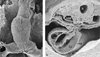
(a) Scanning electron micrograph of ventral view of the heart tube in a chicken embryo at the 10th stage of the classification made by Hamburger and Hamilton. The blood is propelled from the venous pole (V) to the arterial pole (A). (b) Transverse section of the heart tube that is connected to the body wall by the dorsal mesocardium (DM). The myocardium (M) is still thin and separated from the inner endocardial layer (E) by the cardiac jelly (CJ). N: neural tube; DAo: dorsal aortae.

(a) Scanning electron micrograph of a looped heart tube of a chicken embryo at the 17th stage of Hamburger and Hamilton. The dorsal mesocardium has been disrupted in the central part, leaving a connection of the heart tube at the venous pole, and at the arterial pole. In the heart tube there are the first indications of formation of segments: ventricle (V), atrium (A), and transitional zones: atrioventricular canal (AVC) and proximal (POT) and distal (DOT) outflow tract. The inner curvature ([sstarf ]) is remodelled to accommodate transition of the outflow tract over the atrioventricular canal as shown in b, from an embryo at the 22nd stage of Hamburger and Hamilton. An animated view of the process of looping is to be found at: www.medfac.leidenuniv.nl/cask/cadim.htm

Schematic drawings of the ventricle and outflow tract of the heart during looping and septation. Indicated are the transitional zones, specifically the atrioventricular canal (AVC), the primary fold (PF), and the ventriculo-arterial transition (VA). During the process of looping, (b) the arterial segment, or “truncus arteriosus” (TA), will move over the inner curvature to the left (see b, c, and e). The atrioventricular canal becomes partly positioned above the newly formed right ventricular inflow tract (RVI). The primary fold (PF, yellow) forms the embryonic basis of the conduction system, that will differentiate into the definitive atrioventricular conduction system (red in e). Septation of the arterial segment (b, d, e) separates the pulmonary trunk (Pu), connected to the right ventricle (RV), through a long outflow tract from the aorta (Ao) that arises by a short outflow tract from the left ventricle (LV). An animated sequence is available on the website of the Journal at www.greenwich-medical.co.uk AVN: atrioventricular node; MB: moderator band; RAVR: right atrioventricular ring bundle; TO: tricuspid orifice.

Schematic drawings of the sinus venosus (SV) and atrium. The transitional zones are the sino-atrial transition (SA) and the atrioventricular canal (AVC). The sinuatrial transition forms boundaries in both left and right atriums. This transition also forms the basis of the embryonic conduction system, which will differentiate eventually into the sinus node (SAN). Other parts of the transition may play a role in pathology of the conduction system, such as abnormal atrial automaticity. AAVN: embryonic anterior atrioventricular node; CV: caval veins; RVV: right venous valve; LVV: left venous valve; fusion of LVV and RVV in the septum spurium (SP); L: limbus of the oval fossa ovalis; PV: pulmonary veins; CS: coronary sinus; SS: septum secundum; SCV: superior caval vein; ICV: inferior caval vein. An animated sequence is available at: www.medfac.leidenuniv.nl/cask/cadim.htm

Overview of the 4 chambers of the heart. (a) View into the right ventricle (RV) shows the characteristic morphological aspects such as a broad muscle band, the supraventricular crest (CS), between the orifices of the tricuspid (TO) and pulmonary valves (Pu). The crest is the proximal margin of the long muscular subpulmonary infundibulum. The continuity of the muscular profiles of the septomarginal trabeculation (TS), the supraventricular crest, and the ventriculo infundibular fold (IF) form the larger part of the boundary between inflow and outflow of the RV. (b) View into the left ventricle (LV) with a smooth septal surface and a fibrous continuity between the leaflets of the mitral (MV) and the aortic valves. The short left ventricular outflow tract ([sstarf ]) forms the connection with the aorta. (c) View into the left atrium (LA) with the attachment points (→) of the valve of the oval foramen clearly visible. The latter partly overlaps, at this stage, with the limbus of the fossa (L) that forms the free rim of the so-called secondary septum in the right atrium. (d) View into the right atrium (RA), into which the systemic veins (SCV: superior caval vein; ICV: inferior caval vein) and coronary sinus (CS) enter. In the upper left hand side the entrance to the right atrial appendage (RAA) is visible. AVN: site of AV node; TO: tricuspid orifice; VFO: valve of the oval foramen.

Schematic picture of neural crest cells derived from the dorsal margin of the neural tube that migrate towards the heart. These cells enter both at the outflow tract (→) as well as at the venous pole (). Also the cells of the epicardial organ, epicardially derived cells, and endothelial cells of the liver migrate towards the heart, specifically spreading from the venous pole ().
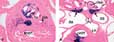
(a) Section of a Wnt1 lacZ reporter mouse at the 15.5 embryonic day, with neural crest cells (blue) in the arterial valves of the aorta (Ao), in the centre of the muscular outflow tract, and surrounding the coronary arteries (CA). (b) Section through the inflow tract, with neural crest cells (→) surrounding the pulmonary veins (PV). CS: coronary sinus; LA: left atrium; LVOT: left ventricular outflow tract; RA: right atrium; Oes: oesophagus; RVOT: right ventricular outflow tract.

(a) Section of the heart of a normal chicken embryo at the 29th stage of Hamburger and Hamilton showing the architecture of the ventricular compact myocardium (M) and the trabecular layer, as well as the overlying epicardium (EP). (b) Section through the ventricular region of an embryo at the same stage in which the outgrowth of the epicardial organ has been inhibited. Septation and valvar formation are deficient, and the compact myocardium is markedly thinned, lacking its covering of epicardial cells. END: endocardium, PERI: pericardium. Staining: anti α smooth muscle actin.
 You have
Access
- 13
- Cited by