Introduction
Canalplasty describes the surgical procedure employed to increase the width of the bony external auditory canal. Indications for canalplasty include chronic stenosing otitis externa, exostoses, congenitally narrow or atretic canals, and the need to improve access for middle-ear surgery. The procedure may be performed in isolation or combined with a soft tissue meatoplasty of the cartilaginous external meatus. Early complications include injury to the facial nerve, temporomandibular joint and tympanic membrane. Re-stenosis of the external canal represents the most common late complication.
We describe two patients who developed a cholesteatoma through the posterior canal wall many years after canalplasty. These cases illustrate a pathophysiological change not previously described. We review the literature and suggest a way of preventing this complication.
Report of two cases
Two patients, aged 28 and 52 years, are presented. Both underwent canalplasty, 14 and 17 years before re-presenting with cholesteatoma evident through a posterior canal wall defect.
The first patient, a 28-year-old woman, presented in 2005 with right-sided otorrhoea. Fourteen years earlier, she had undergone a right-sided myringoplasty for the closure of a large, central perforation. A canalplasty was performed as part of the procedure in order to provide access to the entire tympanic membrane.
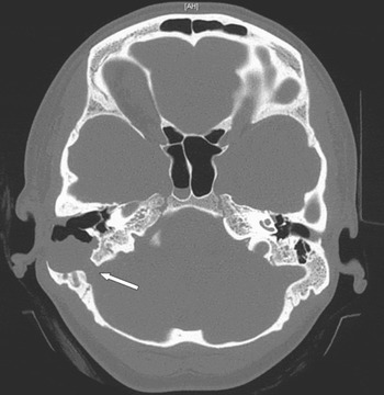
On examination of this patient, there was a defect in the posterior ear canal wall which appeared to be filled with squamous epithelium. The tympanic membrane was intact. A computed tomography (CT) scan demonstrated the presence of an extensive soft tissue mass eroding the mastoid (Figure 1).
Axial computed tomography scan for the first patient, demonstrating a large cholesteatoma eroding through the right posterior canal wall as well as the postero-medial wall of the mastoid cavity, exposing the dura over the sigmoid sinus (arrow). Note the intact tympanic membrane and aerated middle ear.

The second patient, a 52-year-old man, presented in 2007 with persistent, left-sided otorrhoea. Seventeen years earlier, he had undergone an atticotomy for attic cholesteatoma. A canalplasty had been performed as part of the procedure. Following this operation, he had not experienced any complications until shortly before re-presenting.
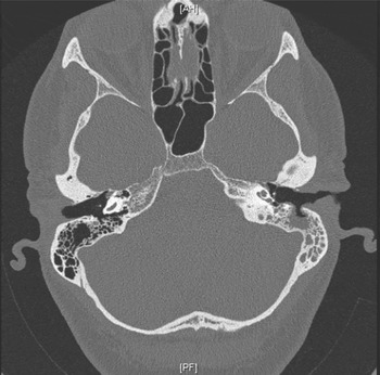
Examination of this patient's left ear revealed a defect in the posterior canal wall which appeared to be filled with squamous debris. The tympanic membrane and reconstructed attic were healthy. A CT scan confirmed the clinical findings of a bony defect in the posterior external auditory canal (Figure 2) with soft tissue and bone erosion in the mastoid region.
Axial computed tomography scan for the second patient, showing a defect in the left posterior canal wall, which contains soft tissue. The tympanic membrane is intact and the mastoid air cells are opacified.

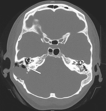
Exploration of the mastoid cavity of the first patient revealed cholesteatoma filling the entire mastoid, eroding the posterior semicircular canal and vertical portion of the Fallopian canal. The cholesteatoma was densely adherent to the sigmoid sinus, middle and posterior fossa dura, and vertical portion of the facial nerve. These relations can be clearly observed in the pre-operative CT scan (Figure 3). Despite the extensive nature of the cholesteatoma, disease had not extended into the antrum, attic or middle-ear cleft.
Axial computed tomography scan for the first patient, demonstrating a large cholesteatoma eroding through the posterior canal wall as well as the postero-medial wall of the mastoid. The cholesteatoma is closely related to the vertical portion of the facial nerve (arrow).

The first patient's cholesteatoma was removed via a cortical mastoidectomy. Disease adherent to the dura was fulgurated using bipolar diathermy and diode laser. The posterior canal wall defect was reconstructed using conchal cartilage. Re-exploration of the mastoid 12 months later confirmed total removal of cholesteatoma.
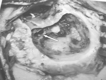
In the second patient, mastoid exploration confirmed the diagnosis of cholesteatoma extending through a bony defect in the posterior bony canal wall (Figure 4). The cholesteatoma was carefully everted through the defect and the canal wall repaired with cartilage.
Operative photograph for the second patient, showing the canal wall defect through the mastoid cavity (arrow).

The first patient was followed up for 24 months after initial presentation. At her last visit, the ear canal was healthy and hearing was normal. The second patient was followed up for three months after surgery. At his last visit, he had a healthy ear canal and normal hearing.
Discussion
The cases described illustrate a late complication occurring after canalplasty performed to provide improved access to the middle ear. The most likely aetiology is the inadvertent opening of a mastoid air cell during the original surgical procedure. The primary operation was performed for the management of chronic middle-ear disease and it is possible that, after successful reconstruction of the middle-ear defect, there was ongoing eustachian tube dysfunction. Over the intervening period, ear canal epithelium retracted into the small defect, causing accumulation of squamous epithelium and cholesteatoma formation. Our first case (Figure 1) had an extensive mastoid air cell system which enabled massive expansion of the cholesteatoma. Another, less likely possibility is that the cholesteatoma in both cases arose as a result of an insertion dermoid from the tympanomeatal flap. Venkatraman and Mattox describe five cases of auditory canal cholesteatomas occurring after prior ear surgery.Reference Venkatraman and Mattox2 In their series, however, the middle ear or mastoid was not directly involved in any of the cases.
The overall prevalence of canalplasty complications ranges from 4.5 to 22 per cent.Reference Lavy and Fagan1, Reference Sanna, Russo, Khrais, Jain and Augurio3–Reference Sharp, Oakley and Padgham5 In a series of 65 canalplasties, Sanna et al. described one case in which the mastoid air cells were opened.Reference Sanna, Russo, Khrais, Jain and Augurio3 Their proposed management for small defects was closure with temporalis fascia; however, they suggested that larger defects would require bone dust or cartilage obliteration. These authors advised that failure to do so may result in an iatrogenic cholesteatoma. In a series of 100 canalplasties, Lavy and Fagan did not describe any cases in which the mastoid air cells were breached.Reference Lavy and Fagan1 The most common complication their patients encountered was a self-limiting discomfort when chewing. Both Sanna et al. and Lavy and Fagan stressed the importance of avoiding opening the mastoid air cells. In contrast, other authors have suggested that the extent of the dissection is dictated by opening the mastoid air cells.Reference Cremers and Smeets6, Reference McDonald, Facer and Clark7 McDonald et al. have suggested that this is a key step in bony canalplasty. Although these authors did not state the length of follow up, they did not encounter complications using this technique.
• Canalplasty is commonly performed to improve access or to widen the external auditory canal
• The post-canalplasty complication rate ranges from 4.5 to 20 per cent
• The most common early complication of canalplasty is discomfort on chewing
• Long-term complications include re-stenosis and cholesteatoma
• If an air cell is opened during the canalplasty procedure, it should be repaired with non-retractable material (e.g. cartilage)
Canalplasty can often result in inadvertent opening of mastoid air cells. In the senior author's experience, breach of the posterior canal wall during mastoidectomy for combined approach tympanoplasty can result in meatal skin retraction through the defect. For this reason, reconstruction of the bony defect is performed before closure. It is our policy to obliterate an inadvertently breached posterior bony canal, following canalplasty or mastoidectomy, with free soft tissue if the defect is small or cartilage if there is a more extensive defect.
Conclusion
This is the first report of iatrogenic posterior auditory canal cholesteatoma following canalplasty. The two cases reported support the view that, when drilling the posterior canal wall during canalplasty, the surgeon should avoid opening a mastoid air cell. If one is inadvertently opened, the defect should be repaired with cartilage or free soft tissue, in order to prevent the formation of a mastoid cholesteatoma which may not present during the usual follow-up period.