Implications
Interest in methanogens from ruminants has resulted from the role of methane in global warming and from the fact that cattle typically lose 2% to 12% of ingested energy as methane (Johnson and Johnson, Reference Johnson and Johnson1995). However, little is known about their distribution in the rumen of Chinese ruminants. To understand their diversity distribution and their abundance, three methanogenic 16S rRNA gene libraries were constructed from different fractions of rumen of Jinnan cattle and their density determined by quantitative real-time PCR. Our results showed that Methanobrevibacter species were predominant in all three libraries, and that there were differences in the methanogenic diversity and abundance in the three different fractions within the rumen. The study for the first time demonstrated that many unidentified sequences were present in epithelium fraction library and that the epithelium and solids fractions had significantly higher density of methanogens than in the liquid.
Introduction
The ruminal methanogenic population has become a focus of increasing scientific attention for the negative effect of methanogenesis on dietary energy and environment (Johnson and Johnson, Reference Johnson and Johnson1995). Many studies have been conducted to investigate the methanogenic diversity in the rumen. Tajima et al. (Reference Tajima, Nagamine, Matsui, Nakamura, Rustam and Aminov2001) found that the predominant species were affiliated with Methanomicrobium mobile in the rumen of Holstein dry cows in Japan. From the rumen of Hereford-Cross cattle in Ontario, almost half of the clones of archaea were related to Methanobrevibacter ruminantium (Wright et al., Reference Wright, Auckland and Lynn2007). In the rumen of merino wethers in Western Australia, more than 97% (713 of 733) of the clones were identified as Methanobrevibacter, while Mbr. ruminantium only accounted for 1.6% (Wright et al., Reference Wright, Williams, Winder, Christophersen, Rodgers and Smith2004). However, the rumen methanogenic population of cattle of Chinese native breeds has received little attention.
Within the complex ecosystem of the rumen, the microbial populations are effectively subdivided into those free in the rumen fluid, those associated with feed particles and those associated with the rumen wall (Cheng and McAllister, Reference Cheng and McAllister1997). Previous studies indicated that these three fractions differ in microbial composition (McCowan et al., Reference McCowan, Cheng, Bailey and Costerton1978; Dehority and Grubb, Reference Dehority and Grubb1981; Yang et al., Reference Yang, Beauchemin and Rode2001), and McAllister et al. (Reference McAllister, Bae, Jones and Cheng1994) suggested that the reason for this was the different growth requirements of microorganisms. Growth requirements of the various rumen methanogens are also different (Bonin and Boone, Reference Bonin and Boone2006). Hence, it might be hypothesized that the methanogenic community differs among these three fractions.
To our knowledge, Shin et al. (Reference Shin, Choi, Lim, Hong, An, Cho, Kim, An, Kang, Lee, Kim and Yun2004) were the first to investigate the methanogenic molecular diversity in the rumen fluid, solid and epithelium. Differences in methanogenic communities among these fractions have been found. In this study, rumen fluid and solid fractions were divided by squeezing through two layers of cheesecloth, and microorganisms on the rumen epithelium were collected by scraping with a spatula. The diversity in methanogenic species was similar for rumen epithelium (20 clones in total) and rumen fluid fractions (45 clones in total), both with Methanomicrobiaceae as predominant species. However, in that study, the purity of each fraction was questionable as the epithelium and solid fractions were not entirely free of rumen fluid.
Jinnan cattle, one of the four major Chinese yellow cattle, is the major beef cattle breed in China. To understand the diversity and distribution of methanogens in the rumen of Jinnan cattle, three 16S rRNA gene libraries were constructed from rumen liquid, solid and epithelium fraction, respectively. In order to better reflect the methanogenic diversity in each fraction in the rumen, large numbers of clones were used for each library.
Material and methods
Sample sources and processing
Four Jinnan cattle, 2 years of age and weighing 400 to 500 kg, were used in the present study. The animals were fed 2 kg of concentrate (corn meal and cottonseed meal in a 6 : 4 ratio, and 5% premix feed) at 0800 h and dried forage (whole corn stalk and wheat stalk) ad libitum. Per kilogram of premix feed contained 32 000 IU of vitamin A, 16 000 IU of vitamin D3, 100 g of sodium chloride, 2.4 g of manganese, 2 g of ferrous, 2.4 g of zinc, 0.5 g of copper, 10.4 mg of iodine, 10.4 mg of selenium and 4 mg of cobalt. All experiments were carried out in accordance with the China Animal Welfare Legislation. Samples of rumen contents and rumen walls were obtained immediately after slaughter that took place before the morning feeding. Rumen fluid for the liquid-associated methanogen (LM) library and feed particle fractions for the solid-associated methanogen (SM) library were obtained through filtration, centrifugation and washing as described by Yang et al. (Reference Yang, Beauchemin and Rode2001). Three ruminal samples per animal were squeezed through four layers of cheesecloth. The solids retained on the cheesecloth were mixed and approximately 10% of the mix was retained for further analysis. The liquid fractions were mixed resulting in a total of around 500 ml, and from the mix 50 ml of the liquid fraction was centrifuged at 800 × g for 15 min at 4°C. The supernatant fraction was stored at −20°C for DNA extraction of the liquid fraction. The precipitate was mixed with the solids obtained above and washed with 400 ml of cold 0.9% NaCl, squeezed twice and then stored at −20°C for DNA extraction of the solid fraction. At the same time, three pieces of the epithelium (each piece about 3 cm2) were cut from ventral rumen (recessus ruminis, saccus ventralis and saccus cecus caudoventralis, which is the front end, the middle and the rear end of ventral rumen, respectively), washed with cold 0.9% NaCl, the liquid was removed and the tissue was stored in a sample bag at −20°C for DNA extraction of rumen epithelium.
DNA extraction, PCR amplification and clone library construction
Prior DNA extraction, samples were thawed. The epithelium samples were homogenized using the Ultra Turrax T25 homogenizer (IKA Werke Labortechnik, Staufen, Germany) and the liquid fraction was washed with phosphate buffer and centrifuged at 27 000 × g for 30 min. Extraction of nucleic acid, based on the bead-beating method described by Zoetendal et al. (Reference Zoetendal, Akkermans and De Vos1998) and 0.5 g of each sample was used for this purpose. The extracted DNA was purified and then checked on agarose gel with most of DNA fragments around 50 kb based on the DNA marker ladder run on the same gel. The DNA was stored at −20°C for PCR to amplify the 16S rRNA gene.
The PCR primers used to amplify 16S rRNA genes were the methanogen-specific primers, Met86F (5′-GCT CAG TAA CAC GTG G-3′) and Met1340R (5′-CGG TGT GTG CAA GGA G-3′) (Wright et al., Reference Wright, Williams, Winder, Christophersen, Rodgers and Smith2004). The amplification conditions were: initial denaturation at 94°C for 3 min, then 40 cycles of 94°C for 30 s, 58°C for 30 s and 72°C for 90 s, and last extension at 72°C for 10 min. The PCR mixture (total volume of 50 μl) contained 200 nM of both primers, approximately 0.35 μg of the purified DNA extract, 1× Taq reaction buffer, 200 μM of each dNTP, 2 mM of MgCl2 and four units of Taq DNA polymerase. After amplification, equal quantities of PCR products from the same fractions of the four cattle were mixed for clone library construction.
Amplified DNA for clone libraries was purified using a PCR Clean-Up system (Promega, Madison, WI, USA) and cloned into Escherichia coli strain TOP10 using the pGEM-T Easy vector (Promega). All the recombinant plasmids were reamplified by PCR using the primers and parameters described above. Different restriction endonucleases (Hae III, Alu I, Hpa II) were used to digest PCR production of the recombinant plasmids. Digested fragments were immediately separated by electrophoresis on 4% agarose gels. The clones with same riboprint patterns digested by all restriction endonucleases were defined as one phylotype, and at least one clone from each phylotype was sequenced in both directions commercially (Invitrogen, Beijing, China). The percentage of each kind of clone (according to the nearest valid taxon and the sequence similarity) in each library was calculated as follows: the number of that clone type divided by the total clone numbers of this library.
Phylogenetic analysis
Phylogenetic analysis consisted of sequences of the best ‘known hits’ from BLAST searches, 16S rRNA gene sequences of validly described rumen methanogens and species belonging to the Euryarchaeota. Two Crenarchaeotas, Sulfolobus acidocaldarius and Thermoproteus tenax, were used as outgroups. Sequences were aligned using Clustalx1.8, and a phylogenetic tree was constructed by MEGA4 (http://www.megasoftware.net/mega4/mega.html) software to illustrate the evolutionary relationships.
Estimation of methanogenic diversity
The Shannon–Weaver diversity index (H shannon) was calculated for each library using the formula
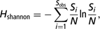
where S obs is the observed number of species, Si is the number of sequences in the ith OTU (the operational taxonomic unit), and N is the number of individuals sampled, with the DOTUR program (http://schloss.micro.umass.edu/software/dotur/downloads.html) to characterize the methanogenic diversity of rumen samples (Schloss and Handelsman, Reference Schloss and Handelsman2005).
Real-time PCR assay for quantification of total methanogens
Real-time PCR was performed on an Applied Biosystems 7300 Real-Time PCR System (Applied Biosystems, California, USA) with the mcrA gene-specific primers, qmcrA-F (5′-TTC GGT GGA TCD CAR AGR GC-3′) and qmcrA-R (5′-GBA RGT CGW AWC CGT AGA ATC C-3′) (Denman et al., Reference Denman, Tomkins and McSweeney2007). A reaction mixture (20 μl) consisted of 10 μl of IQ SYBR Green Supermix (Bio-Rad, California, USA), 0.2 μM of each primer set and 1 μl of the template DNA. The amount of DNA in each sample was spectrophotometrically determined in triplicate using GeneQuant pro RNA/DNA Calculator (Amersham Biosciences, Bath, UK), and the mean values were calculated. A standard curve was generated by using the serially diluted mcrA gene amplicons obtained from two methanogens isolated from goat rumen, Methanoculleus sp. RPS4 (accession numbers of the 16S ribosomal RNA gene in the GenBank: EU544029) and Methanobacterium beijingense strain M4 (EU544027). The PCR was performed under the following cycle conditions: one cycle of 50°C for 2 min and 95°C for 2 min for initial denaturation, 40 cycles at 95°C for 15 s and 60°C for 1 min for primer annealing and product elongation (Denman et al., Reference Denman, Tomkins and McSweeney2007). Fluorescence detection was performed at the end of each denaturation and extension step.
Nucleotide sequence accession numbers
The nucleotide sequences reported in this paper have been deposited in the GenBank database under accession numbers EF055490 to EF055559.
Statistical analysis
Differences of quantification of total methanogens between fractions were tested for significance with an ANOVA program with Duncan post hoc test using the statistical software SAS8.0. The statistical model was as follows: Y = μ + Fi + εik, where Y is the parameter to be tested, μ is the overall mean, Fi effect of fraction i, εik is the error term. Significant differences were declared when P < 0.05.
Results
Sequence analysis of methanogenic 16S rRNA gene clone libraries
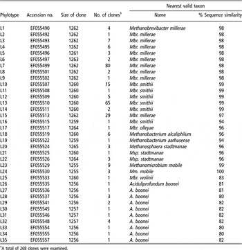
The LM library consisted of 268 clones. Sequence examination of these clones revealed 35 different phylotypes (Table 1). The majority of clones had their sequences closely related to Methanobrevibacter spp. (Mbr. millerae, Mbr. smithii, Mbr. olleyae, Mbr. wolinii) with similarity ranging from 83% to 99%. Seven clones had their sequences closely related to Methanobacterium spp. (Mb. alcaliphilum, Mb. aarhusense) (similarities from 94% to 96%). Seven clones related to Methanosphaera sp. (Msp. stadtmanae) with similarity below 97%. Twelve clones showed a similarity above 99% to Mm. mobile. This library also showed some unique sequences with similarities of only around 80% to Aciduliprofundum boonei, a species that was found in deep sea but has not been found in the rumen before.
16S rRNA gene clones of the liquid-associated methanogen library

†A total of 268 clones were examined.
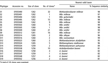
The SM library contained 135 clones, consisting of 19 phylotypes (Table 2). These phylotypes were similar to Mbr. millerae, Mbr. smithii, Mbr. ruminantium, Mbr. gottschalkii, A. boonei, Msp. stadtmanae, Mbr. olleyae, Mb. alcaliphilum and Mb. aarhusense.
16S rRNA gene clones of the solid-associated methanogen library

†A total of 135 clones were examined.
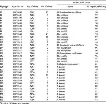
The EM library consisted of 267 clones, representing 33 phylotypes (Table 3). These phylotypes had high degrees of sequence similarity to Mbr. millerae, Mbr. smithii, A. boonei, Mb. alcaliphilum and Msp. stadtmanae.
16S rRNA gene clones of the epithelium-associated methanogen library

†A total of 267 clones were examined.
Overall, a total of 669 clones were examined, representing 70 phylotypes. The majority of clones from the three libraries belonged to the family Methanobacteriaceae, 526 sequences of the 669 clones showed similarities of more than 97% to the genus Methanobrevibacter. Only 12 sequences from LM (1.8%) were associated to Mm. mobile. A minor proportion of the sequences showed a similarity level with known methanogenic sequences in the range from 83% to 96% only. The remaining sequences were closest to A. boonei sequences, but similarity was only between 80% and 83%.
Comparison of the three fractions
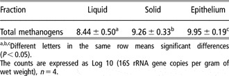
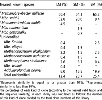
There were differences between the three fractions in methanogenic diversity and abundance. Quantitative real-time PCR showed that the density of methanogens on the epithelium was highest in the rumen. The total methanogenic mcrA gene copies in the epithelium fraction (9.95 Lg (copies per gram of wet weight)) were higher than that in the liquid (8.44 Lg, P < 0.05) and solid (9.26 Lg, P < 0.05) fractions, respectively (Table 4). And the comparison of clones among the three libraries showed that Mbr. millerae was dominant in all three libraries, representing 50.4%, 54.1% and 65.2% of the clones of LM, SM and EM, respectively (Table 5). The second dominant methanogen was Mbr. smithii, with an abundance of 32.8%, 20.0% and 9.4% as based on the LM, SM and EM clone libraries, respectively. Methanomicrobium mobile (4.5%) was found in the LM library only, while Mbr. ruminantium (1.5%) and Mbr. gottschalkii (0.7%) was only present in the SM library. Novel sequences accounted for 12.4% in LM library, 23.7% in SM library and 25.5% in EM library. Methanobacterium alcaliphilum-like, Msp. stadtmanae-like and A. boonei-like sequences were present in all libraries, but their proportion differed in the three libraries. For instance, A. boonei-like clones accounted for 19.9% in EM library, while 6.0% and 15.5% in the LM and SM library. There were Mbr. smithii-like sequences in libraries of LM (0.4%) and EM (2.6%), and Mbr. olleyae-like and Mb. aarhusense-like sequences in libraries of LM (both 0.4%) and SM (both 1.5%), but Mbr. wolinii-like sequences were only found in LM library (0.4%). The Shannon index (a diversity index that accounts for both abundance and evenness of the species present in the community) of LM, SM and EM library were 2.05, 1.73 and 2.12, respectively. This showed that the methanogenic communities of LM and EM were similar and both had higher genetic diversity than that of SM.
Quantitative real-time PCR analysis of total methanogens (mean ± SD) in samples from different rumen fractions of four cattle

a,b,cDifferent letters in the same row means significant differences (P < 0.05).
The counts are expressed as Log 10 (16S rRNA gene copies per gram of wet weight), n = 4.
Comparison of the three libraries with clone percentage and nearest known species

aRepresents similarity is equal to or greater than 97%; bRepresents similarity is less than 97%.
The percentage of each kind of clone (according to the nearest valid taxon and the sequence similarity) in each library was calculated as follows: the number of this kind of clone divided by the total clone numbers of this library.
Phylogenetic analysis of archaeal 16S rRNA gene sequences
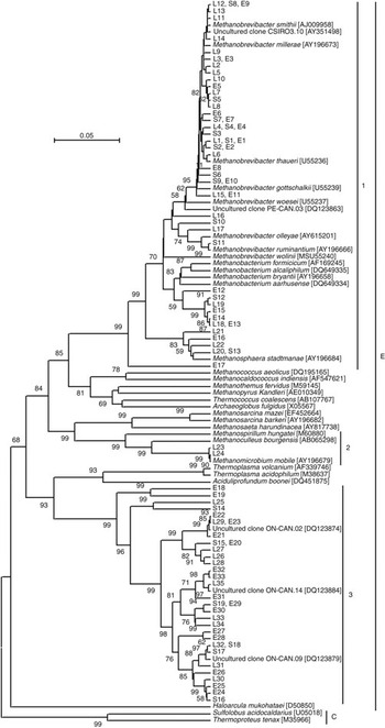
The phylogenetic analysis indicated that archaea found in the three libraries were all clustered into the Euryarchaeota (Figure 1). The data from the tree showed that those sequences belonged to three monophyletic groups. Phylotypes L1–L22, S1–S13 and E1–E17 grouped within the Methanobacteriales. Phylotypes L23 and L24 grouped within the Methanomicrobiales. Other phylotypes grouped together, and were sister group to A. boonei and species of the Thermoplasmatales including Thermoplasma volcanium and Thermoplasm acidophilum.
Phylogenetic relationships of archaeal clones derived from 16S rRNA gene evolutionary distances produced by the Bootstrap model and constructed using the Unweighted Pair Group Method with Arithmetic mean method. The tree was bootstrap-resampled 1000 times. Only bootstrap values greater than 50% are shown on the internal nodes. Higher taxonomic groupings are indicated as follows: E = Euryarchaeota; C = Crenarchaeota; 1 = Methanobacteriales; 2 = Methanomicrobiales; 3 = novel group of uncultivated archaea.

Within the Methanobacteriales, phylotypes L1–L17, S1–S11 and E1–E11 grouped together with Mbr. millerae, Mbr. thaueri, Mbr. gottschalkii, Mb. smithii, Mbr. woesei, Mbr. olleyae and Mbr. ruminantium. Phylotypes L18, L19, S12 and E12–E15 were sister groups to Mb. bryantii, Mb. aarhusense, Mb. alcaliphilum and Mb. formicicum. Phylotypes L20–L22, S13 and E16 were sister groups to Msp. stadtmanaea, and are likely to be new species within the genus Methanosphaera. The genetic distances of phylotype E17 with the methanogens of Methanobacteriaceae (in the range from 0.12 to 0.18) were much less than that with the methanogens of other families (from 0.26 to 0.28). Hence based on its position in the phylogenetic tree, E17 likely represents a new genus within the Methanobacteriaceae.
Within the order Methanomicrobiales, two phylotypes (L23 and L24) branched very close to Mm. mobile.
The novel methanogen-like sequences
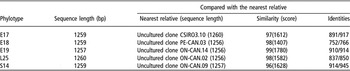
In the three libraries, there were five phylotypes (E17, E18, E19, L25 and S14) that showed similarities between 91% and 99% with uncultured euryarchaeote (Table 6), and they were only 83% to 96% similar to Mbr. smithii, Mbr. wolinii or Mb. aarhusense (Tables 1 to 3). Thus, these sequences may represent novel methanogens. For instance, the nearest relative of phylotype E17 was the uncultured Methanobrevibacter sp. CSIRO3.10 (accession number: AY351498), and the genetic distance between them was 11.6%.
The novel methanogen-like sequence in the three libraries

Discussion
Investigating the diversity and structure of methanogenic communities has been a long-standing challenge in rumen ecology. In this study, PCR-retrieved methanogenic 16S rRNA gene libraries were established from the rumen fluid, solid and epithelium. The results showed that the rRNA gene clones generated in this study exhibited high degrees of sequence similarity to ten methanogenic species, Mbr. millerae, Mbr. smithii, Mbr. gottschalkii, Mbr. olleyae, Mbr. ruminantium, Mbr. wolinii, Mb. alcaliphilum, Mb. aarhusense, Msp. stadtmanae and Mm. mobile. The most predominant species of methanogens in the three groups were related to the genus Methanobrevibacter. This is in agreement with previous studies indicating species belonging to the genus Methanobrevibacter as the major methanogens in the rumen of sheep (Wright et al., Reference Wright, Williams, Winder, Christophersen, Rodgers and Smith2004 and Reference Wright, Ma and Obispo2008; Skillman et al., Reference Skillman, Evans, Naylor, Morvan, Jarvis and Joblin2004; Nicholson et al., Reference Nicholson, Evans and Joblin2007) and cattle (Whitford et al., Reference Whitford, Teather and Forster2001; Skillman et al., Reference Skillman, Evans, Strömp and Joblin2006; Nicholson et al., Reference Nicholson, Evans and Joblin2007; Ouwerkerk et al., Reference Ouwerkerk, Turner and Klieve2008), of which some were associated with the rumen protozoa (Tokura et al., Reference Tokura, Chagan, Ushida and Kojima1999a; Sharp et al., Reference Sharp, Ziemer, Stern and Stahl1998; Chargan and Ushida, Reference Chargan and Ushida2004). The predominance of this genus has also been suggested before based on cultivation studies (e.g. Miller et al., Reference Miller, Wolin, Hongxue and Bryant1986).
In the present study, our libraries uncovered some unidentified rumen euryarchaeotic sequences. Fifteen phylotypes (L16–L22, S10, S12, S13, E12–E17) were 94% to 97% identical to known methanogens, thus they are likely to be unknown methanogenic strains or species. Four phylotypes (L25, S14, E18, E19) were similar to Mbr. wolinii, Mb. aarhusense or Mbr. smithii, however with a similarity level of 83% to 86% only. Twenty-five phylotypes (L26–L35, S15–S19, E20–E33) were 80% to 84% similar and most closely to A. boonei, an Euryarchaeota found in deep sea. Four phylotypes (L25, S14, E18, E19) were also found in this group from the phylogenetic tree (Figure 1). All of them were clustered within Euryarchaeota, in which eight classes can be distinguished (Methanobacteria, Methanococci, Methanomicrobia, Methanopyri, Halobacteria, Thermoplasmata, Thermococci and Archaeoglobi). All classes except for the methanogenic ones are hyperthermophilic or halophilic. Whitford et al. (Reference Whitford, Teather and Forster2001) also reported that the diversity of methanogenic archaea in the sheep rumen was much more elaborate than expected, and suggested that new probes and assays may be required to evaluate the abundance and composition of this diverse population. In the current study, 29 unique phylotypes (29 clones) clustered within a strongly documented phylogenetic group along with other clone sequences from uncultured methanogens. Interestingly, the sister group to this distinct branch is a branch containing two Thermoplasmatales species (T. volcanium and T. acidophilum) and a deep sea Euryarchaeota (A. boonei). The sister group of Thermoplasmatales species was also found in the rumen in other studies (Whitford et al., Reference Whitford, Teather and Forster2001; Wright et al., Reference Wright, Toovey and Pimm2006 and Reference Wright, Auckland and Lynn2007). Although it is unlikely that the uncultured and unidentified methanogens ultimately will be classified as A. boonei, regarding their relatively low similarity, the consistency in our results and the ones of Whitford et al. (Reference Whitford, Teather and Forster2001) is striking. This might indicate that the novel group represents a stable methanogenic group in the rumen. Although Wright et al. (Reference Wright, Toovey and Pimm2006) reported that research is underway in several laboratories attempting to cultivate species of this newly discovered group, to date there have been no formal reports about the success of the isolation of these methanogens.
The results of the present experiment showed differences between the three fractions in methanogenic diversity. For example, the solid fraction contained sequences related to Mbr. ruminantium (S11) and Mbr. gottschalkii (S3), but none of these sequences were detected in the LM and EM libraries. Mm. mobile and Mbr. wolinii-like sequences were found in the LM library only. Moreover, methanogen abundance was different between the three fractions. Quantitative real-time PCR showed significant difference in methanogen density between the three fractions, with the numbers of total methanogenic mcrA gene copies per gram of wet weight being higher in the epithelium fraction than in the liquid and in the solid fractions. This suggests that much hydrogen, an inhibitor of the microbial fermentation (Janssen and Kirs, Reference Janssen and Kirs2008), might be consumed and much methane might be released in the solid and epithelium fraction. The clone library approach further indicated that the abundance of each methanogen differed between the three fractions. The proportion of Mbr. smithii (32.8%) in LM library was much higher than that in the SM (20.0%) and EM (9.4%) library, but that of Mbr. millerae (50.4% to 54.1% and 65.2%), unidentified (12.4% to 23.7% and 25.4%) and A. boonei-like (6.0% to 15.5% and 19.9%) clones was lower in the LM library. Up to now, only Shin et al. (Reference Shin, Choi, Lim, Hong, An, Cho, Kim, An, Kang, Lee, Kim and Yun2004) reported the methanogenic molecular diversity in the rumen fluid, solid and associated to the epithelium. They showed a relatively similar methanogenic diversity among the different fractions. However, the present study revealed more unknown and unique methanogenic species. Overall, the percentage of unidentified methanogenic clones is smaller in the liquid fraction (12.4% compared to 23.7% and 25.4% in solid fraction and epithelium fraction, respectively). Many methanogens in the solid and epithelium fractions have remained unknown though these unknown species may be functionally important in the rumen. However, whether the wide diversity of methanogens with many unknown is important to the rumen function remains unknown.
So far, the main factor affecting the diversity and abundance in the methanogenic community in LM, SM and EM remains unclear. The different turnover rates of multiple fractions (Janssen and Kirs, Reference Janssen and Kirs2008) may be the main influencing factor on methanogenic density. The liquid fraction has the fastest turnover rates while the methanogens generally grow slowly with up to 20 days for a generation, thus the methanogenic density was lowest. The rumen solid fraction has relatively slow turnover rate and the epithelium fraction even the lowest. The methanogens associated with these different fractions can be expected to have different growth rates since they will be removed from the rumen at different rates (Janssen and Kirs, Reference Janssen and Kirs2008). According to the rumen environment, the ruminal epithelium is between the aerobic environment in the bovine tissue and the anaerobic environment in the rumen (Cheng and McAllister, Reference Cheng and McAllister1997). Thus, the environment on the rumen epithelium is more oxygenated and richer in urea than that in the rumen (Cheng and McAllister, Reference Cheng and McAllister1997). This possibly might be one of the reasons for the difference between epithelium and rumen fractions in terms of methanogenic community. Also, differences in distribution of dietary compounds may affect the growth of the methanogens, as compared with the rumen liquid or the rumen solid fraction, which mainly consists of plant fiber. In addition, the methanogenic populations may be affected by the abundance of protozoa and anaerobic fungi, which are mainly associated with the solid fraction. Mbr. ruminantium and Mbr. gottschalkii, which were only detected in solid fraction as observed in the present study, were likely to be associated with protozoa. Sharp et al. (Reference Sharp, Ziemer, Stern and Stahl1998) reported that the order Methanobacteriales was associated with ruminal ciliates, and the order Methanomicrobiales mainly existed in the fluid. The studies of Chargan et al. (Reference Chargan, Tokura, Jouany and Ushida1999) and Chargan and Ushida (Reference Chargan and Ushida2004) further showed that predominant sequences on ruminal ciliates were most related to Mbr. ruminantium or Mbr. gottschalkii. The study of Tokura et al. (Reference Tokura, Tajima and Ushida1999b) even revealed that Mbr. ruminantium was the unique methanogen isolated from ruminal ciliates. However, in another study of the same group (Tokura et al., Reference Tokura, Chagan, Ushida and Kojima1999a), Mbr. smithii was reported to be the dominant sequence of methanogens associated with rumen ciliates. The different results may be caused by the methanogens that came from different parts of rumen ciliate protozoa. Mbr. smithii was dominant in all the methanogens (including the living and the dead) in and on the whole ciliates (Tokura et al., Reference Tokura, Chagan, Ushida and Kojima1999a), while Mbr. ruminantium was the unique living methanogen on the ruminal ciliates (Tokura et al., Reference Tokura, Tajima and Ushida1999b). With rumen anaerobic fungi, it has been widely reported that rumen anaerobic fungi could rapidly colonize and efficiently degrade fibrous plant materials as reviewed by Theodorou et al. (Reference Theodorou, Zhu, Rickers, Nielsen, Gull and Trinci1996). Cheng et al. (Reference Cheng, Mao, Pei, Liu and Zhu2006 and Reference Cheng, Edwards, Allison, Zhu and Theodorou2009) recently showed the natural co-existence of rumen methanogens with anaerobic fungi. The natural co-culture of rumen methanogens with anaerobic fungi enriched from the rumen of cattle could effectively degrade fibrous material and produce methane (Cheng et al., Reference Cheng, Edwards, Allison, Zhu and Theodorou2009). Together with the above, the environment, the feed constitutes or degradation products and the interaction between the methanogens and other microbes may all contribute to differences in the methanogenic community in the three rumen fractions.
The present approach using large clone libraries from relatively pure fractions enabled to reveal many methanogenic species including many unknown sequences and thus could effectively reflect the diversity, abundance and differences of rumen methanogens in the three fractions. Our study also revealed apparent differences among these fractions. This may be due to the sample collection practice. In the Shin et al. (Reference Shin, Choi, Lim, Hong, An, Cho, Kim, An, Kang, Lee, Kim and Yun2004) study, the rumen fluid and solid were separated by filtration, and the microorganisms on the rumen epithelium were collected by scraping with a spatula. In present study, after filtration, the rumen liquid was further centrifuged by low speed in order to remove feed micro-particles remaining in the rumen fluid. The epithelium-associated microorganisms were obtained from tissue parts cut from the epithelium; and the solid and epithelium were washed to remove loosely associated microorganisms. Such marked differences between these two studies are also likely to be explained, in part, by animal species and diet. Lin et al. (Reference Lin, Raskin and Stahl1997) targeting the small subunit ribosomal RNA through oligonucleotide probes demonstrated that animal species and diet affected the quantity and dominance of methanogens in the rumen. The molecular diversity of methanogens also confirmed this (Wright et al., Reference Wright, Williams, Winder, Christophersen, Rodgers and Smith2004 and Reference Wright, Toovey and Pimm2006). Other factors such as the environment, fitness and age of animal could also have some effect on the microbial diversity (Wright et al., Reference Wright, Toovey and Pimm2006).
In conclusion, this study revealed an apparent difference in methanogenic diversity and abundance among the three fractions of the Jinnan cattle rumen, using a diet that is widely adopted in the North China. By quantitative real-time PCR, the present study for the first time revealed the difference in methanogen density between the three fractions, with rumen epithelium and solid fractions having a relatively higher density of methanogens than the liquid fraction. Many unique unknown methanogenic sequences were found on the rumen epithelium and in the solid fraction. Nevertheless, whether these unique sequences represent new taxonomic groups and their role in the rumen need further investigation.
Acknowledgements
This work was supported by a grant from the Natural Science Foundation of China (30530560).