Brugada syndrome is a channelopathy that affects cardiac repolarisation in patients with structurally normal hearts leading to sudden cardiac death due to malignant ventricular arrhythmias. It is electrocardiographically characterised by a coved ST-segment elevation, greater than or equal to 2 millimetres, in the right precordial leads followed by a negative T wave, Type-1 ECG pattern. It represents approximately 20% of sudden deaths in patients without structural heart disease.Reference Antzelevitch, Brugada and Borggrefe1 Brugada Type-1 ECG pattern can also be induced by various medications, electrolyte abnormalities, and disease states other than Brugada syndrome. The term Brugada phenocopy, previously named Brugada-like pattern, was recently introduced in the literature to describe the typical ECG pattern associated with diseases other than Brugada syndrome.Reference Riera, Uchida and Schapachnik2, Reference Arce, Riera, Femenia and Baranchuk3
Rhabdomyomas are the most common cardiac tumours in paediatrics with 80% of cases occurring in patients under the age of 1. They are usually benign lesions that lack mitotic activity and tend to regress spontaneously by age 6 years, resulting in greater similarity to a hamartoma than a neoplasm.Reference Sabatine, Colucci and Schoen4 Rhabdomyomas can present as single or multiple tumours and are frequently located in the interventricular septum.Reference Marx and Flyer5 The tumours can encroach on the ventricular cavities and lead to obstruction of the right and/or left ventricular outflow tracts.Reference Sabatine, Colucci and Schoen4, Reference Marx and Flyer5 A diagnosis is often made with prenatal ultrasound or postnatal echocardiography. In asymptomatic patients, clinical observation is the management choice in 80% of cases. Should obstruction or arrhythmias develop, surgical excision of the tumour has shown excellent prognosis.Reference Sabatine, Colucci and Schoen4
We describe the first case in the literature of a rhabdomyoma of the interventricular septum presenting as a Brugada Type-1 EGG phenocopy.
Case report
At 20 weeks of gestation, evidence suggesting rhabdomyoma was revealed during foetal anatomic ultrasound. Subsequent to an uncomplicated vaginal delivery at 39 weeks of gestation, the female infant was transferred to the cardiac unit and monitored with a Holter and daily echocardiogram for 1 week. The patient tested negative for tuberculosis and has no family history of tuberculosis either. Genetic testing for tuberous sclerosis did not detect any mutations.
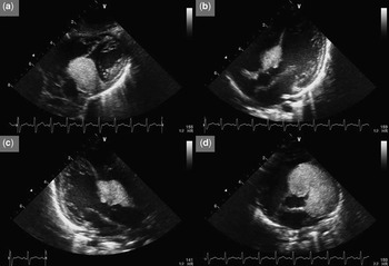
Initial echocardiogram revealed a large rhabdomyoma measuring 23 × 27 millimetres in the anterior interventricular septum, extending into the right and left ventricular free walls anteriorly and into the left ventricular outflow tract posteriorly. There were no signs of right ventricular outflow tract or left ventricular outflow tract obstruction (Fig 1). At 8 months, interventricular septal motion was found to be locally hypokinetic, but still no obstruction was present. The tumour appeared to be stable and of no mechanical or electrical significance, and therefore a plan for continual follow-up with echocardiography and ECG without therapeutic or surgical intervention was recommended.
Echocardiogram performed at 2 months of age showing a large hyperechoic, homogeneous mass extending from the anterior interventricular septum into the right and left ventricular free walls anteriorly and into the left ventricular outflow tract posteriorly. (a) subcostal view, (b) apical five chamber view, (c) apical long-axis view, and (d) modified four chamber view. No appreciable obstruction to outflow tracts – left ventricular outflow tract peak gradient = 9 millimetres of mercury; right ventricular outflow tract peak gradient = 10 millimetres of mercury.

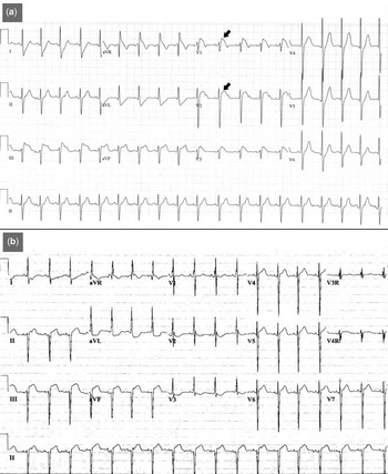
During a follow-up at 17 months, the echocardiogram revealed a significant reduction of the intracardiac tumour with reduction of the gradient; significant obstruction was never present in this case. A new ECG revealed the typical Brugada Type-1 ECG pattern (Fig 2). Vital signs were stable and physical exam findings were unremarkable. A new Holter recording showed no ventricular arrhythmias. No evidence of syncopal episodes and no family history of syncope, sudden death, or coved ST-segment elevation were found either. ECG follow-up at 21 months showed normalisation of the ECG with no evidence of Brugada pattern.
We performed 12-lead ECG at (a) 17 months of age and (b) at birth – sinus rhythm, left anterior fascicular block. Note in panel a: the characteristic Brugada type-1 ECG pattern in leads V1–V3, see arrows.

Genetic testing completed at 19 months did not detect mutations in the SCN5a gene.
Discussion
The term Brugada phenocopy has recently been introduced in the literature to describe a growing list of agents and conditions which induce an acquired Brugada Type-1 ECG pattern, thereby mimicking the true Brugada syndrome.Reference Riera, Uchida and Schapachnik2, Reference Arce, Riera, Femenia and Baranchuk3 As reported by Antzelevitch et al,Reference Antzelevitch, Brugada and Borggrefe1 sodium channel blockers, febrile episodes, vagotonic agents, alpha/beta-adrenergic blockers, tricyclic/tetracyclic antidepressants, insulin and glucose combinations, electrolyte imbalances, alcohol toxicity, and cocaine toxicity can present as Brugada phenocopies.
Rhabdomyomas are the most common primary cardiac tumours encountered in paediatrics. The tumour is believed to develop from mutations in the TSC1 and TSC2 genes and is associated with tuberous sclerosis in 50% of cases.Reference Sabatine, Colucci and Schoen4 Rhabdomyomas are well-demarcated lesions with a diameter ranging from 0.3 to 9 centimetres. A characteristic histological feature is the presence of eosinophilic spider cells – enlarged cells with a clear cytoplasm and rich glycogen supply.Reference Amonkar, Kandalkar and Balasubramanian6 Extension of the tumour into the ventricular cavities can lead to obstruction of the left ventricular outflow tract and/or right ventricular outflow tract. In the event of significant haemodynamic impairment, stillbirth or early postnatal death may result. Arrhythmias are another clinical presentation, which are more commonly observed in adults, but can also manifest in foetal cases.Reference Sabatine, Colucci and Schoen4
Nakamura et alReference Nakamura, Funabashi and Miyauchi7 reported on a case of pericardial haemangioma presenting in association with Brugada Type-1 ECG pattern. Since the patient had a family history of sudden death and the tumour did not extend into cardiac structures, the ECG manifestations were deemed to be unrelated to the tumour and a diagnosis of Brugada syndrome was established.
In this case, there was no history of arrhythmias, syncope, or family history of sudden death, and no mutations were detected in the SCN5a gene, suggesting that the ECG abnormalities were due to a Brugada phenocopy with no other evidence of true Brugada syndrome.
Despite no obstruction of the outflow tracts being found with serial echocardiography, the significant size of the rhabdomyoma and its communication with the interventricular septum and left ventricular outflow tract are most likely responsible for the Brugada Type-1 ECG pattern. Tarin et alReference Tarin, Farre, Rubio, Tunon and Castro-Dorticos8 described a patient with an anterior mediastinal tumour that displaced the right ventricular outflow tract and presented as a Brugada phenocopy. ECG normalisation was observed following tumour excision and follow-up showed no return of ECG abnormalities, implying that the Brugada Type-1 ECG pattern was a consequence of tumoral compression of the right ventricular outflow tract. Our case supports the notion that communication between tumours and cardiac structures may induce an acquired Brugada Type-1 ECG pattern. Junttila et alReference Junttila, Gonzalez and Lizotte9 examined 47 patients who developed Brugada Type-1 ECG pattern during acute events including fever, treatment with certain medications, drug overdose, and electrolyte imbalances. Surprisingly, 51% of the patients developed a malignant arrhythmia and 38% experienced sudden cardiac death, regardless of their genetic predisposition to Brugada syndrome. The risks associated with Brugada phenocopies, which present under non-acute settings, such as in this case, remain unclear and require further characterisation.
Rhabdomyoma of the interventricular septum should be included in the growing list of Brugada phenocopies.
Conclusion
Rhabdomyomas of the interventricular septum can present as Brugada phenocopies. Possible mechanisms include tumoral communication with cardiac structures and extrinsic compression. Currently, the clinical significance of Brugada phenocopies is not completely understood. Larger series may bring light to the natural evolution of these conditions.