Introduction
Mastoid cavities requiring obliteration mostly result from canal wall down tympanomastoidectomy performed for the treatment of chronic otitis media squamous disease. This disease is characterised by the presence of cholesteatoma – a sac derived from an abnormal growth of squamous epithelium in the middle ear and mastoid.Reference Kuo, Shiao, Yung, Sakagami, Sudhoff and Wang1
Canal wall down procedures are often associated with cavity-related problems, such as persistent ear discharge, vertigo associated with a change in temperature or with water exposure, difficulty in hearing reconstruction or hearing aid fitting, change in pinna position, and the need for regular mastoid cavity care. Cavity problems (mainly ear discharge) have in the past been reported to affect as many as 52 per cent of all cavities,Reference Wormald and Nilssen2 and the percentage has remained high even in present-day mastoidectomies (about 27 per cent).Reference Wilkie, Chudek, Webb, Panarese and Banhegyi3
These problems can be minimised by obliterating the cavity, either as a primary procedure in the same sitting as tympanomastoidectomy or in a second-stage surgical procedure. Various autologous materials such as blood clot, cartilage, bone chips and bone pâté, and a range of synthetic materials including inert glass beads, bone wax, hydroxyapatite and bioactive glass, have been used in conjunction with various local musculo-periosteal flaps and grafts for cavity obliteration. In the autologous category, bone pâté has been the most favoured filler material for mastoid obliteration and has shown high success rates in achieving a dry, well-epithelised cavity with low recidivism.Reference Rajan and James4–Reference Deshmukh, Sharma and Dabholkar9 Recently, the use of synthetic biomaterials such as bioactive glass has also gained momentum in mastoid obliteration. There are many variants of bioactive glass, of which S53P4, containing 53 per cent silicon dioxide (silica) and 4 per cent phosphorus pentoxide, has been the most studied.
The available quantity of bone pâté may not be sufficient in many cases. It also loses volume with time,Reference Yung and Bennett10 and harvesting autologous materials increases the operation time and the risk of morbidity.Reference Sarin, Grenman, Aitasalo and Pulkkinen11 Bone pâté may harbour epithelial cells, causing implantation of cholesteatoma in the obliterated cavity, if harvested from subcortical and antral areas.Reference Yorgancilar, Firat, Alabalik, Gun, Yildirim and Bakir12
Bioactive glass, on the other hand, is readily available; hence, no extra time is spent in its harvesting. Bioactive glass cannot be contaminated with cholesteatoma cells, and it has both osteoconductive and osteopromotive properties.Reference Jones13–Reference Silvola15 It retains its volume over time, is resistant to infections because of its antibacterial effect,Reference Zhang, Lepparanta, Munukka, Ylanen, Viljanen and Eerola16 and has shown promising results in obtaining a safe, dry ear with low recidivism in mastoid obliteration.Reference Bernardeschi, Pyatigorskaya, Russo, De Seta, Corallo and Ferrary17–Reference Stoor, Pulkkinen and Grénman20 However, bioactive glass is a costly material, which adds to the overall expenses related to the procedure.
The available literature shows extensive studies on the role of bone pâté and bioactive glass in mastoid obliteration individually. However, no prospective studies comparing these two fillers are available. Therefore, we performed this study to compare the efficacy of bone pâté versus bioactive glass in mastoid obliteration following the canal wall down procedure performed as a single-stage surgery for the eradication of chronic otitis media squamous disease. We assessed both materials in terms of: success in achieving a dry ear, recidivism, hearing outcome and quality of life (QoL) after the procedure.
Materials and methods
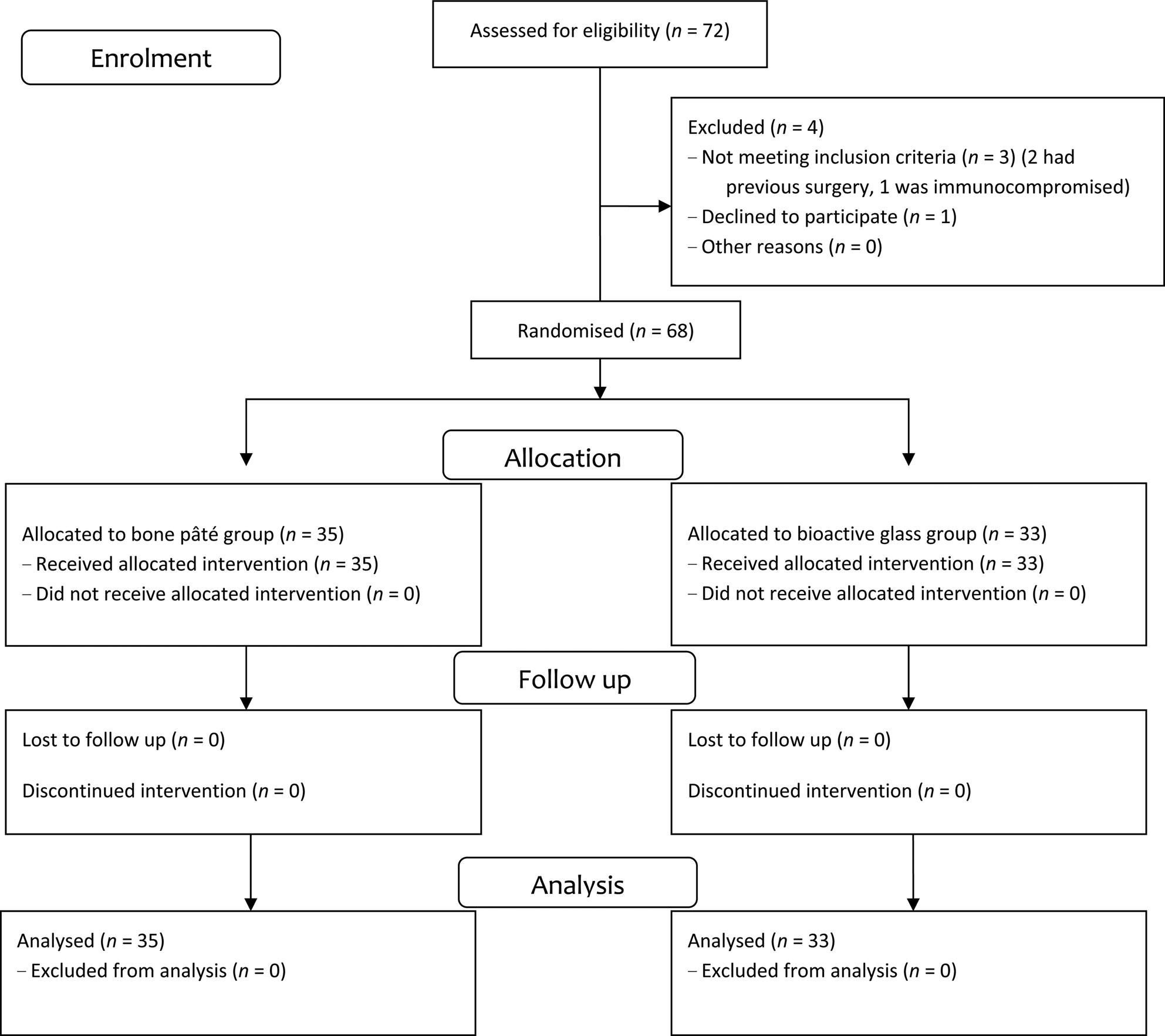
This prospective randomised (1:1) parallel group study was conducted at a tertiary care centre in India between September 2017 and August 2019. Ethical clearance and scientific approval were respectively obtained from the institutional ethics committee and the scientific review committee of our institute. Informed written consent was obtained from all patients regarding undergoing the surgery and participation in the study. A flow diagram of the study is depicted in Figure 1.
Flow diagram of the study.

Inclusion criteria
All patients attending our out-patient department who had active chronic otitis media squamous disease with cholesteatoma were included in the study.
Exclusion criteria
Patients were excluded if they had: previously undergone ear surgery, a history of chronic otitis media complications, a severe systemic illness or an immunocompromised state, or a known allergy to bioactive glass. Patients below 12 years of age were also excluded.
Sample size calculation
The primary objective of the study was to compare the two groups in terms of the success rates in achieving a well-epithelised cavity with a dry ear. Considering this, for an alpha error of 0.05, a beta error of 0.2 and an assumed difference of 25 per cent in the success rates of the two techniques (95 per cent vs 70 per cent) for a two-tailed test, the required sample size was calculated as being a minimum of 33 subjects in each arm.
Patient evaluation
A detailed history was elicited from each patient. A complete ENT examination, including otoendoscopy and otomicroscopy, was performed. Pure tone audiometry was carried out for estimation of hearing thresholds by air conduction and bone conduction. High-resolution computed tomography of the temporal bone was performed to determine the extent of the disease, facial nerve involvement, bony dehiscence, pneumatisation of the mastoid and for surgical planning reasons.
Randomisation and blinding
The patients were allocated to one of the two groups by simple randomisation. The patients were allotted a computer-generated random number between 1 and 100 on enrolment; this number was matched with a pre-concealed computer-generated list of random numbers (1–100) auto-arranged into two groups. This matching was performed in the operating theatre by a resident who had sole access to the concealed list of numbers in the two groups.
All surgical procedures were performed by the same team, comprising two of the most experienced otolaryngologists in our department. The researchers who evaluated the post-operative status of the ear, hearing outcome or adverse events had not operated on the patients and were not aware of the material used in a particular patient.
Intervention
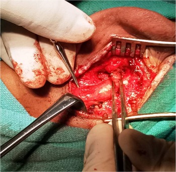
A single-stage procedure involving canal wall down tympanomastoidectomy, hearing reconstruction and mastoid obliteration under general anaesthesia was carried out in both groups via a post-auricular transcortical approach. A temporalis fascia graft and a wide post-auricular musculo-periosteal flap were harvested at the beginning of surgery (Figure 2).
Musculo-periosteal flap raised before tympanomastoidectomy.

The cholesteatoma sac, granulations and diseased mucosa were completely removed. The incus, malleus head and tensor fold were also removed if needed. The key steps included: aggressive saucerisation of the mastoid; removal of bony overhangs; and removal of the external auditory canal posterior wall, down to the level of the facial nerve. The operative area was thoroughly cleaned with saline irrigation, and all areas were inspected using 0 and 30 degree otoendoscopes (Karl Storz, Tuttlingen, Germany) to ensure complete removal of the diseased tissues.
The external auditory canal posterior wall was reconstructed using conchal cartilage, to facilitate hearing reconstruction and mastoid obliteration. The tympanic membrane was grafted using temporalis fascia via the underlay technique. Ossicular reconstruction was performed using a Plastipore™ partial ossicular replacement prosthesis if stapes superstructure was present (type II tympanoplasty) and a Plastipore total ossicular replacement prosthesis if it was absent (type III tympanoplasty). The surgical steps were common to both groups up to this stage.
Mastoid obliteration in bone pâté group
Bone pâté was used to fill the mastoid cavity in this group. The bone pâté was prepared before the start of mastoidectomy. A Sheehy bone dust collector (Otomed, Grace Medical, Memphis, Tennessee, USA) was used to collect bone pâté from the mastoid and squamous cortex of the temporal bone, without exposing air cells. The bone pâté was kept moistened in saline till the completion of mastoidectomy. After completion of mastoidectomy, the bone pâté was used to fill the cavity to the level of the mastoid cortex. The bone pâté was then covered by the post-auricular muscle flap raised at the beginning of the surgery. Temporalis fascia was then spread over the reconstructed posterior canal wall and the muscle flap, thus providing complete soft tissue cover over the bone pâté.
Mastoid obliteration in bioactive glass group
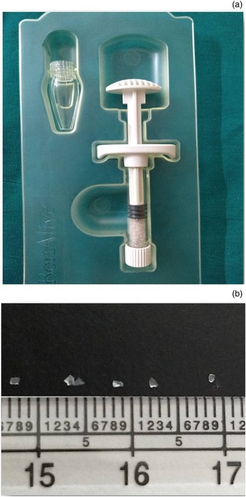
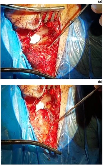
In this group, the cavity was filled with bioactive glass granules (Bon Alive Biomaterials, Turku, Finland), size 0.5–0.8 mm and composition S53P4 (containing 53 per cent silicon dioxide and 4 per cent phosphorus pentoxide) (Figure 3). The granules were first moistened with normal saline solution and then used to fill the cavity to the level of the mastoid cortex. After filling the cavity, excess saline from the granules was suctioned out by gently placing a suction tip over the granules. A fibrin glue (Tisseel; Baxter, Vienna, Austria) was then spread over the bioactive glass granules to bind them together. Subsequently, the bioactive glass granules were covered by post-auricular muscle flap and temporalis fascia, to provide complete soft tissue cover, as in the case of bone pâté (Figure 4).
Bioactive glass granules used in the study. (a) Granules in the pre-filled syringe packaging. (b) Close-up view of a few granules spread on paper to show their size. The smallest marking on the ruler denotes 1 mm.

Mastoid cavity obliteration using bioactive glass granules. (a) Bioactive glass granules filling in the cavity. (b) Musculo-periosteal flap being spread to cover the granules.

Closure and post operative care
The harvesting of conchal cartilage for the posterior canal wall reconstruction resulted in adequate meatoplasty; hence, no additional cartilage was excised for this purpose. Finally, the exterior auditory canal was packed with ribbon gauge impregnated with bismuth iodoform paraffin paste. The post-auricular incision was closed in layers after haemostasis. A standard mastoid dressing was applied.
The patients received amoxycillin clavulanate intravenously, at a dose of 25 mg/kg (maximum 1000 mg) of amoxycillin, after a sensitivity test and before the induction of anaesthesia. This was continued 12-hourly for 48 hours in the post-operative period, followed by oral administration for the next 5 days. In patients allergic to penicillin or beta lactams, azithromycin 10 mg/kg (maximum 500 mg) was administered intravenously before the induction of anaesthesia and once daily for 2 days post-operatively, followed by 500 mg once daily orally for the next 3 days.
Mastoid dressings were removed after 48 hours. Sutures were removed after 7 days. The ribbon gauge pack from the external auditory canal was removed after three weeks. Thereafter, the patients were followed up at 6 weeks, 12 weeks and then 6-monthly. During follow up, patients were asked about symptoms and the ear was examined using an otoendoscope. Hearing assessment by pure tone audiometry and QoL assessment by Glasgow Benefit Inventory scores were performed at 12 months post-operatively. Diffusion-weighted magnetic resonance imaging (MRI) was also ordered in all patients at 12 months’ follow up to detect recidivism.
Main outcome measures
The following main outcome measures were noted: surgery duration (in minutes), measured from the time of skin incision to application of the mastoid dressing; success in achieving a well-epithelised cavity with a dry ear, as assessed using Merchant's grading systemReference Merchant, Wang, Jang, Glynn, Rauch and McKenna21 (grade 0 or 1 was considered a successful outcome, while grades 2 and 3 were considered failure); recidivism, diagnosed based on the presence of squamous epithelium (cholesteatoma) found on otoendoscopic examination or the detection of a high-intensity signal with no post-contrast enhancement on diffusion-weighted MRI at 12 months post-operatively; hearing status, based on pure tone averages by air conduction and mean air–bone gap for four frequencies (0.5, 1, 2 and 4 kHz) pre-operatively and at 12 months post-operatively, performed as per Committee on Hearing and Equilibrium guidelines;22 QoL, assessed using Glasgow Benefit Inventory scores at 12 months post-operatively; and any complications of the procedures.
Statistical analysis
Continuous variables are presented as mean ± standard deviation values, and categorical variables are shown as frequencies and percentages. Depending on the type of data, parametric and non-parametric tests were used to compare the groups. The data were analysed using SPSS statistical software, version 21.0 (IBM, Chicago, Illinois, USA). Level of statistical significance was set at p ≤ 0.05.
Results
Demographics and descriptive data
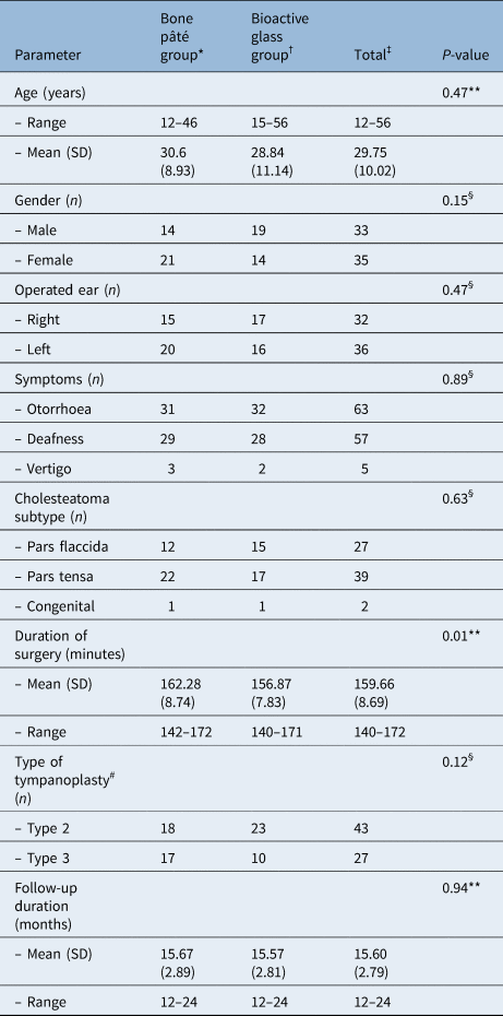
The study population consisted of 68 patients, 33 males and 35 females, aged 12–56 years, with mean age of 29.75 ± 10.2 years. The bone pâté group comprised 35 patients and the bioactive glass group consisted of 33 patients. The patients were followed up for 12–24 months, for an average of 15.6 ± 2.79 months. There was no significant difference between the groups in age, gender, side of operated ear, main symptoms, cholesteatoma subtype or type of tympanoplasty (Table 1).
Demographic and descriptive data

*n = 35; †n = 33; ‡n = 68. **One-way analysis of variance; §chi-square test. #Wullstein classification. SD = standard deviation
Operating time
Duration of surgery was significantly less in the bioactive glass group than in the bone pâté group (156.87 ± 7.83 minutes vs 162.28 ± 8.74 minutes; p = 0.01).
Well-epithelised cavity with dry ear
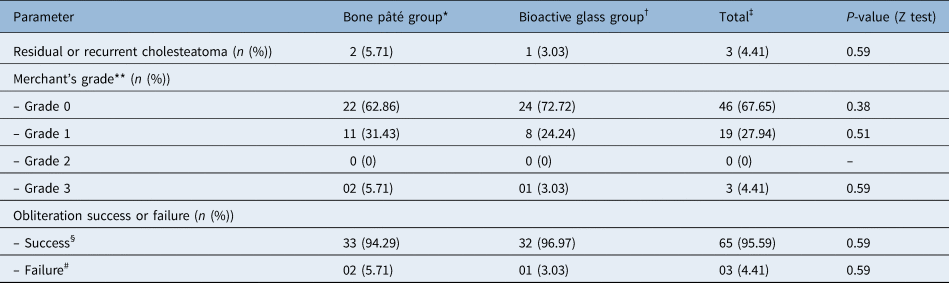
At 12 months post-operatively, the filler materials retained their volume and all patients had shallow or negligible cavities. Success in providing a well-epithelised cavity with a dry ear (Merchant's grade 0 and 1) was achieved in 65 patients (95.59 per cent), while failure (Merchant's grade 2 and 3) occurred in 3 patients (4.41 per cent). There were no statistically significant differences between the two groups in these parameters (Table 2).
Status of mastoid cavity 12 months post-operatively

*n = 35; †n = 33; ‡n = 68. **Adapted from Merchant et al.Reference Merchant, Wang, Jang, Glynn, Rauch and McKenna21 §Merchant's grade 0 and 1; #Merchant's grade 2 and 3. Grade 0 = no episode of otorrhoea, and no pus or granulation tissue on otological examination. Grade 1 = one episode of otorrhoea of less than two weeks’ duration in a three-month period, or no otorrhoea but a subjective feeling of wetness in the ear. Grade 2 = more than one episode of otorrhoea in a three-month period, or an episode of otorrhoea lasting more than two weeks, or an otological examination showing localised granulation tissue or pus that was promptly cured with antibiotic drops, curettage or silver nitrate cautery. Grade 3 = constant purulent otorrhoea on a daily basis, or otological examination showing extensive granulation tissue, or a need for a revision procedure to control infection.
Recidivism
Overall, three patients (4.41 per cent) showed residual or recurrent cholesteatoma, as detected by otoendoscopy and diffusion-weighted MRI. Two of these patients were from the bone pâté group and one was from the bioactive glass group. The difference was not significant (p = 0.59). The cholesteatoma was identified by its hyper-intense signal on diffusion-weighted MRI. Partial osseointegration was also seen on imaging, but no osteolysis of the bone in contact with filler material was noted. All three cases of residual or recurrent cholesteatoma were present in the sinus tympani and not in the obliterated mastoid.
Hearing outcomes
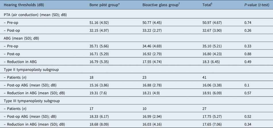
Mean pre-operative pure tone average by air conduction (for all patients of both groups together) was 50.97 ± 4.67 dB, and the mean air–bone gap was 35.10 ± 5.21 dB. The corresponding values were 32.67 ± 3.90 dB and 16.80 ± 4.23 dB, respectively, 12 months after surgery. The pre- versus post-operative differences in pure tone average by air conduction and in air–bone gap were highly significant (p < 0.001 for both measurements). The mean closure of the air–bone gap was 18.3 ± 6.45 dB. There were no statistically significant differences between the two groups for any of these parameters. Further, there was no significant difference in post-operative mean air–bone gap or mean reduction of air–bone gap between the two groups on subgroup analysis according to type of tympanoplasty (Table 3).
Comparison of pre- and post-operative hearing status 12 months post-operatively

*n = 35; †n = 33; ‡n = 68. PTA = pure tone average (mean of thresholds at 0.5, 1, 2 and 4 kHz); SD = standard deviation; pre-op = pre-operative; post-op = post-operative; ABG = air–bone gap
Quality of life
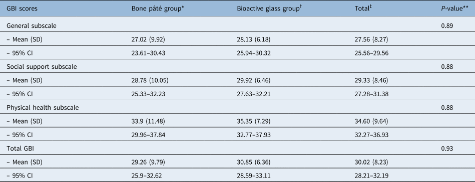
All patients reported good improvements in QoL and benefit from the surgery, as indicated by the total Glasgow Benefit Inventory scores and subscales. Maximum benefit was seen on the physical health subscale. There was no significant difference between the two groups in total Glasgow Benefit Inventory score or any of its subscales (Table 4).
GBI scores after mastoid obliteration 12 months post-operatively

*n = 35; †n = 33; ‡n = 68. **Comparing the means of the two groups (Mann–Whitney U test). GBI = Glasgow Benefit Inventory; SD = standard deviation; CI = confidence intervals
Complications
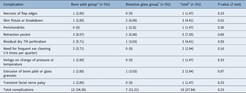
Nineteen patients (27.94 per cent) had complications related to the procedures, the majority of which were minor and could be managed conservatively (Table 5). However, skin flap related issues required antibiotics, local dressings and surgery in one case (excision of tract, temporalis fascia support and rotational skin flap to cover the defect). Tympanic membrane perforations were small and healed after 10 per cent silver nitrate cautery of the edges. There was no significant difference in the number or type of complications between the two groups.
Between-group comparison of complications

*n = 35; †n = 33; ‡n = 68. TM = tympanic membrane
Discussion
The mean operating time was 5.42 minutes shorter in the bioactive glass group than in the bone pâté group. Although this difference is statistically significant, we feel that it is not clinically significant.
We were able to achieve 95.59 per cent success in obtaining acceptably dry, infection-free ears with well-epithelised shallow or negligible cavities (Merchant's grade 0 and 1) after mastoid obliteration in our cases, without any statistically significant difference between the two groups. Merchant's grading was originally described for the control of infection after surgery for chronic otitis media, and included canal wall up and canal wall down procedures, but not obliteration. However, we found it convenient to use this grading system also for obliterated cavities, to compare the success of the two techniques. We could not find an earlier study comparing bone pâté and bioactive glass, but similar or higher success rates have been reported in mastoid obliteration using either bone pâté or bioactive glass individually.Reference Ghiasi5,Reference Sarin, Grenman, Aitasalo and Pulkkinen11,Reference de Veij Mestdagh, Colnot, Borggreven, Orelio and Quak18,Reference Kim, Kim, Lee, Lee, Lee and Lee23
Recidivism was detected in 4.41 per cent of cases in our study, without any significant difference between the two groups. This is similar to the incidence of recidivism reported in cases of canal wall down procedures without obliteration,Reference Pareschi, Lepera and Nucci24 indicating no increase in recidivism following obliteration of the cavity with either bone pâté or bioactive glass. Low recidivism in cases of canal wall down procedures with obliteration of the mastoid cavity has also been reported by other researchers.Reference Mokbel and Khafagy8,Reference de Veij Mestdagh, Colnot, Borggreven, Orelio and Quak18,Reference Harris, Mettias and Lesser25 By obliterating the mastoid and closing the route to the epitympanum, one blocks the route of spread of cholesteatoma. Further, an obliterated mastoid cavity means a smaller area to be epithelised, resulting in faster healing, and less mucosa being available for the absorption of gases, thereby reducing the chances of retraction due to negative pressure.Reference Csakanyi, Katona, Konya, Mohos and Sziklai26 This finding also dispenses with the fear that bone pâté could harbour cholesteatoma, as there was no significant difference in the recidivism in the case of bone pâté compared with bioactive glass.
Both the bone pâté and bioactive glass materials showed partial osseointegration and no bone lysis in our cases. These observations were similar to those of other studies.Reference Bernardeschi, Law-Ye, Bielle, Hochet, Sterkers and Dormont27
The hearing results of the two groups also did not show a significant difference. Good hearing improvement was achieved in both the groups. Overall, a significant reduction in pure tone average by air conduction and closure of the air–bone gap (18.3 ± 6.45 dB) was achieved post-operatively at 12 months. This was in line with other studies.Reference de Veij Mestdagh, Colnot, Borggreven, Orelio and Quak18,Reference Bernardeschi, Pyatigorskaya, Russo, De Seta, Corallo and Ferrary17,Reference Kim, Kim, Lee, Lee, Lee and Lee23 The groups did not show any significant difference on sub-analysis according to type of tympanoplasty. Surprisingly, the results of type III tympanoplasty were similar to those of type II tympanoplasty. This improvement could be a result of the reconstructed posterior canal wall and obliterated mastoid cavity, which provided better restoration of anatomy, allowing a better functional outcome.
It is difficult to say how much the restoration of normal anatomy by obliteration added to the hearing improvement already achieved by ossiculoplasty, because mastoid obliteration and ossiculoplasty were performed in all patients and there were no cases of non-obliterated mastoids in our study for comparison. In an earlier study, hearing neither improved nor deteriorated significantly in obliterated mastoids compared with the non-obliterated state if ossiculoplasty was not performed. However, a significant reduction in the air–bone gap was seen in patients who underwent ossiculoplasty in addition to obliteration.Reference de Veij Mestdagh, Colnot, Borggreven, Orelio and Quak18 Thus, hearing improvement can be attributed to better ossiculoplasty after obliteration, rather than to obliteration alone.
In this study, the total and subscale Glasgow Benefit Inventory scores indicated good improvement in the QoL of patients after the surgery, again without significant differences between the two groups. Developed by Robinson et al., the Glasgow Benefit Inventory is used to determine the change in health status after ENT procedures according to patients’ own assessment.Reference Robinson, Gatehouse and Browning28 It is a generic tool for all ENT procedures. The questions are independent of any specific intervention or health condition, and the subject of the questions remains constant. The mean total Glasgow Benefit Inventory score achieved in our cases was 30.02 ± 8.23, which indicates high satisfaction and good improvement in QoL post procedure. Only a few studies have evaluated QoL after mastoid obliteration. Our results are similar to these studies. Bernardeschi et al. performed mastoid obliteration in 41 cases using bioactive glass S53P4 and reported a mean Glasgow Benefit Inventory score of 28, showing good improvement in QoL.Reference Bernardeschi, Pyatigorskaya, Russo, De Seta, Corallo and Ferrary17 Kurien et al.Reference Kurien, Greeff, Gomaa and Ho29 also showed similar Glasgow Benefit Inventory scores in their patients.
Both the groups had similar rates and types of complications, without any significant differences. However, there was a trend towards reduced complications in the bioactive glass group. Perhaps our study size was too small to pick up the difference. Further studies would be required to elucidate this issue.
The majority of the complications resolved with conservative management. The complications were a result of a combined canal wall down and obliteration procedure, and cannot be attributed to obliteration alone, as we used a single-stage procedure for mastoidectomy and obliteration. Previous studies also have reported a low incidence of major complications in these procedures.Reference de Veij Mestdagh, Colnot, Borggreven, Orelio and Quak18 Serious complications resulting from the use of bioactive glass are not described in the literature.Reference Van Gestel, Geurts, Hulsen, van Rietbergen, Hoffmann and Arts14 The same is true for bone pâté.Reference Stoor, Pulkkinen and Grénman20
• The usefulness of bone pâté and bioactive glass in mastoid obliteration has been reported, but no prospective studies have compared the two materials
• This study prospectively compared the efficacy of bone pâté versus bioactive glass in mastoid obliteration following canal wall down mastoidectomy
• This was performed as a single-stage procedure to eradicate chronic otitis media squamous disease
• At 12 months post-operation, a dry epithelised cavity was achieved in 65 of 68 patients; 3 patients showed recidivism
• Operating time was significantly shorter in the bioactive glass group
• However, there were no significant between-group differences in any outcome measure, including air–bone gap and quality of life
The flap-related problems in our cases occurred only in the initial cases where overzealous filling of the filler material was undertaken to avoid volume loss on account of possible shrinkage of the tissues or filler material in the post-operative period. These factors led to stretching of the flap, resulting in vascular compromise. We soon realised the problem, and no such complications occurred in the subsequent cases.
Limitations
The relatively short duration of follow up (mean of 15.6 months) is the main limitation of our study, especially when comparing the rates of recurrence or residual disease. Ideally, the follow-up duration after such procedures should be 10 years, as recurrence may take up to 10 years to appear.
Conclusion
Both the bone pâté and bioactive glass materials achieved: high success rates in achieving a dry ear, low recidivism, and a good hearing outcome and QoL post procedure, without significant differences between them. The cost of filler material in the case of bioactive glass (approximately US$270 per millilitre) adds to the overall cost of the procedure. However, the limited available quantity of bone pâté in individual cases may restrict its use.
Acknowledgement
The authors are grateful to the Director General, Armed Forces Medical Services, India, for providing financial assistance to this study through the Armed Forces Medical Research Committee Project (number 4975/2017).
Competing interests
None declared