Introduction
Squirrel monkeys (Saimiri spp.) are neotropical primates with a lifespan of approximately 30 years (Helvacioglu et al., Reference Helvacioglu, Aksel, Yeoman, Williams and Abee1994). Female reproductive life starts at the age 2.5 to 3.5 years (Baldwin, Reference Baldwin1969; Taube, Reference Taube1980; Boinski, Reference Boinski1992), and at approximately 8 years old a massive depletion in the number of reserve gametes is initiated, resulting in the complete follicular loss when the females are about 20 years old (Walker et al., Reference Walker, Anderson, Herndon and Walker2009). Monkeys from this genus present a seasonal reproduction and an extremely short ovarian cycle, lasting for 7 to 9 days (Lang Reference Lang1967, Wolf et al., Reference Wolf, O'Connor and Robinson1977; Schiml et al., Reference Schiml, Mendoza, Saltzman, Lyons and Mason1999; Trevino, Reference Trevino2007), that is different from other neotropical primates that present longer ovarian cycles, such as 18–23 days for Sapajus apella (Nagle & Denari, Reference Nagle and Denari1983), 20–24 days for Ateles geoffroyi (Campbell et al. Reference Campbell, Shideler, Todd and Lasley2001), 21 days for Brachyteles hypoxanthus (Strier & Ziegler Reference Strier and Ziegler1994, Reference Strier and Ziegler1997), 24–30 days for Callithrix jacchus (Kendrick & Dixson Reference Kendrick and Dixson1983; Dixson, Reference Dixson2001), 15 days for Aotus trivirgatus (Dixson, Reference Dixson and Hearn1983), and 22 days for Saguinus oedipus (Brand, Reference Brand1981). Also, progesterone peak levels are usually higher in Saimiri species (399 ng ml−1) (Wolf et al., Reference Wolf, O'Connor and Robinson1977) than in other neotropical primates such as Sapajus apella (60–100 ng ml−1) (Nagle & Denari, Reference Nagle and Denari1983), suggesting species-specific differences. As observed, information on ovarian cycle and late folliculogenesis is abundant for this genus. However, knowledge related to the early folliculogenesis focusing on preantral follicles of Saimiri sp. or other neotropical species is still needed.
Preantral follicles are the reserve of female gametes, and will decrease with age until females become senile and enter menopause. These follicles represent 90% of the total ovarian follicle population (Gougeon & Chainy, Reference Gougeon and Chainy1987). Besides nutritional and health status, the total population of preantral follicles in the ovary is species specific and age dependent. For instance, the number of follicles per ovary from adult women ranges of 76,000 (19 years old) to 27,000 (46 years old) (Gougeon & Chainy, Reference Gougeon and Chainy1987), each ovary from an adult female of Sapajus apella contains about 51,000 preantral follicles (Domingues et al., Reference Domingues, Diniz, Furtado, Ohashi, Rondina and Silva2004), while in Macaca nemestrina preantral follicular population was found in the range of 30,900 (8 months old) to 9940 (12–13 years old) (Miller et al., Reference Miller, Charlenston, Battaglia, Klein and Soules1999), and in Macaca mulatta preantral follicular population was found to be 910,000 follicles at birth (Baker & Wai, Reference Baker and Wai1976). Almeida et al. (Reference Almeida, Santos, Scalercio, Leão, Haritova, Oskam and Domingues2012) estimated the population of primordial, primary, and secondary follicles in a senile Saimiri sciureus as 915, 230 and 115 follicles, respectively. Besides the number of follicles, morphometric characteristics of preantral follicles can vary with species (Gougeon, Reference Gougeon1996), which can be used to study female reproduction in different animal species. Furthermore, such information can be used to determine characteristics of follicular atresia, infertility, and menopause (Almeida et al., Reference Almeida, Santos, Scalercio, Leão, Haritova, Oskam and Domingues2012). Knowledge on ovarian morphology and follicular population size may support the development of protocols for the conservation in situ and ex situ of endangered primate species (Mayor et al., Reference Mayor, Bowler and Lópezâ–Plana2013). Moreover, knowledge contributes to the understanding of the processes related to formation, growth and maturation of oocytes enclosed in preantral follicles (Scalercio et al., Reference Scalercio, Brito, Domingues, Santos and Amorim2014).
Information on population estimates, with morphological and morphometric characterization of preantral follicles are limited for neotropical primate species, such as Sapajus apella (Domingues et al., Reference Domingues, Diniz, Furtado, Ohashi, Rondina and Silva2004), Saimiri sciureus (Walker et al., Reference Walker, Anderson, Herndon and Walker2009; Almeida et al., Reference Almeida, Santos, Scalercio, Leão, Haritova, Oskam and Domingues2012), and Alouatta caraya (Lopes et al., Reference Lopes, Lucci, Garcia, Azevedo and Báo2006), and absent for the three Saimiri species recently recognized: Saimiri macrodon, Saimiri cassiquiarensis and Saimiri vanzolinii. The last one is a threatened species (IUCN, 2008). Therefore, in the present study, we aimed to estimate the population of preantral ovarian follicles, as well as to characterize the morphology and morphometry of these three Saimiri species: Saimiri macrodon, Saimiri cassiquiarensis and Saimiri vanzolinii.
Materials and methods
Animals
All animals were captured and collected during field expeditions at the Mamirauá Reserve, Amazonas, Brazil (Lopes et al., Reference Lopes, Brito, Paim, Santos, Queiroz and Domingues2017). Animals were euthanized as part of a large research program to investigate different biological aspects of these primate species. All experimental procedures of this study were approved by the Research Ethics Committee and the Mamirauá Institute for Sustainable Development Ethics Committee, under protocol number 002/2012. The license for collection was granted by the Brazilian Institute of Environment and Renewable Natural Resources, through the System of Authorization and Information on Biodiversity (SISBIO 29906-1). All the biological material used in this investigation are deposited at the Mammals Section of the scientific collection of Mamirauá Institute for Sustainable Development.
The ovarian samples were separately maintained in paraffin sections for histological analysis (Lopes et al., Reference Lopes, Brito, Paim, Santos, Queiroz and Domingues2017). Ovarian sections from four adult squirrel monkeys were evaluated: one pair from an adult pregnant S. macrodon, two ovarian pairs from adult S. cassiquiarensis females (one of them pregnant), and one left ovary from a senile S. vanzolinii. The approximate age of the females was estimated based on the phenotype of dental chronology (Smith, Reference Smith1989), and they were classified as adults (ages ranging from 1.5 to 5 years old) or senile (older than 5 years). No pathologies or abnormal findings regarding the ovarian donors were reported.
Histological analysis
The ovaries were fixed in 10% formalin and subsequently prepared for classical histology. Serial sections (5 µm thick) were cut and every 10th section was mounted and stained with hematoxylin and eosin and examined under a converted light microscope (Leica, Wetzlar, Germany).
Preantral follicles were classified according to Domingues et al. (Reference Domingues, Diniz, Furtado, Ohashi, Rondina and Silva2004) in which the oocyte is surrounded by a layer of flattened granulosa cells (primordial follicle), by one layer of flattened and cuboidal granulosa cells (primordial), by one layer of cuboidal granulosa cells (primary), or by two or more layers of cuboidal granulosa cells (secondary). Preantral follicles were characterized as morphologically normal or atretic as described by Domingues et al. (Reference Domingues, Diniz, Furtado, Ohashi, Rondina and Silva2004) and Brito et al. (Reference Brito, Santos, van den Hurk, Lima, Miranda, Ohashi and Domingues2013), in which the normal follicles presented intact basal membrane, absence of pycnotic bodies in the oocyte nucleus, no signs of oocyte and/or granulosa cells degeneration, as well as no shrunken oocyte, nor detachment between oocyte and granulosa cells.
The total number of each type of follicle was estimated using a correction factor as described by Gougeon & Chainy (Reference Gougeon and Chainy1987):
 $$\begin{equation*}
{\rm{Ni}}\;{\rm{ = }}\;{\rm{No}}\;{\rm{^*}}\;{\rm{St}}\;{\rm{^*}}\;{\rm{Ts/So}}\;{\rm{^*}}\;{\rm{dn}}
\end{equation*}$$
where Ni is the total corrected number of follicles of one class; No is the number of follicles observed in the analyzed sections; St is the total number of sections in the ovary; Ts is the thickness of the section (5 µm); So is the number of observed sections; and dn is the mean diameter of the nucleus of the oocyte for each follicle class.
Follicular, oocyte and oocyte nucleus diameters were measured using a digital camera (Moticam® 5, 5.0) coupled to a computer and a morphometric analysis program (Motic Images Plus 2.0ML, Australia). For this, the follicular dimensions were measured with a micrometric eye lens in an optical microscope (×400 magnification). The largest and the smallest diameters of the oocyte nucleus, oocyte and follicle were measured. The mean diameter of each structure was calculated. The thickness of the granulosa layer was estimated in all preantral follicles categories, subtracting the follicular diameter from the oocyte diameter (Domingues et al., Reference Domingues, Diniz, Furtado, Ohashi, Rondina and Silva2004). Follicles were only considered when the nucleus of the oocyte was visible. At least 400 follicles per animal species were measured.
Statistical analysis
Statistical analysis was performed applying the programs BioEstat 5.3 and StatView 5.0 (SAS Institute Inc., Cary, NC, EUA). Comparisons between the number of follicles per left and right ovary within each species were performed with the Wilcoson–Mann–Whitney test. Morphometric data were compared among different follicular class within a same species using ANOVA and Tukey as a post-hoc test. Results are expressed as mean ± standard error of the means (SEM) and differences were considered significant when the P-value was < 0.05.
Results
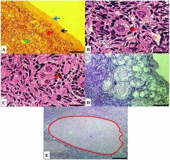
The ovaries were characterized by two regions: cortex and medulla. Most blood vessels were found in the medulla, but the cortical area also presented vascular irrigation, as expected (Fig. 1 A). The ovarian cortex was characterized by the presence of ovarian follicles at different developmental stages (Fig. 1 B, D), including those multi-oocyte follicles (MOFs), which is follicle containing two or three oocytes (Fig. 1 C). This type of follicle was present in all evaluated ovaries (0.4% in S. macrodon, 1.2% in S. cassiquiarensis and 0.2% in S. vanzolinii ovaries), and at all developmental stages of preantral follicles. All ovaries presented a corpus luteum (Fig. 1 E).
Photomicrography of Saimiri ovaries. (A) Histological section of ovarian tissue showing epithelium (blue arrow), tunica albuginea (black arrow), cortex (red arrow), blood vesser (green arrow); ×40 magnification. (B) A transit in follicles is pointed out with a red arrow; ×100 magnification. (C) Multi-oocyte follicle (MOF) enclosing three oocytes (red arrow); ×40 magnification. (D) Preantral follicles at different developmental stages, including secondary follicles (marked with an ‘s’); 100 magnification. (E) A delimited corpus luteum; ×40 magnification. Scale bar: 100 µm.

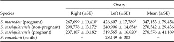
The major population of preantral follicles (mean number of 347,153) was encountered in the ovaries of the S. macrodon, which presented a greater number of follicles in the left ovary (426,607) than in the right one (267,699); thus, a difference >150,000 preantral follicles (Table 1). Both adult non-pregnant and pregnant S. cassiquiarensis females presented similar preantral follicular population, i.e. a mean number of 240,906−299,778 and 237,187–319,565 preantral follicles per ovary respectively. From the senile S. vanzolinii, only the left ovary was recovered and evaluated, presenting the lowest follicular population (28,149); see Table 1.
Mean (±SE) number of ovarian preantral follicles present in the ovaries from S. macrodon (pregnant), S. cassiquiarensis (pregnant and non-pregnant), and S. vanzolinii (senile)

a,b Different letters indicate significant differences between right and left ovaries within the same Saimiri species and category; P< 0.05
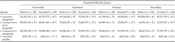
Table 2 depicts the total number of evaluated follicles and the proportions of follicles per class, as well as the number of morphologically normal preantral follicles within each follicular class, with their respective percentages. The ovaries of S. macrodon presented similar percentages of total primordial (32%) and secondary (30%) follicles, being higher than the percentages of transition (20%) and primary (18%) follicles. S. cassiquiarensis females presented ovaries containing mostly primordial (38−39%) and transition (25−27%) follicles, when compared with the percentages of primary (18−19%) and (16−18%) secondary follicles. The ovary from the senile S. vanzolinii female presented the lowest percentages of primordial (15%) and transition (24%) follicles when compared with primary (31%) and secondary (30%) ones. Most preantral follicles (>70%) were morphologically normal in the ovaries from all studied S. macrodon and S. cassiquiarensis females. As expected, the ovary of the senile S. vanzolinii female presented a significant decrease in the percentage of morphologically normal preantral follicles, especially those classified as transition (52%), primary (54%), and secondary (48%) follicles, when compared with the primordial (61%) ones.
Number and mean (±SE) percentage of total and morphologically normal ovarian preantral follicles per class and animal species/category, i.e. in the ovaries from S. macrodon (pregnant), S. cassiquiarensis (pregnant and non-pregnant), and S. vanzolinii (senile)

a–d Different lower-case letters indicate significant differences among the percentages of total follicles per follicular class within the same Saimiri species and category; P < 0.05.
A–D Different upper-case letters indicate significant differences among the percentages of morphologically normal follicles per follicular class within the same Saimiri species and category; P < 0.05.
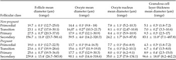
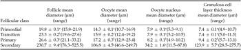
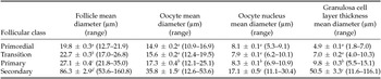
Tables 3, 4 and 5 present the measured mean diameter (µm) of follicles, oocytes, oocyte nucleus, as well as thickness of granulosa cells layer per follicular class when evaluating ovaries from non-pregnant and pregnant S. cassiquiarensis, as non-pregnant S. macrodon and senile S. vanzolinii, respectively. In ovaries from non-pregnant and pregnant S. cassiquiarensis the only remarkable difference observed was the larger follicular and oocyte diameter of secondary follicles from pregnant compared with non-pregnant females (Table 3). However, because only one specimen per group was evaluated, no statistical conclusion can be drawn. Morphometric evaluation resulted in similar results for both females, where follicular diameter increased significantly from primordial to transition, and subsequently from primary and to secondary follicles. Oocyte diameter started to increase significantly only in primary and secondary follicles, and oocyte nucleus increased significantly in diameter only when follicles were at the secondary stage. Thickness of the granulosa cells layer increased significantly in each follicular class after activation (Table 3). In ovaries from S. macrodon (Table 4) and S. vanzolinii (Table 5), follicular and oocyte diameter increased significantly from primordial to transition, and subsequently to primary and to secondary follicles. The same was observed when the thickness of the granulosa cells’ layer was compared within S. vanzolinii follicles (Table 5). In S. macrodon follicles, diameter of oocyte nucleus, as well as the layer formed by granulosa cells increased in thickness from the transition stage to further primary and secondary ones (Table 4). In S. vanzolinii follicles, diameter of oocyte nucleus increased significantly only from the primary to the secondary stage (Table 5).
Mean (± SEM) follicular, oocyte, oocyte nucleus diameter and granulosa cells layer thickness (µm) in primordial, transition, primary and secondary follicles from S. cassiquiarensis (non-pregnant and pregnant)

a–d Different letters indicate significant differences among follicular classes within the same evaluated parameter, i.e. follicular, oocyte, oocyte nucleus, and granulosa cell diameter, and specimen, i.e. non-pregnant or pregnant female; P < 0.05.
Mean (± SEM) follicular, oocyte, oocyte nucleus diameter and granulosa cells layer thickness (µm) in primordial, transition, primary and secondary follicles from S. macrodon (pregnant)

a–d Different letters indicate significant differences among follicular classes within the same evaluated parameter, i.e. follicular, oocyte, oocyte nucleus, and granulosa cell diameter; P < 0.05.
Mean (± SEM) follicular, oocyte, oocyte nucleus diameter and granulosa cells layer thickness (µm) in primordial, transition, primary and secondary follicles from S. vanzolinii

a–d Different letters indicate significant differences among follicular classes within the same evaluated parameter, i.e. follicular, oocyte, oocyte nucleus, and granulosa cell diameter; P < 0.05.
Discussion
Information regarding the reproductive aspects of threatened species is crucial to increase effectiveness of conservation programmes (Andrabi & Maxwell, 2007; Mayor et al., Reference Mayor, Bowler and Lópezâ–Plana2013). Obviously, the paucity of biologic material for evaluation is more accentuated for endangered species (Pukazhenthi & Wildt, 2004). Therefore, analyses must be performed with a limited number of animals. This study is the first to describe morphological and morphometric aspects of initial folliculogenesis in the threatened species S. vanzolinii, as well as of S. macrodon e S. cassiquiarensis, all of them living in the same geographic location.
Follicular population varied between left and right ovaries in each species studied here, as well as among species. The remarkably different follicular population from left and right ovaries found in the S. macrodon female and in the pregnant S. cassiquiarensis female was also previously described in ovaries of Sapajus apella (Domingues et al., Reference Domingues, Diniz, Furtado, Ohashi, Rondina and Silva2004). These later authors studied the ovaries of four adult Sapajus apella females, and recorded more follicles present in the right ovary than in the left one (Domingues et al., Reference Domingues, Diniz, Furtado, Ohashi, Rondina and Silva2004). They suggested that in S. apella the right ovary has more follicles because it is less functional than the left one, since Nagle et al. (Reference Nagle, Digiano, Paul, Terlato, Quiroga and Mendizabal1994) observed the lowest ovulation rates in the right ovaries S. apella. In women, however, no differences in ovulation rates are observed between right and left ovaries (Lass et al., Reference Lass, Croucher, Lawrie, Margara and Winston1997), although some authors describe a more frequent ovulation rate in the right ovary (Potashnik et al., Reference Potashnik, Insler, Meizner and Sternberg1987; Fukuda et al., Reference Fukuda, Fukuda, Andersen and Byskov2000). Although we found the opposite in the present research, our data are restricted to one animal only. However, all together, it can be confirmed that ovarian follicular population is also variable between right and left ovary in different individuals (Miller et al., Reference Miller, Charlenston, Battaglia, Klein and Soules1999).
Importantly, the low number of follicles encountered in the ovary from the senile S. vanzolinii female was most probably a physiologic response of ovarian aging (Tardif et al., Reference Tardif, Richter and Carson1985; Walker et al., Reference Walker, Anderson, Herndon and Walker2009; Almeida et al., Reference Almeida, Santos, Scalercio, Leão, Haritova, Oskam and Domingues2012). Besides aging, reproductive state, nutrition, and genetic factors are also known to affect follicular population (Erickson, Reference Erickson1966; Erickson et al., Reference Erickson, Reynolds and Murphree1976; Cahill et al., Reference Cahill, Mariana and Mauléon1979; Scaramuzzi et al., Reference Scaramuzzi, Adams, Baird, Campbell, Downing, Findlay, Henderson, Martin, McNatty, McNeilly and Tsonis1993; Forman et al., Reference Forman, Hong, Ferry, Tao, Taylor, Levy, Treff and Scott2013).
Primordial, transition, primary and secondary follicles presented morphometric differences as shown before for other species (Domingues et al., Reference Domingues, Diniz, Furtado, Ohashi, Rondina and Silva2004), being indicated to determine follicular morphological quality. Our results confirm that morphometric information supports follicular classification in different developmental stages (Domingues et al., Reference Domingues, Diniz, Furtado, Ohashi, Rondina and Silva2004; Lopes et al., Reference Lopes, Lucci, Garcia, Azevedo and Báo2006). Differences observed among species were previously found when studying other mammals (Koering, Reference Koering1983; Gougeon, Reference Gougeon1996). The changes in the diameters of oocyte nucleus, oocyte, follicle and granulosa cells layer thickness indicate that preantral follicular development follows two distinct phases (Gougeon & Chainy, Reference Gougeon and Chainy1987; Domingues et al., Reference Domingues, Diniz, Furtado, Ohashi, Rondina and Silva2004). In the first phase (activation), the pre-granulosa cells surrounding the oocyte are differentiated to granulosa cells, which is characterized by the change in their flattened to cuboidal structure. In this period, the oocyte size increases (Braw-Tal, Reference Braw-Tal2002). In a second phase, increase in both follicular and oocyte diameters take place together with the proliferation of the granulosa cells (Gougeon & Chainy, Reference Gougeon and Chainy1987; Domingues et al., Reference Domingues, Diniz, Furtado, Ohashi, Rondina and Silva2004). Correlations are difficult to interpret, once herein we have not the possibility to obtain several samples per species.
Multi-oocyte follicles were observed and previously described in other primates (Harrison, Reference Harrison1949; Graham & Bradley, Reference Graham and Bradley1971; Domingues et al., Reference Domingues, Diniz, Furtado, Ohashi, Rondina and Silva2004; Lopes et al., Reference Lopes, Lucci, Garcia, Azevedo and Báo2006). It was suggested that MOFs are a result of an incomplete ovarian organogenesis, i.e. defected follicular formation (Domingues et al., Reference Domingues, Diniz, Furtado, Ohashi, Rondina and Silva2004). It was believed that all those follicles become atretic (Hartman, Reference Hartman1926; Harrison, Reference Harrison1949). However, MOFs can be ovulated (Bysted et al., Reference Bysted, Dieleman, Hyttel and Greve2001; Reynaud et al., Reference Reynaud, Fontbonne, Marseloo, Thoumire, Chebrout, de Lesegno and Chastant–Maillard2005; Silva-Santos & Seneda, Reference Silva-Santos and Seneda2011) as an exceptional phenomenon (Gougeon, Reference Gougeon1981). These follicles present a high concentration of estradiol and low progesterone concentration, which may influence steroidogenesis (Stankiewicz et al., Reference Stankiewicz, Błaszczyk and Udała2009).
In conclusion, for the first time the morphometry and morphological characteristics of preantral follicles were described for three recently recognized squirrel monkey species (S. macrodon, S. cassiquiarensis and S. vanzolinii), and revealed that specific follicular development patterns are present, as well as are age dependent. These differences among species in their early folliculogenesis will be important during the development of reproductive technologies applied to ovarian preantral follicles, especially to protect threatened species such as S. vanzolinii.
Acknowledgements
The study was financially supported by Ministério da Ciência, Tecnologia e Inovação (Mamirauá Institute) and Universidade Federal do Pará. The authors thank Mamirauá Institute and Universidade Federal do Pará for assistance in obtaining research materials.
Conflict of interest
The authors declare that they have no conflict of interest.