INTRODUCTION
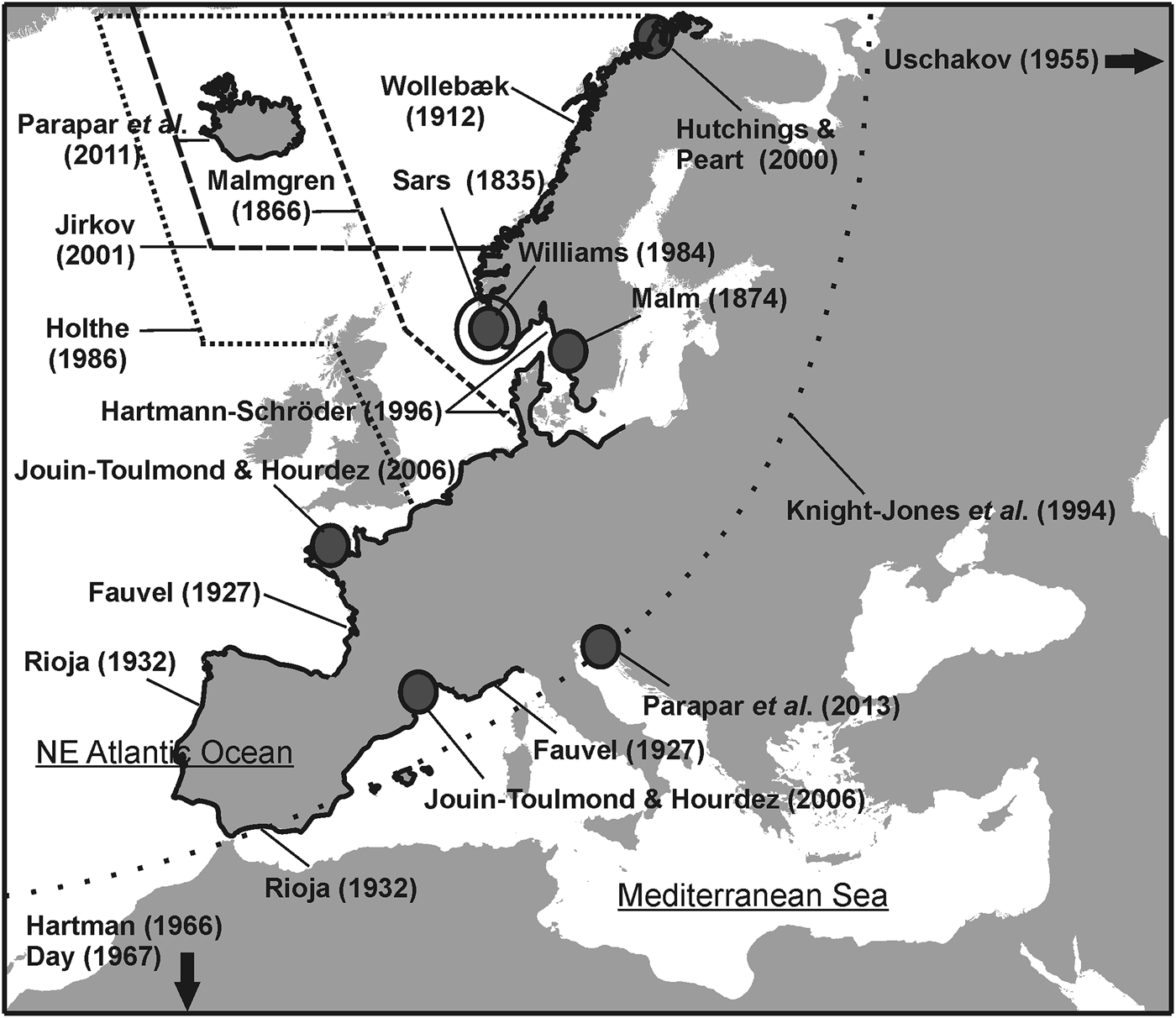
Michael Sars (Reference Sars1835) described Terebellides stroemii from specimens obtained in a locality in the vicinity of the city of Bergen (south-west Norway). Since its description, and probably due to the unique morphology of the branchiae, the taxon has been commonly referred to in the literature from a wide variety of habitats, not only in European waters (e.g. Malmgren, Reference Malmgren1866; Malm, Reference Malm1874; Wollebæk, Reference Wollebæk1912; Fauvel, Reference Fauvel1927; Rioja, Reference Rioja1932) (Figure 1) but also around the world (e.g. Caullery, Reference Caullery1915, Reference Caullery1944; Uschakov, Reference Uschakov1955; Hartman, Reference Hartman1966; Day, Reference Day1967). Throughout the 19th and early 20th Centuries other species of Terebellides were also described worldwide (e.g. Müller, Reference Müller1858; Kinberg, Reference Kinberg1867; McIntosh, Reference McIntosh1885; Caullery, Reference Caullery1915; Hessle, Reference Hessle1917). Later, Williams (Reference Williams and Hutchings1984) revealed the existence of different morphotypes within what was traditionally considered as T. stroemii; over the following years, description of new species of Terebellides around the world increased (e.g. Imajima & Williams, Reference Imajima and Williams1985; Solís-Weiss et al., Reference Solís-Weiss, Fauchald and Blankesteyn1991; Bremec & Elias, Reference Bremec and Elías1999; Hilbig, Reference Hilbig, Blake, Hilbig and Scott2000; Hutchings & Peart, Reference Hutchings and Peart2000; Garraffoni & Lana, Reference Garraffoni and Lana2003; Schüller & Hutchings, Reference Schüller and Hutchings2010, Reference Schüller and Hutchings2012, Reference Schüller and Hutchings2013). Therefore, nowadays, the distribution of taxon T. stroemii is considered to be restricted to North Atlantic waters where it coexists with other species, some of them described recently (e.g. Parapar et al., Reference Parapar, Moreira and Helgason2011, Reference Parapar, Mikac and Fiege2013).
Map of Europe showing areas studied in taxonomic papers with descriptions of Terebellides stroemii.

Polychaetes collected by Michael Sars are located in the Natural History Museum of Oslo (NHMO). Revision of the collection of Terebellides confirmed what Holthe (Reference Holthe1986) already noted, i.e. that M. Sars (Reference Sars1835) did not designate any individuals as type material of this species. The concept of type species was not formalized until 1905 with the publication of the International Code of Zoological Nomenclature (Blanchard et al., Reference Blanchard, von Maehrenthal and Stiles1905). Therefore, a designation of a neotype and a redescription of the taxon becomes necessary because numerous genetically distinct, yet morphologically similar morphospecies to T. stroemii are known to exist. To date, several sibling species have been identified using molecular methodologies; these species will be described and named elsewhere (Nygren et al., in preparation).
In this paper, a description of the adult morphology of T. stroemii from south-west Norway is provided based on preserved material. A neotype was selected from a set of specimens taken by M. Sars at Manger, a locality situated near Bergen. The neotype is compared with material hitherto described as T. stroemii in North Atlantic waters, as well as other species described or reported from the area. By defining the type species, a baseline for a future revision of the Terebellides stroemii species complex is established.
MATERIALS AND METHODS
All the examined specimens are deposited in the collections of the NHMO with accession numbers from NHMO C5896 to NHMO C5908 and NHMO C5955 to NHMO C5969 for type collection and NHMO C5909 to NHMO C5919 for complementary non-type material. Observations and photomicrographs were made with light microscopy using an Olympus BX40 light microscope and an Olympus SZX9 stereomicroscope. Specimens used for examination with scanning electron microscopy (SEM) were prepared by critical point drying, covered with gold in a BAL-TEC SCD 004 evaporator, and examined and photographed under a JEOL JSM-6400 scanning electron microscope at the SAI (University of A Coruña-UDC, Spain). Methyl green (MG) stain saturated in 80% ethanol (ETOH) was used to determine MG staining patterns following the methods of Schüller & Hutchings (Reference Schüller and Hutchings2010). The specimen used for micro-computed tomography (micro-CT) scan at the Marine Biology Station of A Graña (University of Santiago de Compostela, USC, Spain) was originally preserved in ethanol 80% and dehydrated in successive baths of ethanol 90 and 96%, then immersed 2 h in hexamethyldisilazane (HMDS) and allowed to air dry overnight (Alba-Tercedor & Sánchez-Tocino, Reference Alba-Tercedor and Sánchez-Tocino2011; Faulwetter et al., Reference Faulwetter, Dailianis, Vasileiadou and Arvanitidis2013). Scanning was done with a Skyscan 1172 (Bruker, Belgium) microtomograph using the following parameters: 40 kV, 250 µA, unfiltered, and image pixel size of 5 µm. Images were reconstructed with the NRecon software, and cleaned with CT Analyzer software (both Skyscan software). To visualize the data, DataViewer and CTVox softwares (also from Skyscan) were used. One specimen was used for DNA extraction at the Department of Biological and Environmental Sciences (University of Gothenburg, Sweden) following the methodology described by Nygren et al. (Reference Nygren, Eklöf and Pleijel2010).
Abbreviations used in the text
- TC
thoracic chaetiger
- SG
segment.
RESULTS
The evolution of the species illustration
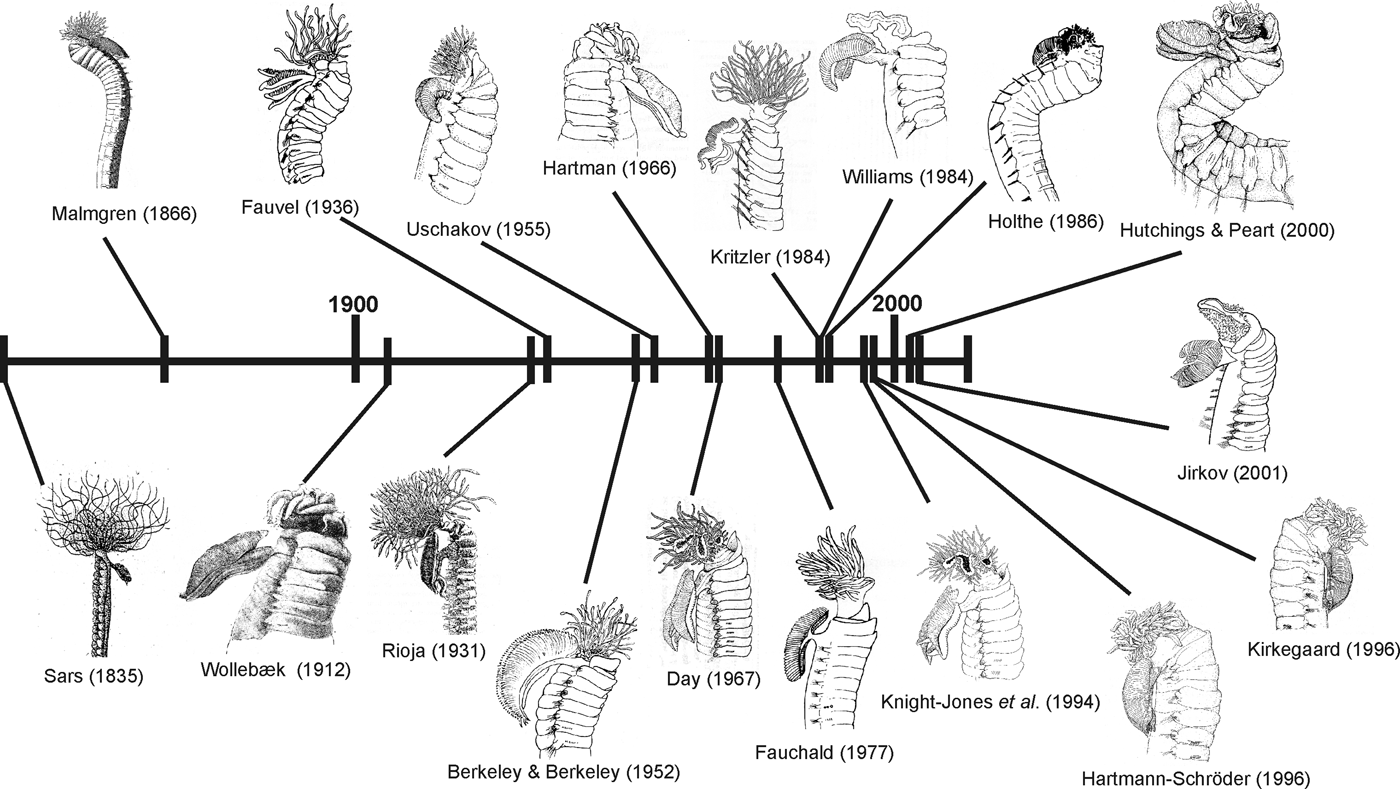
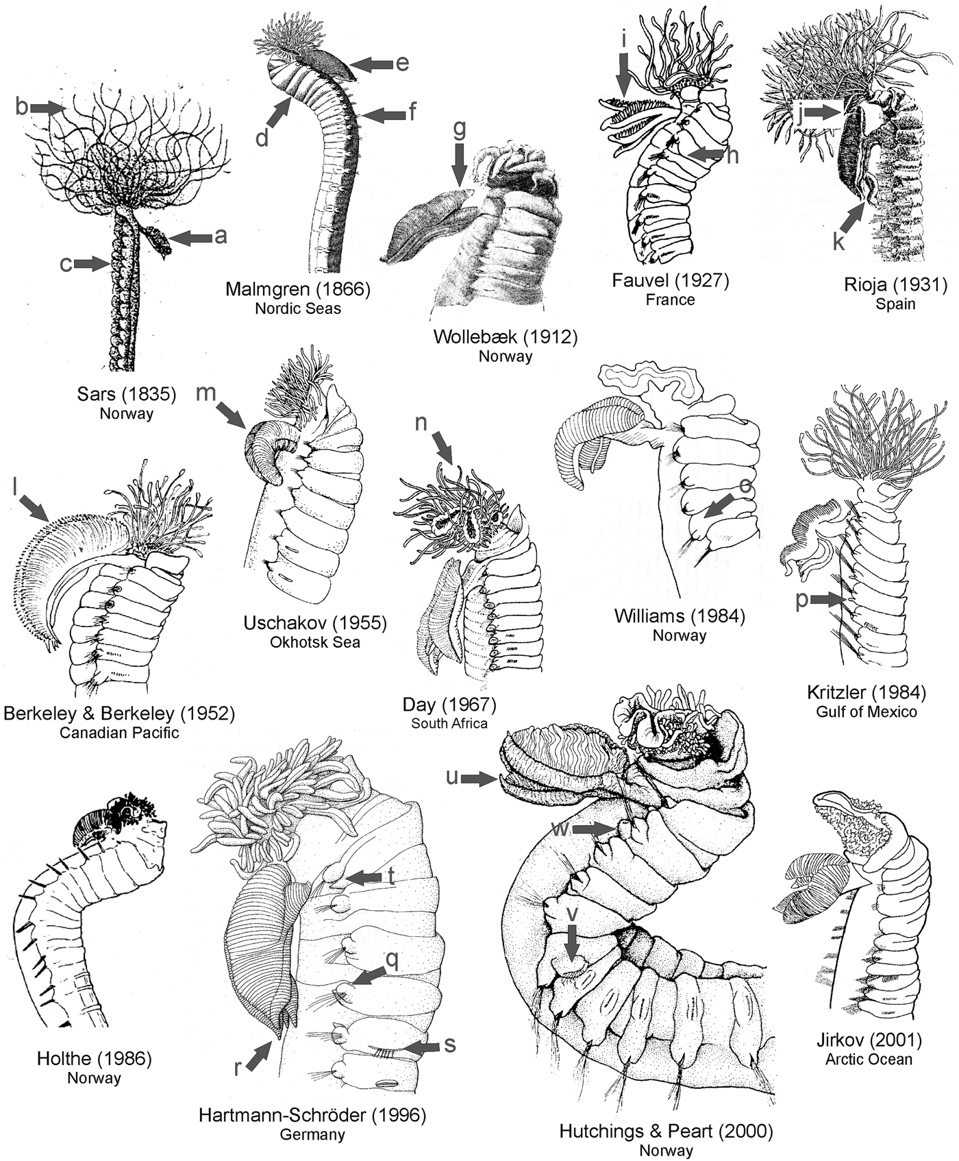
A review of the taxonomic literature provided us with numerous illustrations attributed to Terebellides stroemii, reflecting the evolution of the illustration of this supposedly single species. The largest number of drawings is from the second half of the 20th Century and particularly in the decades of the 1980s and 1990s, including Williams (Reference Williams and Hutchings1984) (Figure 2). Figure 3 shows illustrations from the aforementioned works along with the geographical location of the material. The first drawing corresponds to the original description by M. Sars (Reference Sars1835); in this picture only three characters are clearly depicted: branchiae, an undifferentiated mass of long buccal tentacles, and the thoracic ventral glandular pads (Figure 3A–C). Later, Malmgren (Reference Malmgren1866) provides a more explicit illustration, showing the general body shape, the development of anterior glandular lappets, the actual shape of the branchiae and the position of the chaetigers (Figure 3D–F). Wollebæk (Reference Wollebæk1912) illustrates in more detail the branchiae, particularly the lamellate structure, the stalk and the fifth lobe (Figure 3G). Fauvel (Reference Fauvel1927) proposes a completely different design, although purportedly illustrating the same species, but already showing some ‘modern’ characters, removing the shading of the body surface, reducing the excessive presence of the buccal tentacles and illustrating new characters still considered important today, such as the ‘lateral lappets’ (Figure 3H); the branchiae, with lobes unfused (Figure 3I), are totally different to the ones previously illustrated by Sars, Malmgren and Wollebæk. A few years later, Rioja (Reference Rioja1931) somewhat returns to the style of Sars, with an illustration excessively overloaded and depicting few relevant body characters except the better defined shape of the buccal tentacles, and the general shape of the branchiae; the latter are presented in more detail, with a big anterior fifth lobe (Figure 3J) and long ventral lobes (Figure 3K). The only drawings of the mid-20th Century are the ones by Berkeley & Berkeley (Reference Berkeley and Berkeley1952) and Uschakov (Reference Uschakov1955), who present few body details and show two very different views of the branchiae, i.e. extremely long in Berkeley & Berkeley (Reference Berkeley and Berkeley1952) (Figure 3L) and very small, simplified, and with no distinction between the branchial stalk and the branchial lobes in Uschakov (Reference Uschakov1955) (Figure 3M). The drawing by Day (Reference Day1967) represents further progress, improving the details of the branchiae and especially the anterior region, showing for the first time details of the tentacular membrane surrounding the mouth (Figure 3N). Nevertheless, there are still significant gaps in the information (e.g. shape of chaetigers and chaetae). Day's drawing is subsequently copied by Knight-Jones et al. (Reference Knight-Jones, Knight-Jones, Nelson-Smith, Hayward and Ryland1994) which is not included here because no additional information is provided. The work of Williams (Reference Williams and Hutchings1984), although marking a turning point in the consideration of T. stroemii as a cosmopolitan species, contributed very little to its illustration, showing a general over-simplification of the body, but providing some characteristics worth mentioning, i.e. the presence of dorsal projections in anterior thoracic segments, already illustrated by Fauvel (Reference Fauvel1927), and the position of geniculate chaetae in TC6 (Figure 3O). Kritzler (Reference Kritzler, Uebelacker and Johnston1984) was the first to mark the position of the nephridial openings in TC1, TC4 and TC5 (Figure 3P). The tendency to simplify the drawings is dramatically reflected in Holthe's (Reference Holthe1986) illustration, which is very poor in taxonomically relevant details. In the 1990s, there was already awareness that T. stroemii is not a cosmopolitan taxon but probably limited to northern European waters and maybe cohabiting with other sympatric species. This has led authors to begin to provide more detailed illustrations, such as Hartmann-Schröder (Reference Hartmann-Schröder1996) and Hutchings & Peart (Reference Hutchings and Peart2000) who present the best illustrations of the modern concept of a non-cosmopolitan T. stroemii. The opposite view is the one defended by Jirkov (Reference Jirkov2001), who considers this species as a more widely distributed taxon, probably reflected in a less detailed drawing of the species. The drawing by Hartmann-Schröder (Reference Hartmann-Schröder1996) improves the quality and detail of many characters, e.g. shape of notopodia and neuropodia (Figure 3Q), pointed projection in posterior region of branchial lobes (Figure 3R), shape of geniculate chaetae (Figure 3S) and nephridial openings in TC1, TC4 and TC5 (Figure 3T). Surprisingly, the author is mistaken when placing the geniculate chaetae in TC5 and the first neuropodium with neuropodial hooks in TC6, which is a character not found so far in any species of the genus. This mistake in the position of the geniculate chaetae was later emended by Kirkegaard (Reference Kirkegaard1996) when reproducing Hartmann-Schröder's drawing. Hutchings & Peart (Reference Hutchings and Peart2000) published the last drawing to date of European specimens of T. stroemii, improving the illustration of several characters, e.g. branchial shape (Figure 3U), lateral lappets and dorsal projections of anterior segments, shape of geniculate chaetae and chaetigers (Figure 3V) and relative length of notochaetae, avoiding excessive schematization and showing some new ones such as the presence of nephridial openings in TC2 (Figure 3W). Recent papers by Parapar et al. (Reference Parapar, Moreira and Helgason2011, Reference Parapar, Mikac and Fiege2013) reporting T. stroemii in Iceland and the Adriatic Sea, respectively, are within the current trend of giving more relevance to the SEM images than to line drawings.
Temporal line showing most relevant illustrations of Terebellides stroemii.
Illustrations of Terebellides stroemii since original description by Sars (Reference Sars1835) to Jirkov (Reference Jirkov2001). See text for characters referred by letters.
Even a cursory review of the drawings in the aforementioned papers reveals clear differences between the illustrated specimens, suggesting that several species are probably involved (Figure 3). Thus, focusing only on the best represented character, i.e. the branchial shape, significant differences are observed with regard to the size (e.g. Berkeley & Berkeley, Reference Berkeley and Berkeley1952 vs Uschakov, Reference Uschakov1955), the degree of fusion between lobes (e.g. Wollebæk, Reference Wollebæk1912 vs Fauvel, Reference Fauvel1927) and to the presence/absence of a fifth lobe (e.g. Rioja, Reference Rioja1931 vs Williams, Reference Williams and Hutchings1984).
Search for the type locality
M. Sars (Reference Sars1835, p. IV) mentioned the locality of Glesvær in Bergensfjord (Figure 4) which M.E. Petersen suggested as the probable type locality of T. stroemii (personal communication in Hutchings & Peart, Reference Hutchings and Peart2000); in the actual description (Sars, Reference Sars1835, p. 49, last paragraph). Sars, however, explicitly mentions this locality saying that he found one specimen in sediments there (‘This Annelid is very rare; I have only once met it at Glesvær on a muddy seabed, where it from mud and clay makes itself a short and thick tube, in which it lives. This tube seems not much longer than the animal itself, and very weak and fragile.’).
Map of the Norwegian coast showing localities referred to in the text.
Torleif Holthe, during the preparation of the volume on Terebellomorpha for the Marine Invertebrates of Scandinavia (MIOS) (Holthe, Reference Holthe1986), visited the Natural History Museum of Oslo in June 1975 in order to borrow all Terebellomorpha specimens. According to a note by a Research Assistant of the 1970s, he wrote a list of the studied material but unfortunately this list is today missing (A.-H. Rønning, personal communication). In a letter to the NHMO (then Zoological Museum of Oslo, ZMO) and dated 7 February 1978, J.A. Sneli (editor of the MIOS series) required information about Terebellomorpha type material (T. stroemii included) given the lack of information about where the types of several species in Holthe's manuscript were stored. In this letter, Sneli also suggests Glesvær as the type locality for T. stroemii. The reply letter probably written by Kari Krog (then Collection Technician at the ZMO), while confirming suggested type localities for several other species, states for T. stroemii that ‘According to Holthe's computer list, we do not have Glesvær material. We have two samples from Manger [locality north of Bergen in Hordaland county] and Bergen (Kvernevik, Sand, Evje)’. This letter was probably the reason why a less explicit locality (Hordaland, south-west Norway, which includes both Bergensfjord and Hardangerfjord) is used for T. stroemii in Holthe (Reference Holthe1986) (see Figure 4).
Concurrently with Holthe's studies, S. Williams (then Assistant Curator of the University of Southern California) contacted the NHMO in September 1983 in order to study Norwegian specimens of T. stroemii (Williams, Reference Williams and Hutchings1984). Four lots of specimens supposedly collected by Sars from Oslofjord, Manger, Raftsund and Øksfjord were reported by I. Winsness, at that time Research Assistant of the NHMO, in a letter to Williams of September 1983. It is likely that this material was not actually sent to her, since in her paper of 1984, she only refers to specimens from Hardangerfjord (Williams, Reference Williams and Hutchings1984, p. 119).
After several years without any reference to the type material, Hutchings & Peart (Reference Hutchings and Peart2000, pp. 256–258), following Holthe (Reference Holthe1986, p. 170), refer to Hordaland county and, following Petersen (personal communication), also propose Glesvær as the type locality. However, the specimens used for describing the taxon (deposited in the British Museum of Natural History) in this paper were from Porsangerfjord, which is a locality in Finnmark County (northern Norway) and therefore distant from Bergensfjord (see Figure 4).
Recently, Parapar et al. (Reference Parapar, Moreira and Helgason2011) compared Icelandic material with some Norwegian specimens identified as T. stroemii from Bunnefjord (Oslofjord, southern Norway) (see Figure 4) and loaned by the NHMO. They propose that neither Icelandic nor the Norwegian specimens examined by Hutchings & Peart (Reference Hutchings and Peart2000) really correspond to T. stroemii.
The NHMO collection and neotype selection
The NHMO collection of T. stroemii is composed of 16 jars. Most of them have material collected outside the Bergen area, and after Sars' times (e.g. specimens collected by the RV ‘Hvitfisken’ in the early 20th Century; specimens collected by the RV ‘G.M. Dannevig’ in the mid-20th Century and specimens also collected in the mid-20th Century by Marit and Bengt Christiansen in the Drøbak Marine Biological Station, Oslofjord). Two large jars do, however, have material examined by M. Sars, and were revised by Holthe in the 1970s (in one of them the label by Holthe is preserved). The first jar has five vials and two of them are from the same locality (label: ‘Terebellides stroemii, Sted: Helle, Manger, Bergensfj., 30–60 f., Sars, Sandbund’). Each vial has two labels, one in pencil and one in ink. The word ‘Sars’ on these labels means that they were sampled by M. Sars, but his original labels have now disappeared, probably because someone removed them as they were unreadable and someone else has copied the information onto new labels later; which has been the case for many of M. Sars' labels (A.-H. Rønning, personal communication). Neither on the ink label, written by Kari Krog in the late 1970s, nor on the earlier pencil label is there any date (probably this was not included in the M. Sars original label, as he hardly ever included dates on his labels). Other vials in the same jar have more specimens labelled as T. stroemii, but from localities different to Bergensfjord, such as the Christianiafjord (=Oslofjord), and Øxfjord (=Øksfjord, Finnmark county)—locality reported for T. stroemii in M. Sars (Reference Sars1851)—and Raftsund, Nordland County (northern Norway) (Figure 4). Interestingly, in both vials old black-framed labels still remain, which were most likely done by M. Sars (Oug et al., in press). The exact dates when the specimens from Manger were collected are not known. In 1829–1830 Michael Sars was in Bergen; in 1831 he got the position of vicar of Kinn near Florø, ~200 km north of Bergen, from 1840–1855 he was vicar at Manger and in 1855 he moved to Oslo as a professor (A.-H. Rønning, personal communication). The sampling at Glesvær (and Solsvik also mentioned in his 1835 paper) was most probably done while he was in Bergen, and sampling at Manger was probably done after 1840 (E. Oug, personal communication).
The second jar labelled as ‘Terebellides stroemii; rev. T. Holthe 1975; Norsk materiale: Blant annet M. Sars og G.O. Sars materiale’ has 11 vials with specimens collected from different Norwegian localities and by different collectors, both M. Sars and his son, Georg Ossian Sars, as well as more recent material.
In conclusion, it seems that Michael Sars never specified any specimen/s of the museum collection used in the original description of T. stroemii. Therefore, there are no available syntypes from which to select a lectotype and paralectotypes. If this material ever existed, it is no longer in the NHMO and has left no trace of its past presence (most Terebellides specimens of the NHMO collection have never been allocated accession numbers).
Therefore, a description of the taxon T. stroemii Sars, Reference Sars1835 is provided below, from specimens collected by M. Sars in the locality of Manger in the Bergensfjord.
Description of the taxon
Family TRICHOBRANCHIDAE Malmgren, Reference Malmgren1866
Genus Terebellides Sars, Reference Sars1835
Terebellides stroemii Sars, Reference Sars1835
(Figures 1–10; Table 1)
Terebellides stroemii M. Sars, Reference Sars1835: 48–50 pl. 13, figures 31a–e.
Stereomicroscope images of several specimens of type collection of Terebellides stroemii: (A) neotype (NHMO C5896); (B) neotype, detail of anterior end; (C) neoparatype NHMO C5898 used in micro-computed tomography; (D) neoparatype NHMO 5901; (E) neoparatype NHMO 5899 (F) neoparatype NHMO 5902. Abbreviations: bl, branchial lobe; gr, glandular region; PER, peristomium; PRT, prostomium; SG, segment; TC, thoracic chaetiger.
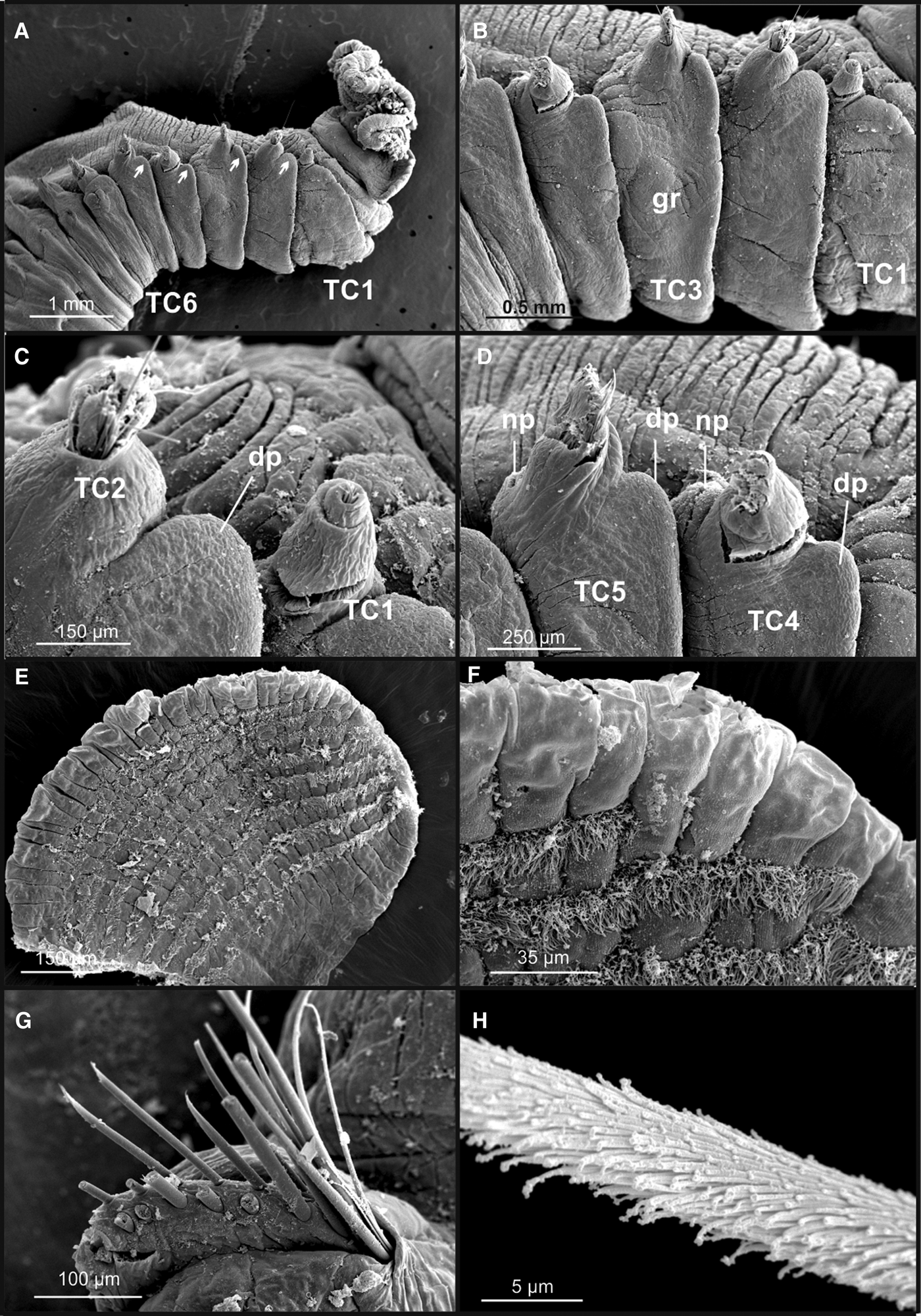
Scanning electron microscopy micrographs of Terebellides stroemii. Neoparatype NHMO C5897: (A) anterior end, right lateral view, branchiae missing; (B) TC1–TC5; (C) TC1 and TC2; (D) TC4 and TC5; (E) branchial lamella showing rows of cilia; (F) edge of branchial lamella; (G) disposition of thoracic notochaetae in two rows, (H) detail of covering of notochaetae. Abbreviations: dp, dorsal projection; gr, glandular region; np, nephridial pore; TC, thoracic chaetiger. Arrows showing position of dorsal projections from TC2 to TC5.
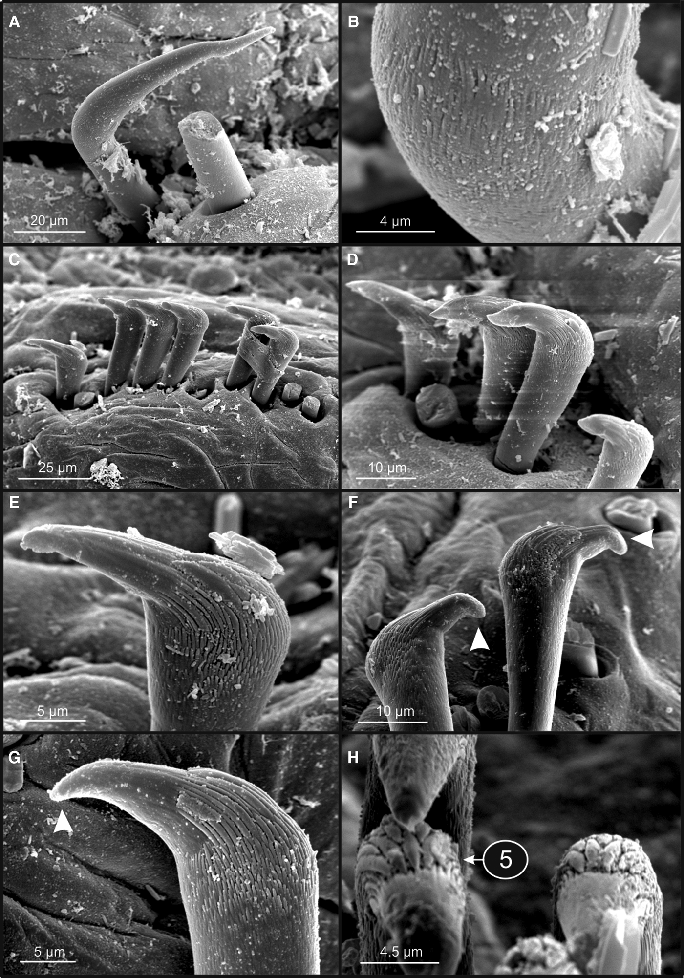
Scanning electron microscopy micrographs of Terebellides stroemii. Neoparatype NHMO C5897: (A) geniculate chaeta of TC6; (B) detail of denticles in capitium of geniculate chaetae; (C–H) different views of thoracic uncini. Arrow in (F) and (G) showing bent distal end of rostrum; encircled number in (H) showing number of teeth upper to rostrum.
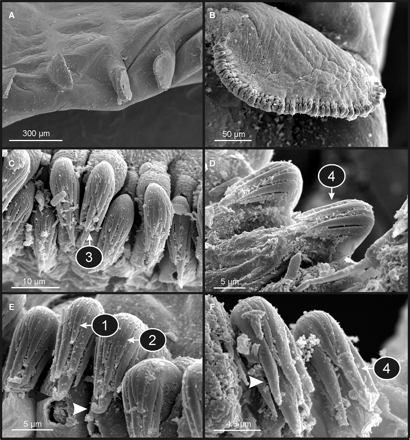
Scanning electron microscopy micrographs of Terebellides stroemii. Neoparatype NHMO C5897: (A) abdominal neuropodia; (B) detail of one abdominal neuropodium; (C–F) different views of abdominal uncini. Arrows in (E) and (F) showing bent distal end of rostrum and first row of capitium denticles respectively; encircled number in (E) showing number of teeth in second row of teeth, and in (F) showing number of teeth upper to rostrum.
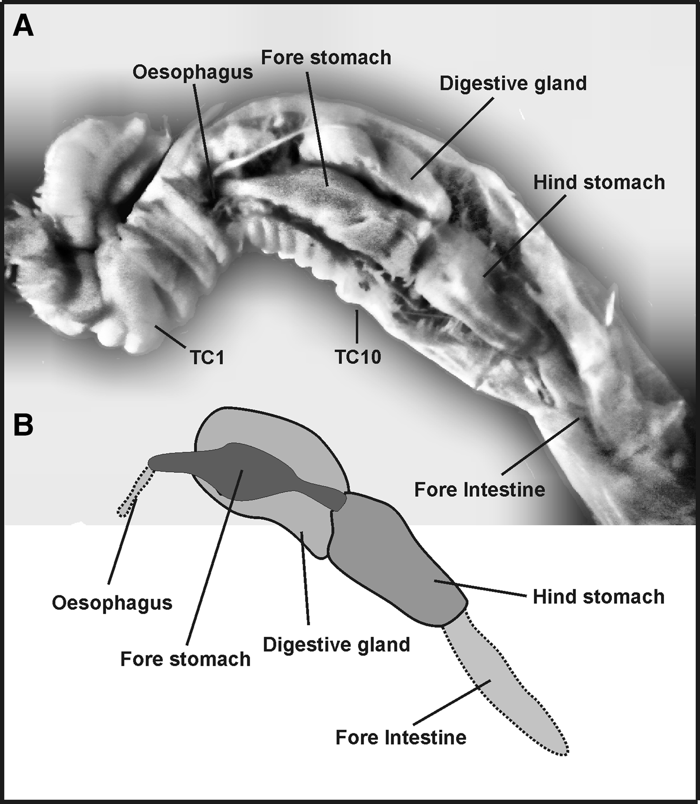
Internal anatomy of Terebellides stroemii: (A) stereomicroscope image of a dissected specimen, later used for scanning electron microscopy (NHMO C5897); (B) schematic representation of the stomach parts.
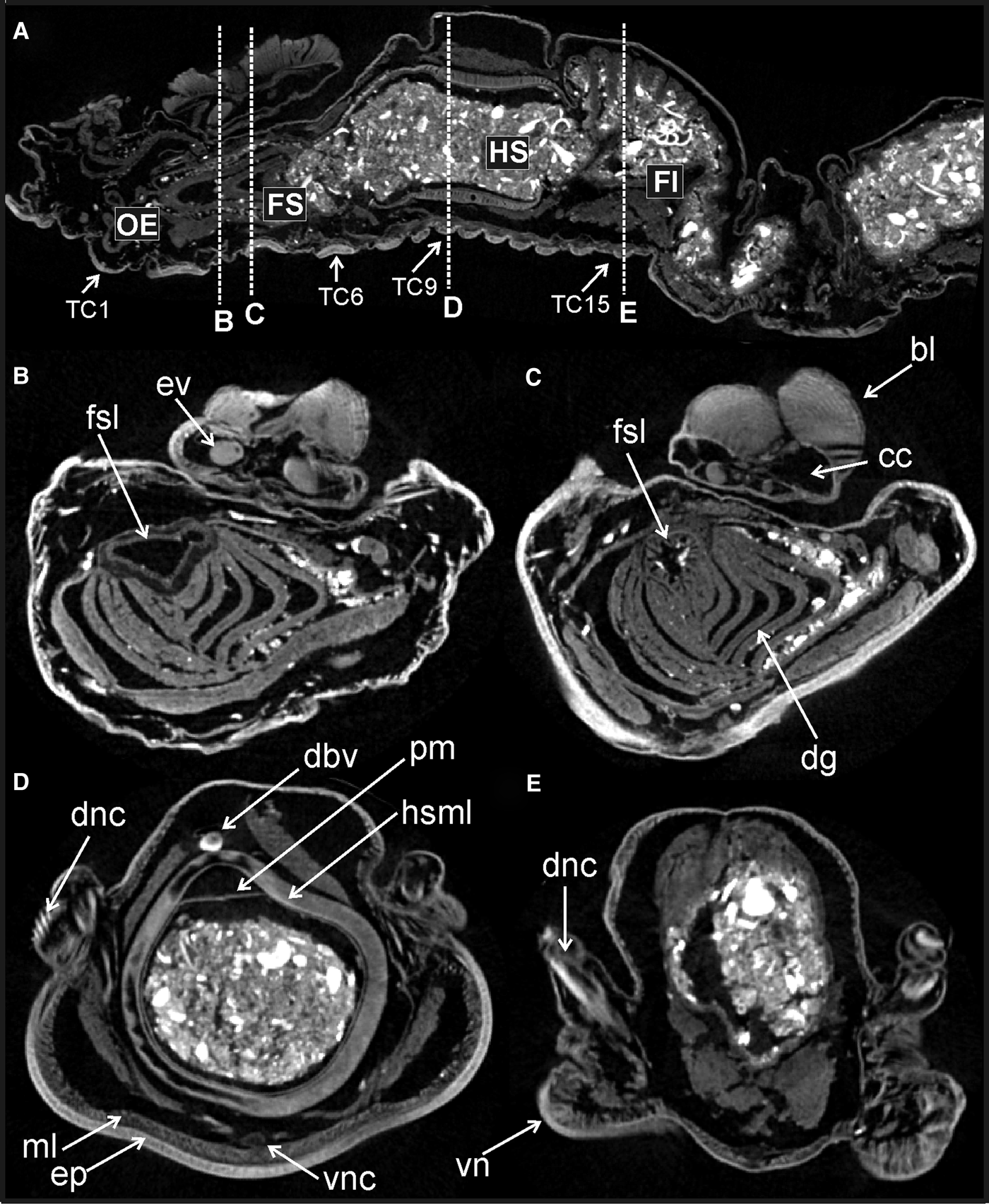
Micro-computed tomography sections of Terebellides stroemii neoparatype NHMO C5898: (A) sagittal section; dotted lines (B–E) marking level of transversal sections showed in figures (B–E). Abbreviations: bl, branchial lamella; cc, coelomic branchial cavity; dbv, dorsal blood vessel; dg, digestive gland; dnc, dorsal notochaetae; ep, epidermis; ev, efferent branchial vessel; FI, fore intestine; FS, fore stomach; fsl, fore stomach lumen; HS, hind stomach; hsml, hind stomach muscle layer; ml, muscle layer; OE, oesophagus; pm, peritrophic membrane; TC, thoracic chaetiger; vn, ventral neurochaetae.
MATERIAL EXAMINED
Type material
As was noted above, given the lack of specimens in the NHMO identified by M. Sars as type material of T. stroemii, we selected the neotype and neoparatypes from specimens of this collection which meet the requirements of being collected by Michael Sars in the Bergensfjord. These specimens belong from two vials with identical labels: ‘Helle, Manger, Bergensfjord (about 60°37′15″N; 05°02′00″E), 30–60 f (=55–110 m). Sandbund (=sandy seabed), Sars’.
The reference numbers for type specimens are the following: Neotype (NHMO C5896) and 27 neoparatypes (NHMO C5897 to NHMO C5908 and NHMO C5955 to NHMO C5969); all specimens are presumed to be always preserved in ethanol. Among those paratypes, NHMO C5897 was initially dissected for study of digestive tract and was posteriorly used for SEM study; NHMO C5898 was used for micro-CT analysis, NHMO C5899 was used for MG staining, and NHMO C5900 was used for molecular analysis.
Non-type material
Other specimens, which also seem to match the original description here proposed for T. stroemii, but not collected in the Fjord of Bergen, were also separated from the NHMO general collection of polychaetes. Genetic analyses that are being carried out with specimens belonging to this genus in Norwegian waters are revealing a high presence of cryptic species, not described so far. It is therefore a risk that morphological variation described from specimens from areas out of the Bergenfjord actually reflect differences between separate species. For this reason, these specimens were identified as Terebellides af. stroemii and appear as non-type material. This material belongs to two groups (see localities in Figure 4):
(1) Specimens presumed to be collected by M. Sars
• ‘Bollærne, Christianiafjord, Sars, 8–10 faths’ (15–18 m), 2 spec. (NHMO C5909).
• ‘Øxfjord’, 3 spec. (NHMO 5910).
• ‘Raftsund (mund.), 250–300 m’, 1 spec. (NHMO C5911).
(2) Specimens collected after M. Sars times
• Material collected during cruises of the R.V. ‘Hvitfisken’
– ‘Hvitfisken, Kristianiafjord, Hvitsten 7-10-VII-1914, 50–80 f. (91–146 m), ler (=clay) og fjeld (=rock)’, 5 spec. (NHMO C5912).
– ‘Hvitfisken, Kristianiafjord, OSTÖEN 1911 9/5’, 1 spec. (NHMO C5913).
– ‘Hvitfisken, Filtvedt-Brendtangen. Kristianiafjord 1910, 18/8’. 1 spec. (NHMO C5914).
• Material collected during cruises of the R.V. ‘G.M. Dannevig’
– ‘Dannevig 1953, St. nr. 57, Svenør – Jomfruland’; 4 spec. (NHMO C5915).
– ‘GM Dannevig, Stn. 6, Stangholmen, Møkkalasset’ 3 spec. (NHMO C5916).
• Material collected from the Drøbak Marine Biological Station by Marit and/or Bengt Christiansen
– ‘Lauer, Hvaler; 59°0.15′N; 11°1.4′E; 06.08. 1969, skrap 3, Christiansen, 15–10 m’; 3 spec. (NHMO C5917).
– ‘Froungen, 23-VII-1953, Skrap l., Leg. MEH-BCh’ 3 spec. (NHMO C5918).
– ‘Rammeboen, Hvitsten; 05-XII-1952, skrap 3, Leg. M.E.H. – B. Ch.’ 3 spec. (NHMO C5919).
DESCRIPTION OF THE NEOTYPE (FIGURE 5A, B)
Complete specimen, 52 mm long and 7.0 mm wide; body tapering posteriorly with segments becoming increasingly shorter and more compacted towards pygidium; gravid, body cavity with oocytes. Prostomium compact; large tentacular membrane surrounding mouth provided with many buccal tentacles with expanded tips and some not tapered. First segment (SGI) forming an expanded structure below tentacular membrane. Lateral lappets on SGIII–VII, thoracic chaetigers (TC) 1–5, largest on TC1–3 and declining in size posteriorly on TC4–TC5. Conspicuous dorsal rounded projections on TC1–5 and oval-shaped glandular lateral region on TC3.
Branchiae arising as single structure from TC1 and reaching TC3, consisting of a single elongate and annulated stalk situated mid-dorsally, and made up of two pairs of lobes fused along half of their lengths; lower pair thinner. Dorsal (anterior) lobes provided with about 80 tightly packed lamella. Posterior region of both upper and lower lobes with very short terminal pointed projections, a large anterior branchial projection (fifth lobe) present.
Eighteen pairs of thoracic notopodia (SGIII–XX; TC1–18). Notopodia present from TC1 well-developed, but slightly smaller in size than subsequent notopodia; notochaetae of TC1 almost similar in length than following notochaetae and vertically aligned. Neuropodia present as sessile pinnules from TC6 to pygidium and with uncini arranged in single rows throughout from TC7 (SG IX). First thoracic neuropodia (TC6) provided with 11 sharply bent acute tipped, geniculate chaetae on both sides. Second and all subsequent thoracic neuropodia with around 30 uncini per torus arranged in one irregular row. Thirty-six abdominal neuropodia as erect pinnules paddle shaped with an entire margin provided with about 70 uncini per torus.
Three pairs of nephridial openings, located dorsal to each notopodium of SGIII, SGVI and SGVII (TC1, TC4 and TC5). Pygidium blunt, as funnel-like depression.
Colour in alcohol pale orange. MG staining pattern 5, compact green colouration from TC1 (SGIII) to TC3 (SGV), becoming a striped pattern from TC4 (SGVI) to TC12 (SGXIV) and fading in following segments.
MORPHOLOGICAL VARIABILITY AND CHAETAE MORPHOLOGY
The study of neoparatypes under stereo microscope (Figure 5C–F) and SEM (Figures 6–8) allowed to establish the range of variability of several body characters as well as the detailed structure of chaetae.
Complete individuals ranging from 23–60 mm in length and 2.5–6.0 mm in width. Dorsal rounded projections more conspicuous on TC2–5 (Figure 6A–D). Branchiae showing slightly different shapes due to different degrees of ventral contraction (Figure 5C–F). Both sides of branchial lamellae provided with several parallel bent rows of cilia (Figure 6E, F); no tufts of cilia on outer edge present (Figure 6F). Thoracic notochaetae simple capillaries arranged in two rows (Figure 6G) and with textured surface (Figure 6H). Geniculate chaetae ranging from 6–9 in number, provided with minute teeth forming a capitium (Figure 7A, B) and showing different bent degree depending on size, being almost straight in larger specimens (sensu Figure 9b in Hutchings & Peart, Reference Hutchings and Peart2000) and sharply bent in smaller ones (sensu Figure 9d in Hutchings & Peart, Reference Hutchings and Peart2000). Subsequent thoracic neuropodia with 20–37 uncini per torus showing uncini irregularly arranged in posterior thoracic uncinigers in larger animals (e.g. NHMO C5899 and C5901). Uncini as shafted denticulate hooks provided with long, thin and pointed main fang (rostrum) appearing bent terminally giving an ‘eagle head’ appearance (Figure 7C–G). Five teeth located above main fang (Figure 7H) surmounted by a row of 4 denticles and an upper crest of several smaller denticles; dental formula MF:5:4:∞. Abdominal neuropodia ranging from 29–36; provided with about 35–80 uncini per torus (Figure 8A, B); uncini with three to four teeth above main fang (Figure 8C, D), also both appearing with distal point bent (Figure 8E, F), and surmounted by a row provided with an irregular number of 1–5 teeth (Figure 8E) and an upper crest of minute teeth (Figure 8C); dental formula MF:3–4:1–5:∞.
GROSS INTERNAL ANATOMY (FIGURES 9 and 10)
Prior to being prepared for the SEM, neoparatype NHMO C5897 was dissected in the thoracic region in order to get a general view of the gross internal anatomy, in particular that of the digestive system (Figure 9) as Williams (Reference Williams and Hutchings1984) had suggested that such structures could be useful in distinguishing between species of the genus. The dissection showed a highly regionalized anterior part of the digestive tract occupying most of the thoracic region of the body, with two well-defined areas which belong to the fore stomach and hind stomach. The anterior stomach is characterized by being covered by a voluminous digestive gland. The posterior stomach is of similar length as the anterior, has no digestive gland and its wall is much thicker than that of the fore stomach.
Neoparatype NHMO C5898 (Figure 10) was scanned with a micro-CT in order to obtain a non-destructive, complementary view of this part of the digestive tract. Images showed again the same pattern of digestive regionalization as was revealed by the dissection, this time also showing that the stomach is slightly shifted further forward (Figure 10A). The fore stomach is located more anteriorly in the thoracic region, in this case below the branchiae, with the twisted oesophagus limited to TC1 and TC2. Transversal body sections B and C reveal the presence of a massive and layered digestive gland surrounding the fore stomach, with a wider lumen at its anterior part (Figure 10B) than at the posterior part (Figure 10C); the latter is apparently provided with a different inner lining. The hind stomach is characterized by a thick muscular wall and is filled with detritus (Figure 10D). Finally, a thin-walled, accordion-shaped, fore intestine (Figure 10A, E) is present, also filled with sediment and foraminiferans.
DISTRIBUTION
M. Sars' label of vial selected for neotype collection reports the locality of Manger in Bergensfjord, which is about 30 km north of the city of Bergen (Figure 4).
ECOLOGICAL NOTES
Sandy bottoms of shallow shelf depths (55–110 m).
DNA EXTRACTION
Given the likeliness that the specimens of the type series had never been in contact with formaldehyde, a DNA extraction from one of the specimens was attempted. Unfortunately, this attempt, done by Arne Nygren (Department of Biological and Environmental Sciences, University of Gothenburg, Sweden), has been unsuccessful, which could be due to the long time elapsed since the time of collection (Nygren, personal communication).
DISCUSSION
Seven species of the genus Terebellides have been hitherto described or reported in Arctic and north-east Atlantic waters (Parapar et al., Reference Parapar, Moreira and Helgason2011; Jirkov & Leontovich, Reference Jirkov and Leontovich2013): T. stroemii Sars, Reference Sars1835; T. gracilis Malm, Reference Malm1874; T. atlantis Williams, Reference Williams and Hutchings1984; T. williamsae Jirkov, 1989; T. irinae Gagaev, Reference Gagaev2009; T. bigeniculatus Parapar et al., Reference Parapar, Moreira and Helgason2011; and T. mediterranea Parapar et al., Reference Parapar, Mikac and Fiege2013. Two of these species were later proposed as being junior synonyms: T. williamsae of T. gracilis (Hansson, Reference Hansson1998; Parapar et al., Reference Parapar, Moreira and Helgason2011; Jirkov & Leontovich, Reference Jirkov and Leontovich2013) and T. irinae of T. stroemii (Jirkov & Leontovich, Reference Jirkov and Leontovich2013). We do not follow this last synonymy proposed by Jirkov & Leontovich (Reference Jirkov and Leontovich2013) because of the very different shape of the branchiae of T. irinae (see Gagaev, Reference Gagaev2009, figures a and b).
Terebellides atlantis, T. bigeniculatus, T. gracilis, T. irinae and T. mediterranea are well characterized by the possession of at least one given character absent in the other species. These characters are: the unique shape of the branchiae provided with four unfused lobes in T. atlantis and T. irinae; the presence of geniculate chaetae in two chaetigers (TC5 and TC6) instead of one (TC6) in T. bigeniculatus; the whitish ventral coloration of TC1–TC4 in T. gracilis; and the larger size and longer notochaetae of TC1 in comparison to the following ones in T. mediterranea. Terebellides irinae is well characterized against T. atlantis by the possession of very short ventral lobes in branchiae and very long thoracic notochaetae.
In T. stroemii, there is so far no unique morphological feature which allows characterizing the species apart from a set of characters none of which is unique among the north-east Atlantic species of Terebellides. In an attempt to overcome this problem, Parapar et al. (Reference Parapar, Moreira and Helgason2011, Reference Parapar, Mikac and Fiege2013) proposed several anatomical features that might be useful to characterize this species: (1) MG staining pattern; (2) relative size and number of teeth of the capitium of the thoracic uncini; (3) ciliation of branchial lamellae; and (4) dorsal papillae in thoracic and abdominal segments. Unfortunately, many of these characters were not or poorly reported in earlier descriptions of T. stroemii and other Terebellides species.
In order to better characterize our material against other descriptions of T. stroemii, we propose the following diagnostic set of characters: (1) body large, reaching at least 60 mm in length in gravid specimens; (2) TC1 slightly smaller than subsequent thoracic chaetigers; (3) five branchial lobes, being lobes 1–2 and 3–4, respectively, fused for half of their length; (4) lateral folds with conspicuous dorsal projections on TC1–TC5; (5) oval-shaped glandular region on TC3; and (6) geniculate chaetae of TC6 mostly sharply bent but being almost straight in larger specimens.
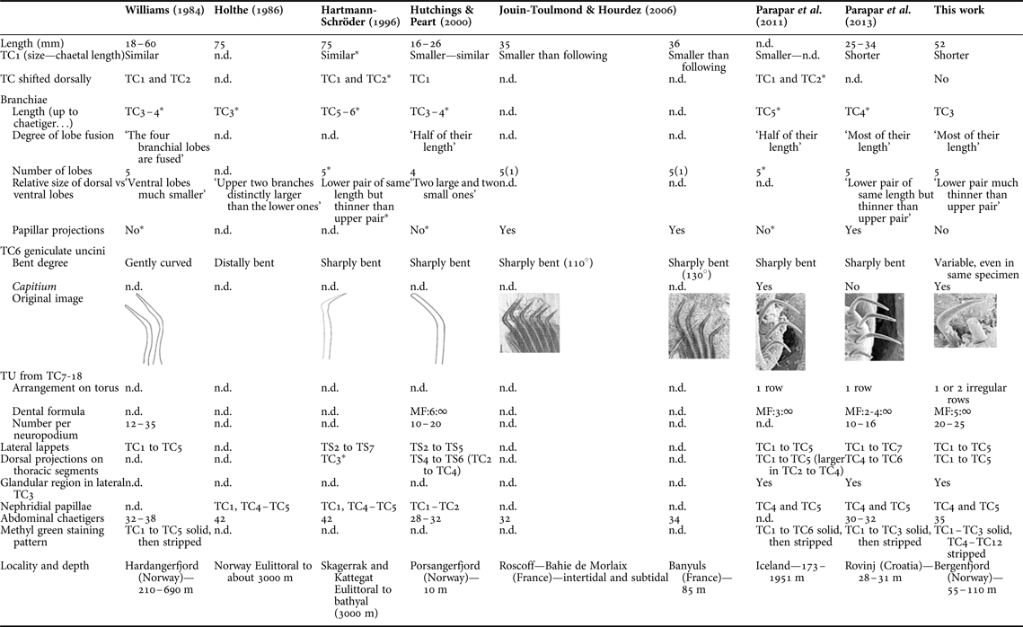
When recent descriptions of T. stroemii are compared, slight morphological differences are also observed, which seem to reveal that, most likely, several sibling species are confused under the same name. Specimens referred to T. stroemii have been recently described and illustrated from material collected in four areas of the north-east Atlantic: Norway (Hutchings & Peart, Reference Hutchings and Peart2000); Iceland (Parapar et al., Reference Parapar, Moreira and Helgason2011); Roscoff and Balyuls-sur-mer (Jouin-Toulmond & Hourdez, Reference Jouin-Toulmond and Hourdez2006); and the Adriatic Sea (Parapar et al., Reference Parapar, Mikac and Fiege2013) (see Figure 1). A comparative study of the morphological characteristics attributed to each of them (Table 1) reveals differences that suggest cryptic species are present, and difficult to be identified using only a traditional morphological study of external characters.
Comparison of some morphological characters of Terebellides stroemii from different sources. Abbreviations: MF, main fang; TC, thoracic chaetiger; TS, thoracic segment; n.d., no data; *, data extracted from drawings because they were not explicitly referred to by the author. (1) Author point 4 in Table 1 but in figures a fifth lobe could be seen
Williams (Reference Williams and Hutchings1984) was the first to suggest the gross stomach morphology as a character valid to differentiate species of Terebellides. This was done in order to distinguish Terebellides distincta Williams, Reference Williams and Hutchings1984 from the other species, and particularly from T. stroemii. The dissection of one specimen of Sars' collection confirmed Williams' observation about the same length of anterior (lamellate) and posterior (muscular) stomach in this species, which is clearly different to the one of T. distincta (see figure 6 in Williams, Reference Williams and Hutchings1984). The micro-CT technique which is still in the early stages of being used in the anatomical reconstruction and taxonomy of polychaetes (Faulwetter et al., Reference Faulwetter, Dailianis, Vasileiadou and Arvanitidis2013), allows a rapid, non-aggressive study of old type material. The possibility of using new characters of the internal anatomy for species discrimination applying this technique may be evaluated in the future, once it becomes more widely used.
In summary, we believe that all previous accounts of T. stroemii, largely from boreal and temperate localities, should be re-evaluated looking for subtle differences other than those related to other traditionally considered characters. To achieve a better characterization of the material, it is essential to address the simultaneous use of different kinds of characters—not solely related to gross morphology—which will help to strengthen the group of characters hitherto used to separate this species complex as a whole, against other species. Those characters will certainly come from a multidisciplinary approach, which will be composed of both a more in-depth study of the traditional characters of the macro-morphology (e.g. general body and branchial shape), but also using other tools, as is the case of the SEM and the micro-CT analysis, the ecology (e.g. bathymetric distribution) and the genetic structure (e.g. COI and ITS analysis).
ACKNOWLEDGEMENTS
The authors wish to thank T.A. Bakke, P.D. Harris and M. Christiansen (Department of Research and Collections, NHMO), for providing information and specimens of Terebellides, and specially A.-H. Rønning (Department of Technical and Scientific Conservation, NHMO) for her help and friendship during J.P.'s visit to the museum. Ann-Helén's knowledge on history of the museum invertebrate collections, was of extraordinary help in clarifying the historical information. Thanks are also due to M. Candás (Marine Station of A Graña-Ferrol, University of Santiago de Compostela, Spain) for the micro-CT analysis and J. Veloso (University of A Coruña, Spain) for the stereomicroscope pictures. A. Castro and C. Sueiro (Servicio de Apoio á Investigación, University of A Coruña, Spain) helped with the SEM, and N. Sánchez (University of Vigo, Spain) provided the maps. Special thanks to A. Nygren (University of Gothenburg, Sweden) who attempted to extract the DNA from one of the neoparatypes, and to J. Moreira (Universidad Autónoma de Madrid, Spain), E. Oug (NIVA Grimstad, Norway), I. Jirkov (Moscow State University, Russia) and Myriam Schüller (Ruhr Universität Bochum, Germany) for their valuable suggestions that greatly improve the quality of this work. Comments of one anonymous referee are also deeply acknowledged.
FINANCIAL SUPPORT
Thanks to A. Sikorsky and Akvaplan-niva (Tromsø, Norway) for partially financing J.P.'s stay in Norway,