Case
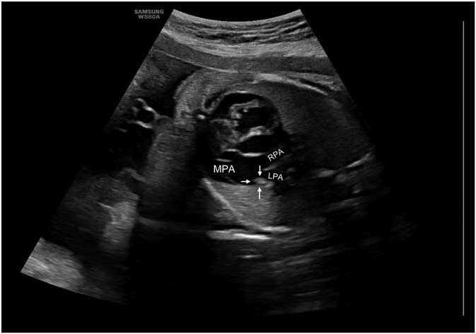
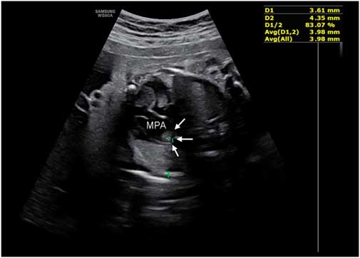
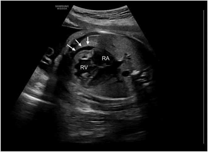
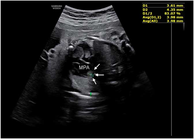
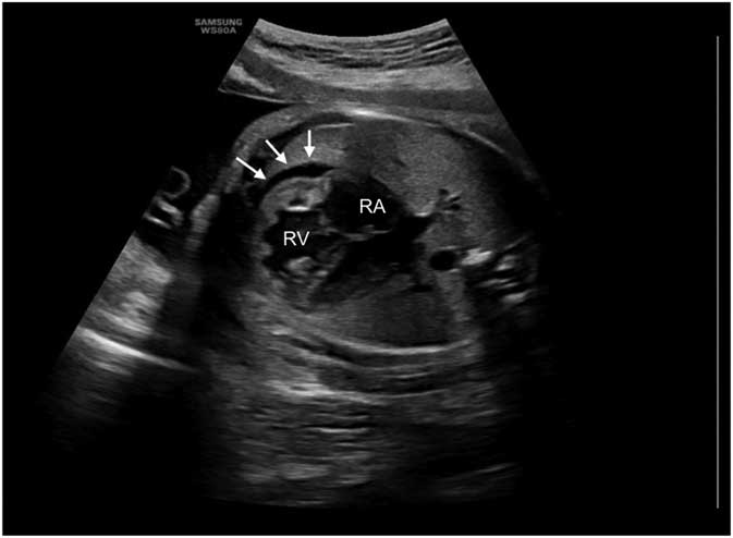
We present a 34-week gestation female, weighing 1750 g, with a prenatal diagnosis of thrombus in the ductus arteriosus. Her mother, a 28-year-old, gravida 7, para 4, had history of gastric ulcer. No history of antenatal anti-inflammatory drug intake or diet rich in polyphenols. At 33 weeks, fetal ultrasound revealed polyhydramnios and intrauterine growth restriction. Fetal echocardiogram showed hypertrophy of the right ventricle with ventricular dysfunction and an echogenic image in the proximal portion of the left pulmonary artery (Figs 1–3).
Fetal echocardiogram – the thrombus is visible in the proximal portion of the left pulmonary artery, occluding the ductus arteriosus. RPA = right pulmonary artery. MPA = main pulmonary artery.

Fetal echocardiogram – the thrombus (3.61 mm × 4.35 mm) is visible in the proximal portion of the left pulmonary artery (white arrows). MPA = main pulmonary artery.
Fetal echocardiogram – pericardial effusion (white arrows), right ventricle hypertrophy, and dilatation. RA = right atrium.
Delivery was through an emergency caesarean section due to non-reassuring fetal status. Apgar score was 9 at minute one and 10 at minute five, with no need for reanimation. Physical examination was normal and oxygen saturations with room air were 95%.
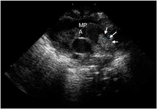
The echocardiogram performed at 6 hours of life revealed dilation, hypertrophy, and moderate dysfunction of the right ventricle, and presence of a completely occlusive thrombus at the pulmonary end of the ductus arteriosus, with no blood flow visible in the ductus (Fig 4).
Echocardiogram – echogenic thrombus (white arrows), 4 mm diameter by 6 mm length, occluding the pulmonary extremity of the ductus arteriosus. MPA = main pulmonary artery.

Chest radiography and electrocardiography were normal. She had low levels of protein-C activity of 17%, with normal range 70–130%, and a moderate deficit of antithrombin-III activity of 43%, with normal range 80–120%. Serial echocardiogram imaging revealed stability of the thrombus dimensions. Six months after discharge the thrombus is limited to the ductus arteriosus and the infant is asymptomatic.
Management of ductus arteriosus thrombosis in the neonatal period remains controversial. In our case, a conservative approach with clinical and echocardiography surveillance provided an excellent outcome.
Acknowledgements
The authors acknowledge Margarida Abrantes, Antónia Santos, Mónica Rebelo.
Financial Support
This research received no specific grant from any funding agency, or from commercial or not-for-profit sectors.
Conflicts of Interest
None.