Depression is prevalent among pregnant womenReference Bennett, Einarson, Taddio, Koren and Einarson 1 , Reference Marcus, Flynn, Blow and Barry 2 and exerts significant neurobehavioral effects on the developing child.Reference Field 3 , Reference Glover 4 Research guided by the Developmental Origins of Health and Disease (DOHaD) model demonstrates that maternal prenatal depressive symptoms are associated with risk for psychopathology, as evidenced by indices of children’s behavioral, physiological and – most pertinent to the current study – neural functioning.Reference Glover 4 – Reference Field, Diego and Hernandez-Reif 6 Evidence suggests that maternal prenatal depression is associated with infant resting brain electrical activity, as measured by electroencephalography (EEG): 2 weeks to 6-month-old infants of women who met criteria for depression during pregnancy exhibit greater activation in the alpha band in the right-frontal cortex relative to the left (referred to as ‘greater relative right-frontal alpha asymmetry’),Reference Field 3 , Reference Field, Diego and Hernandez-Reif 6 a pattern that has been associated with impaired emotional processing, disposition to express negative affect and risk for psychopathology.Reference Allen and Kline 7 – Reference Davidson 9 In contrast, infants of non-depressed mothers typically exhibit approximately equal amounts of frontal activation across right and left hemispheres. However, questions remain regarding the level of depression symptom severity associated with infant brain development and when in development the impact may occur.
Specifically, it is not clear if these offspring neural differences are apparent only when comparing women with and without frank clinical depression, or if the magnitude of the right-frontal asymmetry varies as a function of the severity of maternal depressive symptoms. The existing literature almost exclusively has examined the EEGs of infants of women in depressed v. non-depressed groups. Yet some studies show that the effect of maternal depressive symptoms on other dimensions of child functioning is directly proportional to the severity of depressive symptomatology, even when these symptoms are subclinical.Reference Campbell, Morgan-Lopez, Cox and McLoyd 10 , Reference McCarty and McMahon 11 To our knowledge, the only study that examines maternal depressive symptoms in relation to infant EEGReference Lusby, Goodman, Bell and Newport 12 used data from a sample of women who met DSM-IV criteria for a major psychiatric disorder, most of whom were taking psychotropic medication during the perinatal period. In that report, maternal prenatal and postnatal depressive symptoms interacted to predict 3-month olds’ EEG asymmetry such that prenatal depressive symptoms were associated with greater right-frontal asymmetry only when mothers also endorsed high levels of depression postpartum. Although these findings provide compelling evidence that maternal depressive symptoms during pregnancy may be associated with infant EEG asymmetry, it remains unclear (a) whether these findings extend to samples of women whose psychiatric histories are less severe and who are not taking psychotropic medication, and (b) at what point in infant development this association emerges. It is possible that prenatal exposure to maternal depression increases the child’s susceptibility to the effects of the postnatal environment, but in itself is not associated with EEG asymmetry. Studying newborn EEG in relation to the range of maternal prenatal depressive symptoms may help determine the relevance of subclinical levels of maternal depression for infant brain development, and whether this effect begins in utero, consistent with the DOHaD model. Examining EEG at birth also offers interpretive advantages, as it removes many confounded postnatal exposures such as parental sensitivity, which in the context of maternal depression can be significant.Reference Murray 13
Using data from a group of women who were at-risk for postpartum depression but who varied in the amount of depressive symptomatology during pregnancy, this study addressed the following research question: Are maternal prenatal depressive symptoms associated with greater relative right-frontal alpha asymmetry in the newborn? We predicted that maternal depressive symptoms would be associated with greater right-frontal alpha (3–13 Hz) asymmetry.
Methods
Participants
Data used in the current study came from a small randomized control trial (RCT) that compared enhanced treatment as usual (ETAU) to Practical Resources for Effective Postpartum Parenting (PREPP; NCT02121496), a brief intervention program aimed at preventing postpartum depression.Reference Werner, Gustafsson and Lee 14 This intervention consists of (1) teaching mothers behavioral techniques to help soothe their crying baby (e.g. swaddling, carrying), (2) providing psychoeducation about the postpartum period (e.g. hormone changes, normative infant crying patterns) and (3) teaching mindfulness meditation techniques to help mothers fall back asleep after waking to care for their infant. This RCT began late in the 3rd trimester of pregnancy, and targeted maternal postnatal behaviors, thus no prenatal effects were hypothesized; intervention group membership (PREPP v. ETAU) was not correlated with newborn EEG metrics.
Pregnant women ages 18–45 were recruited through the Department of Obstetrics and Gynecology at Columbia University Medical Center (CUMC) during their 2nd or 3rd trimesters of pregnancy. Enrollment in the RCT (n=54) required that the woman be identified as at-risk for postpartum depression, defined as scoring above 24 on the Predictive Index of Postnatal Depression.Reference Cooper, Murray, Hooper and West 15 Women who reported smoking tobacco or using recreational drugs, lacking fluency in English, currently receiving psychological/psychiatric treatment, taking medication or having a medically complicated pregnancy were not eligible for enrollment. All study procedures were approved by the Institutional Review Board of the New York State Psychiatric Institute/CUMC.
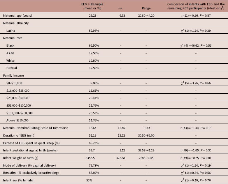
Data included in the current study came from a subsample of women (n=18) enrolled in the overarching study. Because of the relatively immobile nature of the EEG equipment and the strict hospital-level approvals necessary to conduct EEG assessments in the hospital shortly after birth, we were able to obtain EEG data only from some participants who delivered at CUMC, and never for those delivered elsewhere. EEG sessions were not attempted for newborns who were born prematurely or who required medical intervention during the early postpartum period. Importantly, the subsample used in the current study did not differ from the complete RCT sample on any of the variables reported in the current manuscript (see Table 1).
Sample demographics

EEG, electroencephalographic; RCT, randomized control trial.
Procedures
Between 36 and 38 gestational weeks, before RCT randomization, participants completed a battery of self-reported and rater-administered measures, including assessments of prenatal mood and household demographics. Shortly after the birth of the child, two research assistants visited the mother and her newborn in the Labor and Delivery Unit at the main hospital at CUMC. They conducted the EEG session in a room adjacent to the newborn nursery, typically on the 2nd day of life (mean=1.39 days, s.d.=0.78 day). Newborn respiration and electrocardiogram were monitored during the EEG session. Information relevant to the child and mother’s medical history was culled from medical charts.
Measures
Maternal depressive symptoms
When women were 36–38 weeks pregnant, maternal prenatal depressive symptoms were measured using the clinician-administered Hamilton Rating Scale for Depression (HRSD). This 15–20 min rater-administered measure indexes depressive symptoms over the previous 2 weeks.Reference Williams 16 HRSD values of 8–13 indicate mild depression (n=3 in our sample), 14–18 moderate depression (n=2) and 19+ severe depression (n=6). The continuous depressive symptoms score was used in all analyses.
EEG acquisition and sleep state coding
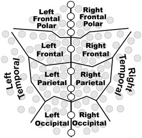
The data collection procedures used in this study have been described previously.Reference Welch, Myers and Grieve 17 , Reference Grieve, Isler and Izraelit 18 EEG was recorded for ~60 min using a 128-electrode data acquisition systems (EGI Inc, Eugene, OR, USA), however, only 125 leads are used in infants (see Fig. 1). Following consultation with the manufacturer of the EEG system, we used special methods to maintain electrode/scalp contact, including covering the electrode net with plastic wrap to diminish evaporation of the saline in the electrode sponges and covering the net and plastic wrap with an elastic material (Surgilast; Derma Sciences, Princeton, NJ, USA). These procedures generally allowed impedances to be kept below 50 kΩ per manufacturer’s recommendation. The EEG was sampled at 1000 samples/second using a vertex reference. The voltage from each lead was band-pass filtered from 0.1 to 400 Hz and then digitized with 16 bits per sample at the rate of 1000 samples/s. Then, in software, data were re-referenced to the average of the 124-lead EEG montage and processed to obtain measures of spectral power (microvolts2) at specific frequencies for each electrode. The negative of the average reference approximates the voltage at the vertex lead as referenced to a point at infinity, thus producing the 125 leads. See Grieve et al.Reference Grieve, Isler and Izraelit 18 for additional information about EEG acquisition and signal artifact rejection.
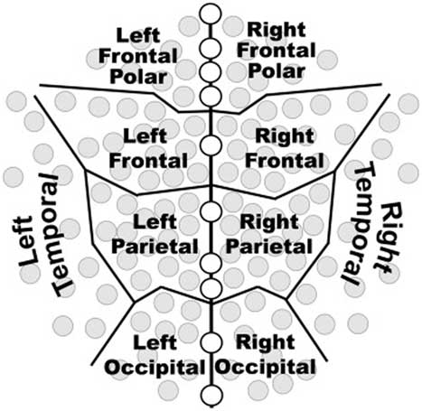
Visual depiction of high-density electroencephalographic (EEG) net. The 125 electrodes in the EEG net are distributed within 10 standard brain regions for the regional analyses of the study. Note 10 midline leads (open circles) were excluded from statistical analyses by region.
Newborn sleep state was determined via infant respiration patterns during the EEG study, as described in Harper et al.Reference Harper, Schechtman and Kluge 19 Before applying the EEG net, an Inductotrace (Ambulatory Monitoring Inc, Ardsley, NY, USA) inductive plethysmograph was placed around the infant’s rib cage and abdomen; infant respiration was measured throughout the EEG session. The coefficient of variation of respiratory cycle time was used to determine sleep state. In a small number of cases (n=4), the respiration belt did not yield usable data due to an equipment malfunction. In these cases, sleep state was determined by examining infant heart rate and heart rate variability using procedures described by van Laar et al.Reference van Laar, Peters, Vullings, Houterman and Oei 20 Periods of quiet sleep were characterized by low heart rate variability and small heart rate oscillation bandwidth, whereas active sleep was characterized by high heart rate variability and wider heart rate oscillation bandwidth. Both of these methods of determining sleep state have been validated against one another and against behavioral measures of determining sleep state.Reference Isler, Thai, Myers and Fifer 21 , Reference Werth, Long and Zwartkruis-Pelgrim 22
EEG power analysis and calculation of right-frontal asymmetry
EEG power was computed using 1-s fast Fourier transforms for each of the 125 leads; average power was computed for each electrode for 30-s epochs throughout the session. Power in each electrode in the frontal polar region (Fig. 1) was averaged to create a single score for the right-frontal polar region and one for the left-frontal polar region. Following standard calculation procedures,Reference Diego, Jones and Field 23 , Reference Dawson, Frey and Panagiotides 24 frontal asymmetry was calculated by subtracting the natural log of the average power in the alpha band (3–13 Hz) in the left-frontal polar region from the natural log of the average power in the right-frontal polar region. Asymmetry was calculated separately for active and quiet sleep.
Analytic strategy
Our research question was tested using multiple linear regression. Specifically, infant alpha asymmetry was regressed on maternal 3rd trimester HRSD scores. Separate regressions were estimated for active and quiet sleep.
Results
Sample descriptives
Table 1 presents descriptive information about the participants included in these analyses. As indicated, this subsample did not differ from the complete sample on any of these variables. Seven infants did not transition into quiet sleep while EEG was being recorded.
Results of regression analysis
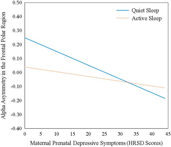
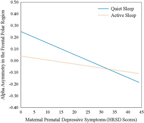
Consistent with expectation, maternal prenatal depressive symptoms were associated with greater right-frontal asymmetry in the alpha (3–13 Hz) band during quiet sleep (β=−0.66, s.e.=0.004, P=0.028). Maternal depressive symptoms were not significantly associated with alpha asymmetry in active sleep, β=−0.23, s.e.=0.004, P=0.37, though the effect was in the predicted direction. See Fig. 2 for a visual depiction of these results.
Visual depiction of the association between maternal prenatal depressive symptoms and infant alpha asymmetry in the frontal polar region during quiet and active sleep. The above figure is a visual depiction of the results of the regression analyses. The lines in this figure correspond to the unstandardized regression coefficient relating maternal prenatal depressive symptoms to alpha asymmetry in the frontal polar region. As can be seen in this figure, greater maternal depressive symptoms are associated with more negative asymmetry scores in quiet sleep (β=−0.66, B=−0.01, P=0.03), indicating greater relative right-frontal asymmetry in the context of maternal depression. This association was not significant for active sleep (β=−0.23, B=−0.003, P=0.37), though the association was in the predicted direction. HRSD, Hamilton Rating Scales of Depression.
Discussion
Using EEG data from a sample of infants assessed in the hospital within hours of delivery, the current study is the first to our knowledge to examine prospectively maternal prenatal depression in relation to newborn EEG. Results suggest that maternal prenatal depressive symptomatology – and not simply categorical, frank, DSM diagnosed depression, as has previously been investigated – is associated with greater relative right-frontal alpha asymmetry during quiet sleep based on EEG recording.
Our findings replicate those of several previous studies that report greater infant and child right-frontal asymmetry in the alpha band (3–13 Hz) in the context of maternal depression; this pattern has been associated with less adaptive emotional processing and risk for psychopathology.Reference Field, Diego and Hernandez-Reif 6 , Reference Davidson 25 Uniquely, we found that greater right-frontal asymmetry varied as a function of depressive symptoms, indicating that maternal prenatal depression, even if below clinical thresholds, may exert a significant effect on the developing brain. These asymmetry differences were present at birth, suggesting in utero origins independent of postpartum influences.
This is the first study to document that these EEG differences are apparent during infant sleep, a more homogenous resting state than has been investigated by previous research, which has been collected while infants were awake when infant attention and orientation is not controlled. We report a significant association between maternal prenatal depressive symptoms and greater relative right-frontal alpha asymmetry during quiet but not active sleep, though the latter association was in the expected direction.
The weaker association between maternal depression and infant EEG during active sleep may reflect methodological differences (e.g. infants move less during quiet sleep, which may result in more accurate data; missing data during quiet sleep may have played a role), or it may represent meaningful differences in infant brain functioning. Given the small sample size of the current study (and thus the possibly reduced power to detect such an effect), future research should replicate these findings before we further interpret these results.
In addition, future research also should investigate the biological mechanisms through which maternal depression may influence the developing brain (e.g. elevated maternal cortisol, inflammation, placental methylation) with regard to alpha asymmetry in particular.
The current study had a number of strengths. First, this prospective, longitudinal study used a rater-administered measure of depression, which offers a more objective account of maternal depressive symptoms than self-report measures. Second, we assessed newborn neural activity using a 125-lead EEG net while newborns slept for an hour. This is far more electrodes and a much longer duration of time than has previously been utilized with an infant sample; previous research in this area has typically relied on 4–16 electrodes (often with only one electrode in each brain region of interest) and has been based on EEGs that lasted only a few minutes (e.g. 3 min). This more detailed measurement may have allowed for the detection of more subtle effects (e.g. asymmetry differences that vary as a function of maternal depressive symptoms, rather than simply diagnostic category). Fourth, this is the first study to our knowledge to examine maternal depression as it relates to newborn EEG, assessed before they have left the hospital after birth, which greatly minimizes the confounding influence of the postnatal environment and points to prenatal influences.Reference Murray 13
These findings should be interpreted in light of the study’s limitations. The sample size in the current study is small, reflective of the challenges of obtaining non-clinically indicated EEG data while infants are in the well-baby newborn nursery. In addition, not all children transitioned into quiet sleep during the EEG session. Though samples of this size are not uncommon in the EEG literature, these results should be considered preliminary until replicated with a larger sample. Though the methods we used to determine sleep state have been validatedReference Isler, Thai, Myers and Fifer 21 , Reference Werth, Long and Zwartkruis-Pelgrim 22 there are other, more comprehensive ways of assessing sleep state that should be used in future research. In addition, the current study targeted a specific population of women due to the goals of the overarching study design (i.e. women at-risk for postpartum depression who lived in a large urban area), and thus these findings may not generalize. Future research should examine these associations using data from populations dissimilar to this one. Finally, similar to the burgeoning research on the influence of maternal prenatal depression on children’s neurobehavioral development, this one cannot rule out shared genes as the basis of these associations.
The current study supports and extends previous research examining the potential impact of maternal prenatal depression on children’s neural development – effects are evident closely following birth, suggesting in utero origins independent of postnatal factors. Consistent with the DOHaD model, these findings suggest that interventions aimed at ameliorating the effects of maternal depression on child development should target prenatal depressive symptoms, and be aimed at those experiencing a range of symptomatology.
Acknowledgments
The authors would like to thank the women and children who participated in this research, as well as the many research assistants involve in the project, without whom this work would not be possible.
Financial Support
This research was supported by a grant from the National Institute of Mental Health (R21MH092665), National Center for Advancing Translational Sciences (TL1TR002371) and the Herbert H. and Ruth S. Reiner fellowship fund.
Conflicts of Interest
None.
Ethical Standards
The authors assert that all procedures contributing to this work comply with the Helsinki Declaration of 1975, as revised in 2008, and has been approved by the Institutional Review Board of the New York State Psychiatric Institute/Columbia University Medical Center. All participants gave informed consent.