Introduction
Stridor in a neonate potentially implies a compromised airway and therefore requires urgent medical attention. The stridor may be mild, with no respiratory distress and good feeding, or it may be associated with severe airway compromise, which requires immediate intervention.Reference Daniel and Cheng 1 We present two cases of severe airway compromise in neonates necessitating emergency airway and surgical intervention.
Case report one
A 5-day-old pre-term male baby, born at 36 weeks, was referred for repeated failure of extubation because of apnoeic spells and intermittent stridor. The neonate's airway was maintained using a 2.5 mm internal diameter, plain nasotracheal tube. Congenital choanal atresia was ruled out as a size 4 transnasal feeding tube could be passed through either nostril. A bedside examination revealed features of laryngomalacia. A computed tomography scan of the neck revealed diffuse opacification of the nasopharynx, oropharynx and posterior aspect of the nasal cavities, which were suggestive of mucosal hypertrophy and thick secretions. Repeated trials of tracheal extubation were futile.
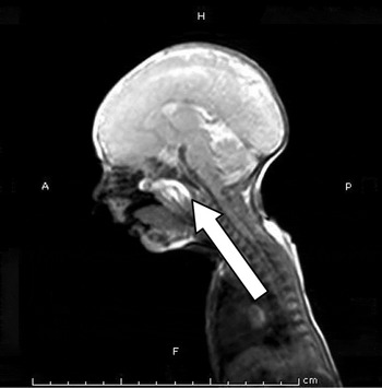
During tracheal re-intubation on the 11th day of life, the neonate was noticed to have a pearly white, solid swelling protruding from the nasopharynx from behind the soft palate, which pushed the uvula and soft palate anteriorly. Magnetic resonance imaging (MRI) revealed a 32 × 22 × 19 mm multiloculated cystic lesion, with predominantly fat and fluid components in the nasopharynx, which extended superiorly to the posterior choana and markedly obliterated the air passage (Figure 1). No dehiscence was seen in the skull base, thereby ruling out a meningoencephalocoele.
Sagittal magnetic resonance image of the neck showing the lesion causing significant narrowing of the nasopharyngeal airway. H = head; A = anterior; P = posterior; F = feet

Nasal endoscopy was performed under general anaesthesia. Anaesthesia was induced by inhalation of 4 per cent sevoflurane in 100 per cent oxygen, and intravenous administration of fentanyl 5 μg. After neuromuscular blockade with atracurium 1 mg was administered intravenously, the nasotracheal tube was removed and a 2.5 mm oral endotracheal tube was inserted. Anaesthesia was maintained with an inhalational mixture of oxygen 50 per cent and nitrous oxide 50 per cent, and sevoflurane 1–2 per cent. Pressure controlled ventilation was delivered, with the peak inspiratory pressure set at 12 cm H2O and a respiratory rate of 50 breaths per minute.
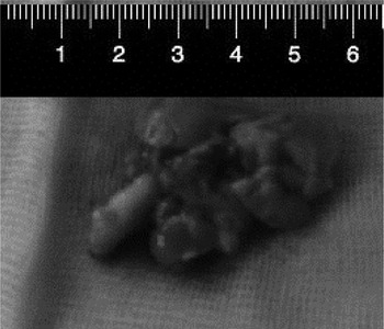
Endoscopy revealed a multiloculated solid lesion originating from the lateral wall of the nasopharynx on the left side, which extended into the whole nasopharynx and partially into the oropharynx. Transnasal endoscopic excision of the lesion was performed and the specimen was delivered through the oral cavity (Figure 2).
Photograph of the nasopharyngeal tumour after excision.

After surgery, the neonate was shifted to the neonatal intensive care unit and mechanically ventilated. Histopathological examination was suggestive of nasopharyngeal teratoma. Tracheal extubation was achieved after 48 hours. The neonate received oxygen supplementation with a hood for 24 hours, and was discharged after 2 days. Repeat nasal endoscopy was carried out a month later and showed no residual lesion. The neonate was observed to be doing well at the four-month follow up.
Case report two
A 5-day-old post-term male baby, born at 42 weeks, was referred with diagnoses of birth asphyxia and seizures. The lungs were ventilated in view of worsening respiratory distress. The trachea was extubated after 72 hours. Otolaryngologist consultation was sought as the neonate continued to have respiratory distress and noisy breathing.
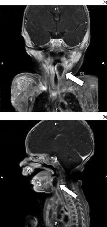
The neonate had significant stridor, and endoscopic evaluation under sedation revealed a large cyst involving the left arytenoid, aryepiglottic fold and pyriform fossa, completely obstructing the laryngeal inlet. The right vocal fold was visualised and appeared to be normal. Magnetic resonance imaging of the neck showed a well-defined 14 × 11 × 17 mm, non-enhancing cystic lesion. The lesion had lobulated margins in the left supraglottic-paralaryngeal region that extended cranially from the level of the glottis to the superior margin of the epiglottis, with significant compression of the supraglottic laryngeal passage (Figure 3).
(a) Coronal and (b) sagittal magnetic resonance images of the neck showing the saccular cyst. H = head; R = right; A = anterior; P = posterior; F = feet

Microlaryngeal excision of the cyst was scheduled. Anaesthesia was induced with 8 per cent sevoflurane in 100 per cent oxygen delivered with a face mask. Tracheal intubation was attempted without muscle relaxation. Laryngoscopy revealed a large, left-sided lesion, which distorted the laryngeal inlet to a slit-like opening and pushed the vocal folds towards the right. An oral 3 mm internal diameter endotracheal tube was inserted. Anaesthesia was maintained with a mixture of oxygen 50 per cent and nitrous oxide 50 per cent, and sevoflurane 1–2 per cent, with manually assisted ventilation.
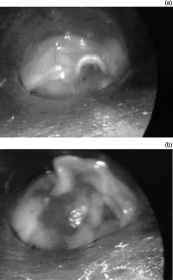
Microscopic laryngeal examination revealed the cyst extending cranially up to the tip of the epiglottis, medially overlying the glottis and laterally occupying the entire pyriform fossa (Figure 4a). The cyst was incised gently. Minimal gelatinous white fluid leaked out and the cyst wall collapsed. The roof of the cyst was excised. A ventriculostomy was performed, excising the false vocal folds and bringing both vocal folds into view (Figure 4b) in order to minimise the chances of recurrence.
Laryngoscopic view of the saccular cyst (a) before and (b) after excision; both vocal folds can be clearly seen following excision of the cyst wall (b).

Tracheal extubation was achieved after 48 hours. The neonate received oxygen supplementation with a hood for 24 hours, and was discharged after a further 48 hours. Histopathological examination revealed features suggestive of a laryngeal saccular cyst. One month later, repeat laryngeal endoscopy under sedation revealed no residual lesion. The child was observed to be doing well at the three-month follow up.
Discussion
Laryngotracheomalacia is the most common cause of congenital chronic stridor. Around 1 in 10 affected infants have lesions in more than one anatomical site of the upper aerodigestive tract.Reference Claes, Boudewyns, Deron, VanderPoorten and Hoeve 2 Congenital anomalies of the upper respiratory tract are relatively rare. The differential diagnosis includes laryngeal cysts, atresia and stenosis, vocal fold immobility, and subglottic haemangiomas.Reference Ahmad and Soliman 3
Teratomas are the most common congenital tumour, but teratomas of the nasopharynx are rare, and are seen almost exclusively in infants, usually in neonates.Reference Hong, Kuo, Wu, Lin, Lo and Juan 4 Teratomas occur in 1 of 4000 live births and show a female predominance of 5:1.Reference Ulger, Egemen, Karapinar, Veral and Apavdin 5 Head and neck teratomas account for 1–10 per cent of the total reported cases, and are most likely to arise in the neck with the nasopharynx being the second commonest location.Reference Cay, Bektas, Imamoglu, Bahadir, Cobanoglu and Sarihan 6 They are composed of tissues that are derived from all three germinal cell layers. The treatment for nasopharyngeal teratomas is complete surgical excision.Reference Rybak, Rapp, McGrady, Schwart, Myers and Orvidas 7 Emergency surgical excision should be undertaken if the airway is compromised. Recurrences are rare and do not occur with complete excision.
In case report one, a nasopharyngeal tumour was not immediately suspected due to its rarity and the presence of features of laryngomalacia. The ease of introduction of the transnasal feeding tube, and unimpeded passage of the endotracheal tube through the nasopharynx during intubation, also precluded the diagnosis of a nasopharyngeal mass. Once the diagnosis of nasopharyngeal tumour was made, however, surgery was undertaken and endoscopic transnasal tumour excision was achieved without any sequelae.
Congenital saccular cysts are rare and represent 25 per cent of all laryngeal cysts. The annual incidence of this condition has been reported to be 1.82 per 100 000 live births.Reference Martin, John and Hasselta 8 Cysts of the aryepiglottic fold are the rarest of the laryngeal cysts and form just 2.2 per cent of all laryngeal cysts.Reference Arens, Glanz and Kleinsasser 9 Endoscopy is the gold standard for diagnosis of these laryngeal cysts. Endoscopic removal of cysts can be an effective treatment that minimises recurrence. Endoscopic deroofing is as effective as endoscopic excision, but is technically simpler and is therefore recommended as the treatment of choice.Reference Arens, Glanz and Kleinsasser 9
In case report two, a diagnosis of saccular cyst was established by endoscopic examination, and an MRI scan was subsequently carried out to examine the origin and extent of the lesion. We decided on microscopic laryngeal excision of the saccular cyst with removal of the cyst wall, which successfully ameliorated the respiratory distress.
Stridor in a neonate should alert the anaesthetist of potential airway difficulties. A difficult airway management cart and a person skilled in fibre-optic intubation must be available. The airway should be maintained on spontaneous ventilation to minimise airway compromise until it is secured.
As described in the current paper, the airway of the neonate with a nasopharyngeal mass (case report one) had been secured pre-operatively with a nasotracheal tube. However, this needed to be replaced with an orotracheal tube to facilitate surgical intervention. The change of tracheal tubes was carried out under neuromuscular blockade. On the other hand, the airway of the child with the laryngeal cyst (case report two) was not secured; anaesthesia was therefore induced by inhalation of sevoflurane, and the trachea was intubated without administration of a neuromuscular blocker. The trachea was atraumatically intubated with the neonate breathing spontaneously. The laryngeal inlet obstruction was incomplete, but did not worsen on induction of anaesthesia, possibly because neuromuscular blocking drugs and positive pressure ventilation were not used. We did not extubate the trachea immediately after surgery because of the potential occurrence of post-operative airway complications such as laryngeal oedema, bleeding and laryngospasm.
-
• Neonatal airway lesions are rare, and a high index of suspicion is necessary
-
• Clinical examination, endoscopy and radiological imaging are essential for diagnosis
-
• Airway management in neonates requires a multidisciplinary team comprising an otolaryngologist, anaesthesiologist and neonatologist
Nasal obstruction in neonates can cause significant airway compromise and may be life-threatening. In general, otolaryngologists are familiar with the management of a neonate born with choanal atresia. However, there are few reports and no guidelines for the management of a neonate or infant presenting with nasal obstruction and airway compromise without choanal atresia.Reference Derkay and Grundfast 10
Conclusion
Neonatal airway compromise indicates an impending medical emergency and the determination of its cause requires experience and a high index of suspicion. An oronasopharyngeal mass is rare in a neonate or infant presenting with intermittent stridor and difficulty in feeding. Neonates with stridor should be managed by an experienced multidisciplinary team comprising an otolaryngologist, a paediatric anaesthesiologist and a neonatologist in order to effectively manage neonatal airway lesions.
Acknowledgements
The authors would like to thank Dr Dilip Singh and Dr Bijender Singh (Consultants in the Department of Neonatology, Pushpanjali Crosslay Hospital) for their active role in the management of these neonates.