Introduction
Ivory is a highly esteemed material that has been traded for centuries, widely used for sculptures, book covers, piano keys and all kinds of art objects and jewelry. This valuable material comes from elephants’ tusks and is, therefore the main product from the hunting and killing of elephants. The increase in illegal elephant poaching forced the establishment of an international law prohibiting the trade of such material. The Convention on International Trade in Endangered Species of Wild Fauna and Flora (CITES) set the baseline to 1975, stating that trading whole or modified tusks after this year would be considered a crime.
In the literature, several studies address ivory samples, involving the use of different isotopes, such as the work of Schmidberger et al. (Reference Schmidberger, Durner, Gehrmeyer and Schupfner2018). Additionally, collagen extraction protocols have been employed in forensic dating, as demonstrated by Uno et al. (Reference Uno, Quade, Fisher, Wittemyer, Douglas-Hamilton, Andanje and Cerling2013), Cerling et al. (Reference Cerling, Barnette, Chesson, Douglas-Hamilton, Gobush, Uno and Xu2016), Quarta et al. (Reference Quarta, D’Elia, Braione and Calcagnile2019), Wild et al. (Reference Wild, Kutschera, Meran and Steier2019), Quarta et al. (Reference Quarta, Molnár, Hajdas, Calcagnile, Major and Jull2021), and Valášek et al. (Reference Valášek, Pachnerová Brabcová, Kufnerová, Molnár and Světlík2022). Based on these studies, we conducted dating tests on some modified ivory pieces with the aim of determining their chronology.
Differentiating the animal source of ivory is a crucial issue and, in forensic science, the Schreger pattern in the tusk is used for this purpose. In the 2021 study by Kufnerová et al., the researchers aimed to validate the modernity of the samples based on prior age data provided by specialists who claimed that the samples were old. They asserted that the ivory pieces were from elephants, using the Schreger patterns as evidence (Espinoza et al. Reference Espinoza and Mann1993). Additionally, radiocarbon dating techniques were used for forensic purposes.
A collaboration between the National Institute of Criminalistics, the forensic unit of Brazilian Federal Police, and the Radiocarbon Laboratory at the Fluminense Federal University (LAC-UFF), was initiated to determine the age at which the seized material entered the market. LAC-UFF is the only complete carbon-14 accelerator mass spectrometry (14C-AMS) facility in South America, housing an NEC 250 kV Single Stage Accelerator Mass Spectrometer (SSAMS) installed in 2012 (Macario et al. Reference Macario, Oliveira, Carvalho, Santos, Xu, Chanca and Rodrigues2015).
In general, for dating the time of death of the elephants based on the ivory tusks, we would need to date the part lastly grown. However, within the partnership with the Brazilian Federal Police, the available material was from modified ivory, such as bracelets, rings, necklaces, and various netsuke (small-carved ivory figures). In this context, our first challenge was to devise a sampling method to obtain a reliable age for these forensic cases. Although we would like to determine the time of death of the animals, depending on the sampling spot we would be limited to measure the carbon-14 concentration during some of the years during the life of the animal. Therefore, understanding the growth pattern of the tusks was essential.
The objectives of this study are as follows: 1) to maximize the amount of collagen extracted, thereby reducing the quantity of material required for accurate dating; 2) to gain a comprehensive understanding of tusk growth patterns, enabling the selection of chronologically ordered sampling points; 3) to calibrate the dating results by ensuring that the pairs of dates obtained are sequential; and 4) to consider and address potential uncertainties inherent in the dating process.
Elephant tusks and their growth pattern
The tusk is composed of approximately 30 wt% organic matrix, which contains collagen, and about 70 wt% inorganic matrix, comprising hydroxyapatite crystals and their compounds. Due to the potential carbon exchange with the environment, which can influence dating, we will use collagen for the analysis of the samples in this study (Cui et al. Reference Cui, Wen, Zhang, Ma and Li1994; Raubenheimer et al. Reference Raubenheimer, Dauth, Dreyer, Smith and Turner1990).
The permanent tusk of the African elephant (Loxodonta africana) emerges after the first year of life, facilitating breastfeeding. Initially, the elephant is born with what is called a primary tusk, or tush, which is considered a deciduous and non-functional tooth. The tush does not erupt, but is displaced by the permanent tusk, which continues to grow throughout the elephant’s life. Elephants can live up to 60–70 yr, with the loss of their last molar indicating the end of their lifespan (Raubenheimer Reference Raubenheimer2000; Steenkamp Reference Steenkamp, Emily and Eisner2021). The elephant tusk grows from the outer part to the inner one and from the tip to the base. For this reason, the most recent part of the sample would be ideally the inner part and close to the animal´s jaw. Dealing with modified ivory requires estimating the growth direction so that the most recent material can be sampled. Additionally, it is important to take into account that using the bomb peak curve can produce two possible dating due to the shape of the curve. On the one hand this may be a drawback, since a single sampling may not be enough to determine the desired date. On the other hand, sampling two spots in chronological order could provide precise fitting within the bomb peak curve.
The elephant is the only tusked mammal with a distinctive growth pattern of mineralized collagen fibers (MCF), the so-called Schreger pattern (Espinoza et al. Reference Espinoza and Mann1993). In principle, the Schreger pattern is highly visible, and its observation requires no special apparatus or microscope; it can be discerned with the naked eye. In the 3D pattern developed by Albéric, there are a total of four patterns based on the orientation in which the tusk was cut. Through this 3D pattern (Figure 1a), we can determine whether the piece was made with a longitudinal or transversal pattern, as an example. Those patterns can be visualized in detail using a scanning electron microscope (SEM). The study of this morphology was accomplished using two different samples cleaved from the same netsuke. These samples were coated with a thin gold layer to facilitate imaging and analyzed using a field emission scanning electron microscope JEOL JSM-7100F. In Figure 1b and 1c, it is possible to visualize two features: one resulting from the fractures at the cleavage process and the other from the growth structure of the tusk as Alberic’s findings. This structure was interpreted as growth in the direction in the RL plane.
(a) Alberic’s Modified Schreger Pattern (2018). The red arrow (b–c) indicates the direction of the RL plane. Illustrated by Ana Beatriz Pinheiro (architect), (b) and (c) SEM images of two different parts of the same ivory netsuke.

Albéric et al. (Reference Albéric, Gourrier, Wagermaier, Fratzl and Reiche2018) observed that the region with the highest concentration of mineralized collagen fibers is in the RL plane, indicating the longitudinal segment of the tusk corresponding to its growth region. This observation is relevant in this context because the tusk grows longitudinally. In this region, ivory exhibits continuous lines in a single direction, identifiable in modified pieces. This is the pattern we look for in our samples during sampling, a crucial step in the protocol. These lines correspond to MCF. By sampling these lines, we are directly targeting the collagen of interest. As illustrated in Figures 1b and 1c, the red arrow indicates the direction of the RL plane. This process enables us, upon completion of the treatment, to obtain a significant amount of collagen, allowing for sample replication and comparison of the resulting data. The amount of collagen extracted can vary depending on the size of the tusk, considering a longitudinal growth rate of approximately 5 cm/yr (e.g. Uno et al. Reference Uno, Quade, Fisher, Wittemyer, Douglas-Hamilton, Andanje and Cerling2013), decades of growth can be expected, compromising the possibility of relating to the year 1976. At the end of the measurement, the result indicates the modernity of the sample and not the age of death, since only the collagen in the piece can be ensured, not the correct location from where part of the tusk was taken to make the object. The time of death would be related to the part that ceased carbon exchange with the atmosphere, which cannot be ensured in modified pieces.
The pattern exhibits multiple orientations, varying depending on the plane in which the tusk is cut. This pattern forms as the tusk grows in a spiral, allowing us to infer, for example, that the inner layers develop before the outer ones. This is based on the Schreger pattern’s plane, specifically the RL plane. Future studies are being conducted based on the pattern’s angulation and radiocarbon dating to support the proposed growth order.
Given the importance of sampling in this study—which involves aligning the pattern lines to increase collagen yield at the end of the treatment—we can observe in Figure 2 that several strategic samplings were conducted near the RL plane, and when the feature was not very clear, we sampled the part closer to this plane, such as the SL plane, which is closer but is the diagonal part. Samples a-d correspond to Test 1, while samples e-f are associated with Test 2, which will be explained later.
Sampling locations for each sample in the study. Illustrated by Ana Beatriz Pinheiro (architect).

Materials and methods
A collagen extraction was performed for subsequent radiocarbon measurement; in this protocol, since the samples were not found in soils, sediments, or similar environments, we opted not to use ultrafilter for collagen cleaning due to the limited quantity of available samples material, such as in case of netsuke.
For example, if we can sample both the inner and outer parts of a piece cylindrical, like we did in the first test of this study, as shown in Table 1, the outer part formed first, followed by the inner part—a sequence confirmed by dating. By analyzing the ascending and descending portions of the bomb curve, the most recent date serves as a parameter to estimate the elephant’s time of death.
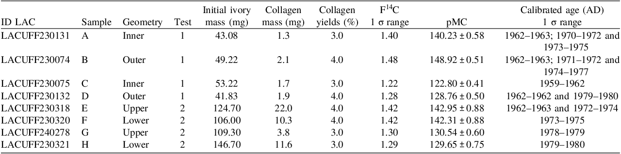
Radiocarbon dating results for ivory samples (and sampling spots) presented as pMC and calibrated age (AD) mass before and after pretreatment chemical. The collagen yield was calculated from the ratio between the obtained collagen mass and the initial ivory mass.

In order to develop a protocol for dating the modified ivory pieces, a total of three tests were conducted. In each test, at least two samples were taken from the same piece for comparison purposes and to ensure replicability at the end of the treatment. All samples were collected using a micro grinder, and the ivory mass of each sample is detailed in Table 1. Before the objects arrived at LAC-UFF, analyses were carried out in the laboratory of the Brazilian Federal Police to determine if the pieces were indeed from elephant ivory or another animal, such as the mammoth, which thrived north of Panama in the Americas during the Glacial and is now extinct. Additionally, isotopic analyses of δ13C and δ15N were performed at the National Institute of Criminalistics (INC), using the Ivory ID system, to determine the geographical origin of the elephant in question.
In the first Test, we utilized the marks already present on the pieces from these preliminary Tests mentioned earlier. In the second Test, we sampled what we believed to be the growth region of the tusk, specifically the longitudinal part. In the third and final Test, we sampled using the Schreger Pattern as a guide, focusing on the part of the piece that had more collagen according to this pattern.
Collagen extraction was done following a modified Longin (Reference Longin1971) procedure used by Oliveira et al. (Reference Oliveira, Macario, Carvalho, Moreira, Alves, Chanca and Fernandes2021) and was conducted on all samples. It is worth noting that in this protocol, the sampling stage is the most critical because the collagen concentration is variable across the sample, as it determines whether we will have a sufficient sample at the end of the treatment. In this protocol, in addition to the crucial role of sampling, the use of an ultrafilter cleaning step was omitted to increase sample yield.
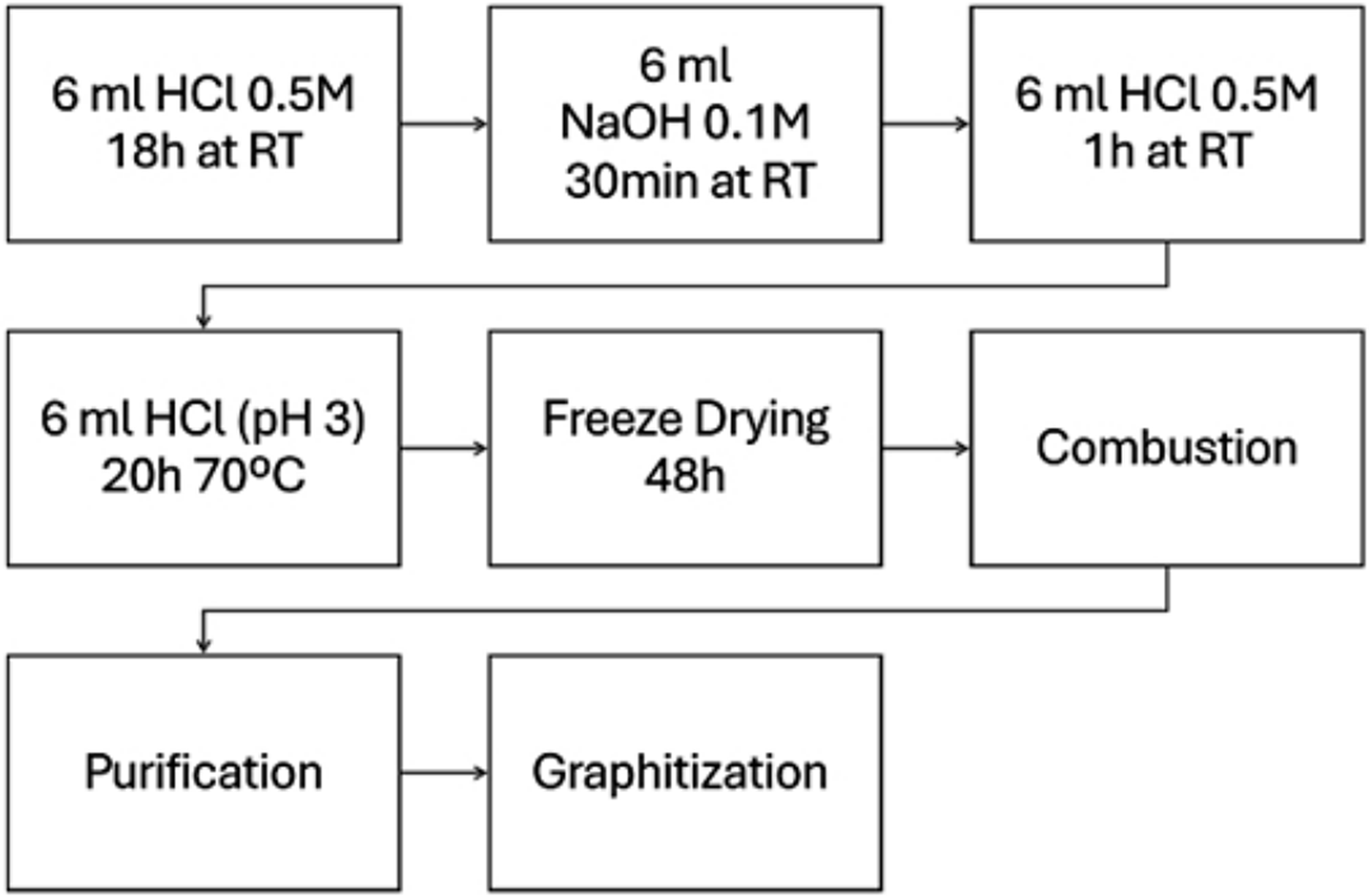
Figure 3 shows an overview of how the chemical treatment of the sample was conducted until it was converted to graphite, the suitable matrix for the ion beam to be extracted in the accelerator.
Protocol for collagen extraction from modified ivory.

Physical pretreatment
Before performing chemical process to remove any visible impurity, the surfaces of all ivory samples are cleaned using a paper moistened with 70% alcohol. This step is crucial for eliminating contaminants prior to sampling.
In all Tests performed, the variable was the approach used in sampling, particularly the selection of the correct part of the plane. It was observed to significantly impacted the results by determining the portion capable of confirming the modernity. All samples underwent the same physical and chemical pretreatment procedures. It is noteworthy that ivory powder is extremely lightweight, which poses a challenge in obtaining the required sample quantity. The initial estimate, based on previous collagen extraction protocols at LAC UFF, ranged from 100 to 400 mg. However, we began with approximately 40 mg. Due to the modified nature of the samples and the need for preservation of the artifacts, it was crucial to minimize the initial sample amount until the correct region of the pattern could be sampled with the smallest possible drill. First, the inner/outer parts of the objects were sampled for comparison. The Schreger pattern structure was then used to identify the most suitable region, where a higher collagen yield was expected, specifically the RL plane, resulting in a more enriched sampling.
In the first Test, we used the two larger samples, which already had marks from previous police analyses. We collected from these marks and also from the unmarked part, that is, the lower and outer portion of the sample, for the second test. To do this, we drilled the piece using a Dremel, collecting approximately 5 mg of powder from each hole. In total, approximately 40–50 mg of ivory was collected, divided into four samplings, two from each piece, as illustrated in Figures 2a and 2c for the inner parts of the pieces, and Figures 2b and 2d for the outer parts.
In the second test, we used an even finer drill, based on previous studies (Quarta et al. Reference Quarta, Molnár, Hajdas, Calcagnile, Major and Jull2021; Valášek et al. Reference Valášek, Pachnerová Brabcová, Kufnerová, Molnár and Světlík2022), who suggested a need for at least 100 mg of sample material to ensure a satisfactory amount of collagen at the end of the treatment. The use of the finer drill was effective to obtaining a larger sample quantity, minimizing sample waste.
After two Tests, where we adopted sampling criteria based on parts of the sample that were assumed to be more recent (i.e., areas in the growth line of the tusk). However, such methods were not completely satisfactory, being applicable only to very specific samples, such as bracelets or circular objects without seams. Faced with this challenge, we sought a methodology that would enhance our work and yield more precise results, as elaborated in the subsequent sections.
The Schreger pattern, developed and refined around 1800 (Espinoza et al. Reference Espinoza and Mann1993), is currently recognized and utilized in forensic science to distinguish between elephant ivory and mammoth ivory based on the angle of the pattern. Since the pattern is visible to the naked eye, in our study, the pieces had been previously analyzed at the INC, confirming that all of them were exclusively from elephants.
Therefore, in the third test, sampling was based on the pattern of the tusk, that is, we looked for the RL pattern in the longitudinal direction, which is the growth direction. We conducted the sampling believing it to be the part that contained more collagen, according to previous studies (Albéric Reference Albéric, Gourrier, Wagermaier, Fratzl and Reiche2018). By combining the experiences of previous tests, we were able to carry out a more precise sampling with a smaller drill, and in the region with more collagen. This way, we obtained a larger amount of collagen at the end of the treatment, as will be described below, allowing for replicates and final comparisons.
Collagen extraction
The sampled material underwent a chemical pretreatment process to extract the collagen from the ivory, initiating the dating process for the pieces. Due to the low quantity obtained during the sampling process, a new protocol was necessary to adapt to this smaller-sized and processed sample. Following collection, the samples were stored in an Eppendorf and throughout the chemical pretreatment as illustrated in Figure 3, they were placed in 0.5 molar (M) hydrochloric acid for about 18 hours at room temperature, initiating the decalcification process. This process involves an acid-base-acid (ABA) treatment. Subsequently, the sample was placed in a 0.1M sodium hydroxide solution for only 30 min at room temperature, aiming to decontaminate any impurities that might have occurred during the decalcification step according to Longin (Reference Longin1971) and Quarta (Reference Quarta, Molnár, Hajdas, Calcagnile, Major and Jull2021). This stage continues with 0.5M HCl, in which the sample is added and left for 1 hour, also at room temperature. Following this process, the sample undergoes rinsing with ultra-pure (UP) water and proceeds to the gelatinization stage, where it is placed in pH 3 HCl for 20 hr at 70°C. During this step, collagen in the sample undergoes gelatinization, a process where the triple-helical, H-bonded polypeptide molecules break their H-bonds, transforming into single-stranded polypeptide collagen molecules that can dissolve in acidified water. After this heating period, each sample is transferred to individual falcon tubes and frozen for approximately 72 hr.
Upon completing the stages of physical and chemical pretreatment, the samples are placed in a lyophilizer and subjected to a drying process for up to 48 hr. During this period, all water content in the sample evaporates, leaving only the present collagen, appearing as a whitish fluffy, snow-like solid inside the tube. It’s crucial to note that the transfer of samples to the combustion tube (CT) containing cupric oxide and a silver wire must occur immediately after removal from the lyophilizer to prevent sample loss, due to the high humidity in Brazil, preserving samples during the analysis process poses a significant challenge. The moisture can cause samples to become damp quickly when exposed to air, compromising their integrity and potentially leading to the loss of valuable information. Before transferring, the samples are weighed, and the results can be found in Table 1, displaying the post-drying mass (“collagen mass”).
Once the samples are placed inside the CT tube, the latter is connected to a vacuum line to be pumped out, and the CO2 conversion is achieved through flame-sealed, closed tube combustion with cupric oxide (Oliveira et al. Reference Oliveira, Macario, Carvalho, Moreira, Alves, Chanca and Fernandes2021). Following this, the samples are baked in a furnace at 900°C to promote combustion and produce CO2 gas, which is then purified in a vacuum line by removing water and other gases and finally stored in graphitization tubes (GT), using the TiH2-Zn graphitization (Macario et al. Reference Macario, Alves, Oliveira, Moreira and Chanca2016, Reference Macario, Oliveira, Moreira, Alves and Carvalho2017; Xu et al. Reference Xu, Trumbore, Zheng, Southon, McDuffee, Luttgen and Liu2007). These tubes are part of the graphitization line that controls the process, converting the samples into graphite form for pressing and analysis in the accelerator. Graphite is the final state of the sample, allowing for measurement in the accelerator.
Given the initially low sample quantity, it was anticipated that multiple divisions in the CT/GT, which require a minimum mass or gas support, would be unfeasible due to the limited gas generated; however, should a sufficient amount of CO2 be obtained after combustion, it would then be possible to prepare two or three graphite targets. This was true only for the samples in the first test. From the second test onwards, sample divisions were possible. All samples with a mass after drying exceeding 10 mg were divided into two CTs, consequently leading to a division in the GT as well.
In the GT, samples are in graphite, their final state before heading to the accelerator system. The AMS isotopic separation and measurement were carried out at LAC-UFF (Anjos et al. Reference Anjos, Macario, Gomes, Linares, Queiroz and Carvalho2013; Linares et al. Reference Linares, Macario, Santos, Carvalho and Dos Santos2015) using the Single Stage Accelerator Mass Spectrometry (SSAMS) of 250 kV, built by the National Electrostatics Corporation (NEC).
Results and discussion
To implement the protocol, preliminary tests (Tests 1 and 2) were conducted all for forensic purposes. Following the Schreger pattern in the ivory during the sampling process, we were able to complete the treatment with a sufficient amount of collagen, adequate for replication and yielding compelling results in compliance with the Convention on International Trade in Endangered Species of Wild Fauna and Flora (CITES) regulations established in 1975 for illegal ivory trafficking.
In Table 1, we can observe the significance of the Tests in implementing this new protocol. Despite altering the sampling process, it remained a pivotal stage for obtaining satisfactory results in terms of comparison. By comparing the mass before and after chemical treatment, it is evident that a smaller pre-treatment sample quantity results in a more significant loss of sample during the transformation into graphite. Previous studies (Alkass et al. Reference Alkass, Saitoh, Buchholz, Bernard, Holmlund, Senn and Druid2013; Oliveira et al. Reference Macario, Oliveira, Moreira, Alves and Carvalho2020) have explicitly indicated the need for approximately 400 mg of sample for collagen extraction from bone and teeth samples to yield satisfactory results. According to Quarta (Reference Quarta, Molnár, Hajdas, Calcagnile, Major and Jull2021), for ivory samples, they use 400–600 mg. However, this work demonstrates that for ivory samples, an initial mass of 40–150 mg is sufficient for analysis, even when following the pattern-oriented collagen extraction process. This result is due to our different method for sampling.
Additionally, it is crucial to highlight the density of ivory, which poses a challenge during sampling, necessitating a careful choice of tools for the process.
The collagen yield from the samples ranged between 0.8% and 2%wt. This signifies a satisfactory yield, as collagen contents exceeding 1%wt are recognized as indication of well-preserved bone and enough collagen quantity (Ambrose Reference Ambrose1990).
From Table 1, we derive a wealth of information about the samples even before considering the calibrated calendar year ages. Apart from the relevance of mass factors in implementing the protocol to achieve satisfactory replicates, a primary goal accomplished in this work, the table displays all percentage of modern carbon (pMC) values greater than 100%, i.e. containing carbon from the post-bomb period. This result indicates the modernity of the samples, presenting the outcomes of this case. As modern samples with a high probability of post-1975 origin, their commercial trade is prohibited under the CITES regulation, which bans ivory dated after 1975. However, if there is any indication of these samples predating the regulation, it still implies illegal importation. This metric is used in software programs to convert the age into calendar years, utilizing the calibration curve from the Northern Hemisphere. Our samples are from Africa, as confirmed by previous Tests at INC. This determination is made by comparing the measured radiocarbon concentration to reference values, such as atmospheric 14C content in different hemispheres from the last 80 yr (post-bomb-period). Nonetheless, solely the findings from the second Test onward can be deemed pertinent for the conclusive evaluation, given that the method of sampling markedly affects the outcomes. It is from the second Test onwards that we commence incorporating the application of the Schreger pattern into our assessments focusing on the section with the highest collagen concentration, as suggested by Alberic et al. (Reference Albéric, Gourrier, Wagermaier, Fratzl and Reiche2018) because in forensic cases, it is crucial to consider sampling methods that preserve the integrity of the specimen.
In Figure 4, the estimated calendar ages of the analyzed samples are presented. Initially, we considered sample pairs, using two samples for each Test and performing two extractions on each, resulting in a total of approximately four samples for the initial Tests.
Calibration of the measured 14 C concentrations into calendars ages using the bomb-peak curve from Test 1 (a) sample pair A–B and (b) sample pair C–D.

In Figure 4, we present the results of Test 1, divided into pairs and plotted against the Northern Hemisphere 3 curve (in blue) and the Southern Hemisphere 3 curve (in green). These curves represent the likely regions of the African elephant. Notably, using both calibration curves, we observe that the results do not change significantly. Figure 4A displays the pair of samples A-B, while Figure 4B shows the pair of samples C-D. These results indicate that the inner part of the tusk is the most recent, whereas the outer part is older, evidenced by the tusk growth premise that establishes an ordered sequence, confirmed by radiocarbon dating and higher F14C content in the outer part and lower F14C content in the inner part.
In the first pair of Test 1, we have the samples A and B, both from the same piece. While similar results were expected, it’s essential to note that, according to previous studies (Cui et al. Reference Cui, Wen, Zhang, Ma and Li1994; Su et al. Reference Su and Cui1999), an elephant’s tusk grows longitudinally at a rate of approximately 8 cm per year. This growth directly impacts the age results. The initial Test’s random sampling was primarily to Test the collagen extraction technique rather than seeking accurate age results. As a result, the estimated ages ranged from 1962 to 1975 for one sample and from 1962 to 1977 for the other. While these results comply with CITES regulations, they cannot be further considered due to the sampling method employed to study growth. In the second samples, “C” and “D” the results confirmed that the inner part grows before the outer part. As shown in Figure 4 and Table 1, the ages for the first extraction of this second piece ranged from 1959 to 1962, while the second extraction ranged from 1962 to 1980. Since the pieces have a probability of being after the CITES regulations, the presence of material attributable to the post-1975 period indicates that their commercial trade would be prohibited under current legal frameworks.
Moving to Test 2, in which sampling was conducted in the tusk growth region while accounting for the Schreger pattern, we successfully obtained scientifically validated results through the analysis of collagen microfibers throughout the ivory. With precise sampling techniques, we anticipated consistent results among the samples. In this section, we analyze a pair of samples.
For the first pair, E and F, we observe a correspondence in pMC values, which are used to calculate radiocarbon age, within the margin of uncertainty. Upon conversion to calendar years, we obtain an age range of 1962 to 1974 for one sample and 1973 to 1975 for the other—both indicating modern ages. For the second pair, G and H, the pMC values are also consistent, with calibrated age ranges of 1978 to 1979 for the first sample and 1979 to 1980 for the second, both of which are modern and post-1975.
Consequently, these pieces cannot be commercialized under CITES regulations. Given the accuracy of the sampling, which targeted the collagen-rich regions, and the satisfactory outcomes achieved, we conclude that the protocol has successfully addressed the analysis of modified ivory samples.
By mere visual inspection, it is not possible to determine the growth sequence of the Schreger pattern in an elephant tusk when the complete tusk is unavailable. However, the angles formed in the transverse (RT) plane, shaping the spiral structure, can be analyzed for this purpose. One of the future objectives of this study is to conduct such an analysis.
It is noteworthy that the two pieces from the same sample were used for both Tests 1 and 2, allowing for a direct comparison of the results between the pieces for the two Tests. The only variation was the sampling method. In both Tests 1 and 2, conducted with the same chemical treatment, the sample in collagen form exhibited nearly a tenfold increase in mass in the second test compared to the first. For the first two Tests, the sample ages were consistent, differing by only one year. This accuracy is paramount in this forensic case in collaboration with the Brazilian Federal Police, necessitating a higher level of precision in the final analysis.
Conclusion
This study represents a notable foray into forensic science through the application of nuclear physics, employing the radiocarbon dating technique in conjunction with a particle accelerator. Initially absent at LAC-UFF, this field required preliminary Tests, demanding solid scientific foundations for its implementation, which were based on previous studies by Albéric et al. (Reference Albéric, Gourrier, Wagermaier, Fratzl and Reiche2018). In forensic settings, the robustness of these foundations is crucial, and upon analyzing the results, we found that Test 2 achieved the initial goal of the study: introducing the dating of forensic samples and modified ivory in our laboratory. This study enabled us to collect and date specific regions of interest using reliable scientific methods. Consequently, the results clearly indicated that both ivory samples violate CITES legislation, making their commercialization prohibited.
This work signifies a substantial advancement in the use of applied nuclear physics in forensic science, particularly in the analysis and dating of modified ivory samples. It required the adaptation and implementation of existing radiocarbon dating techniques, paving the way for the identification and verification of samples in forensic cases, as well as ivory artifacts, which could potentially be illegally traded.
Throughout the process, careful sample selection, combined with the refinement of collection methods, was pivotal in ensuring result accuracy. This approach offers a valuable tool in combating the illegal trade of ivory and other associated criminal activities.
Based on the specific objectives presented in the introduction, we conclude that: 1) We successfully maximized the amount of collagen extracted, allowing for more precise dating of the material; 2) The first test confirmed that the internal part of the samples is older than the external part, along with the prior understanding of spiral growth. These results support the growth pattern from the inside out.; 3) Although the exact sequence of modified samples is not always clear, the obtained results allowed for the adjustment of ages to calibrate the pairs sequentially; 4) Calibration curves from both hemispheres covering the African continent were used, which is essential when the exact origin of the ivory pieces is uncertain.
These results successfully fulfill the study’s objectives, offering a solid foundation for the dating and authentication of ivory samples. A key future consideration for this work is to account for the time-lag in the sample, particularly the interval between the incorporation of carbon into the ivory and its eventual measurement.
Supplementary material
To view supplementary material for this article, please visit https://doi.org/10.1017/RDC.2025.10164
Acknowledgments
We are grateful for the research award granted to K. D. Macario (Brazilian National Council for Science and Technology (CNPq) 317397/2021-4; FAPERJ E-26/200.540/2023; INCT-FNA 464898/2014-5), which funded this study and Brazilian Federal Police for providing ivory samples that made this work possible.