




Introduction
The mammary gland of female mammals is a specialized organ responsible for milk synthesis and secretion, thereby supporting neonatal growth and development. It undergoes a highly dynamic and complex developmental process spanning embryogenesis, puberty, and the pregnancy–lactation–involution cycle (Hannan et al. Reference Hannan, Elajnaf and Vandenberg2023; Shi et al. Reference Shi, Zhao and He2023; Visvader and Stingl Reference Visvader and Stingl2014). These processes are tightly regulated by hormonal signals, genetic factors, environmental exposures, and lifestyle influences (Li et al. Reference Li, Zhang and Luo2025; Northey et al. Reference Northey, Hayward and Yui2024). Therefore, a comprehensive understanding of mammary gland biology and the mechanisms underlying its developmental dynamics is essential for improving lactation performance, enhancing neonatal survival, and preventing breast-related diseases (Rauner Reference Rauner2024). In particular, elucidating the regulatory networks governing mammary gland development and lactation holds significant agricultural values for improving the productivity of dairy animals (Bhat et al. Reference Bhat, Ahmad and Ibeagha-awemu2019). Consequently, the development of physiologically relevant in vitro models has become a central challenge in mammary biology research.
Traditional two-dimensional (2D) culture systems of mammary epithelial cells have long been widely used to investigate mammary biology. However, their limitations, particularly the lack of three-dimensional (3D) biomechanical interactions, often result in the loss of cellular polarity and the suppression of mammary gland-specific functions (Bissell and Hines Reference Bissell and W.c2011; Debnath et al. Reference Debnath, Muthuswamy and Brugge2003; Petersen et al. Reference Petersen, Rønnov-jessen and Howlett1992). To overcome these limitations, 3D organoid systems have emerged as more physiologically relevant models of the mammary gland. Mammary organoids are typically established in hydrogels or extracellular matrix (ECM)-based scaffolds that mimic key features of the native microenvironment, allowing cells to self-organize and recapitulate important structural and functional properties of the mammary epithelium (Clevers Reference Clevers2016; Li and Izpisua Belmonte Reference Li and J.c2019). Compared with conventional 2D cultures, mammary organoids more faithfully recapitulate in vivo physiology while remaining experimentally tractable. When derived from human tissues, they can also help mitigate the interspecies differences inherent in animal models (Debnath et al. Reference Debnath, Muthuswamy and Brugge2003). Mammary organoids represent a powerful platform for investigating mammary development, regulatory mechanisms, and related diseases, thereby advancing research in both normal physiology and pathological conditions (Temime et al. Reference Temime, Djoual and Chiche2025).
This review provides a comprehensive overview of the progress, applications, and future directions of mammary organoids. We outline the methodologies for generating mammary organoids derived from humans, mice, and non-model species. The key factors influencing organoid culture, including cell sources, ECM components, and growth factors, are discussed. Furthermore, we highlight the applications of mammary organoids in developmental and physiological research, as well as their emerging potential in combination with advanced technologies. Finally, we address current technical challenges and provide perspectives for future research in this rapidly evolving field.
Establishment of mammary organoids
Mouse mammary organoids
Mouse mammary organoids are among the earliest and most well-established systems (Fig. 1). As early as the 1980s, a 3D culture method for mouse mammary epithelial cells using a laminin-rich Engelbreth–Holm–Swarm (EHS) matrix was developed. This work showed that the ECM is essential for maintaining cell polarity and enabling milk protein secretion (Barcellos-hoff et al. Reference Barcellos-hoff, Aggeler and Ram1989; Li et al. Reference Li, Aggeler and Farson1987). However, early Matrigel-based systems mainly supported the formation of simple acinar structures and failed to reproduce the ductal branching observed in vivo. Later studies found that collagen I was critical for inducing invasive protrusions and branching morphogenesis, whereas basement membrane components alone were insufficient to drive ductal elongation (Nguyen-Ngoc and Ewald Reference Nguyen-ngoc and A.j2013; Nguyen-ngoc et al. Reference Nguyen-ngoc, Shamir and Huebner2015). Although these models improved structural features, they lacked proper hormone responsiveness. In 2017, Jamieson and colleagues established a long-term culture system that retained sensitivity to steroid hormones (Jamieson et al. Reference Jamieson, Dekkers and Rios2017). Following studies further optimized culture conditions to support prolactin-induced milk secretion and even mimic postweaning involution in vitro (Charifou et al. Reference Charifou, Sumbal and Koledova2021; Sumbal et al. Reference Sumbal, Chiche and Charifou2020). This marked a shift from structural modeling to functional modeling for mammary organoids.
A brief history of mammary organoid development. The upper timeline illustrates major advances in the mouse mammary organoid model, whereas the lower timeline shows representative milestones in human mammary organoid development. Created with BioRender.com.

Figure 1 Long description
The timeline illustrates key developments in mammary organoid research. For mouse mammary organoids: 1980 marks the 3D culture with EHS matrix (Barcellos-Hoff et al., 1989), 2013 shows ECM inducing branching morphogenesis (Nguyen-Ngoc and Ewald, 2013), 2017 highlights long-term culture and hormone responsiveness (Jamieson et al., 2017), 2021 features functional reconstitution (Charifou et al., 2021) and 2023 introduces the mini-gland system and physiological cycle (Yuan et al., 2023). For human mammary gland organoids: 1992 introduces the first 3D human epithelial spheroids (Petersen et al., 1992), 2003 shows stromal-epithelial crosstalk in 3D (Dontu et al., 2003), 2015 presents organoids with TDLU features (Linnemann et al., 2015), 2020 highlights a long-term culture system (Rosenbluth et al., 2020) and 2026 features functional reconstitution and hormone response (Wang et al., 2026).
Recently, Yuan and colleagues developed the “mini-gland” system, which represents a more advanced culture approach (Yuan et al. Reference Yuan, Xie and Bai2023). Upon hormone stimulation, this mini-gland can model different physiological stages of mammary gland development, such as puberty, estrous cycling, pregnancy, and lactation (Yuan Reference Yuan2024) (Table 1). This platform also allows tracking of stem cell dynamics and early tumorigenic events, making it one of the most comprehensive mouse mammary organoid systems available.
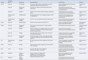
Established mammary organoid culture systems. Summary of representative mammary organoid models derived from humans, mice, cats, goats, sheep, and cattle, organized by cell/tissue source, matrix/extracellular matrix (ECM), key growth factors, and functional characteristics

Table 1 Long description
Columns list species, cell or tissue source, matrix or ECM, key growth factors, characteristics, and reference. Human entries include stem cell, single cell, and patient derived tissues grown in EHS Matrigel, Matrigel with collagen I, collagen I, or Matrigel like gel, with growth factors such as EGF, insulin, hydrocortisone, PRL, FGF7, FGF10, R-spondin 1, Noggin, and B27, and characteristics including acinar morphogenesis, lumen formation, epithelial polarity, branching morphogenesis, ER responsiveness, tumor modeling, and hormone responsive lactation. Mouse entries use stem cells in Matrigel like gel, Matrigel with collagen I, or Matrigel, with factors including insulin, hydrocortisone, PRL, FGF2, FGF10, Wnt3a, R Spondin2, and Y27632, and characteristics including alveolar morphogenesis, ECM dependent branching and invasion, clonality, bilayer organization, bipotency, lipid secretion, myoepithelial contraction, involution modeling, and mammary regeneration. Cat patient derived tissues are cultured in Matrigel with factors including NIC, NAC, A8301, L-WNR, EGF, FGF7, FGF10, and TGF alpha, with characteristics including tumor heterogeneity, xenograft tumorigenicity, and LMTK3 FADS2 pathway modeling. Sheep stem cells in Matrigel include hydrocortisone, penicillin, streptomycin, FGF2, EGF, PRL, ITS, and heparin, with characteristics including lactation, milk component secretion, budding morphogenesis, PRL responsiveness, and genetic manipulation. Goat tissue in Matrigel includes R spondin, NRG, FGF7, FGF10, SB202190, B27, FGF2, EGF, A8301, Y27632, and PRL, with characteristics including long term culture and sustained lactation with milk fat and protein production. Cattle single cells use a floating hydrogel of collagen I with laminin, fibronectin, and HA, with factors including Gastrin I, A 8301, FGF10, R spondin, EGF, Noggin, Wnt 3A, Nicotinamide, SB202190, and Y27632, and characteristics including branching morphogenesis, ductal lobular modeling, and alveolar like differentiation. Pig has a single cell entry where the matrix or ECM cell lists PRL responsive lactation, milk fat and protein production, and sustained lactation, with other columns not shown for that row.
Human mammary organoids
Compared with mouse models, the development of human mammary organoids has focused more on preserving cellular heterogeneity and clinical relevance (Fig. 1). Early efforts primarily focused on establishing stable 3D culture systems capable of supporting epithelial organization. These initial models demonstrated that human mammary epithelial cells can form luminal structures in a matrix-dependent manner (Bissell and W.C Reference Bissell and W.c2011; Petersen et al. Reference Petersen, Rønnov-jessen and Howlett1992). Attention shifted toward enriching stem and progenitor cell populations, leading to the development of non-adherent culture systems such as mammosphere assays. These approaches enabled the expansion of cells with regenerative potential but lacked structural organization (Dontu et al. Reference Dontu, Abdallah and Foley2003; Visvader and Stingl Reference Visvader and Stingl2014). More recent studies have highlighted the critical role of the microenvironment in shaping tissue architecture. In particular, co-culture systems incorporating epithelial cells and stromal components, such as fibroblasts, have been shown to promote epithelial self-organization and support the formation of ductal and acinar structures (Debnath et al. Reference Debnath, Muthuswamy and Brugge2003; Krause et al. Reference Krause, Maffini and Soto2008; Qu et al. Reference Qu, Han and Yu2015).
Human mammary organoids have evolved from simple spheroid structures into more organized, tissue-like models (Fig. 1 and Table 1). A key advance was the development of single-cell-derived organoid systems capable of generating structures resembling human terminal ductal lobular units, bringing these models closer to native tissue architecture (Linnemann et al. Reference Linnemann, Miura and Meixner2015; Nguyen-ngoc et al. Reference Nguyen-ngoc, Shamir and Huebner2015). Despite this progress, maintaining cellular heterogeneity during long-term culture remained a major challenge. Early systems often showed a gradual loss of specific epithelial subpopulations, particularly estrogen receptor-positive (ER+) cells. To address this, optimized culture conditions including R-spondin 1/Noggin-based Wnt–bone morphogenic protein modulation, EGF/FGF7/FGF10/heregulin β1 growth-factor support, and transforming growth factor β/p38 mitogen-activated protein kinase pathway inhibition were developed to support sustained expansion (Gray et al. Reference Gray, Girnius and Kuiken2023; Rosenbluth et al. Reference Rosenbluth, Schackmann and Gray2020) (Table 1). These improvements have significantly enhanced the stability and physiological relevance of human mammary organoids.
At the same time, advances in ECM and hydrogel design have contributed to improved functional performance in human mammary organoids (Kozlowski et al. Reference Kozlowski, Crook and H.t2021; Porter et al. Reference Porter, Murray and M.h2020; Rauner Reference Rauner2024). Current organoid systems can respond to hormonal cues. For instance, prolactin stimulation can induce lobular expansion and lipid droplet accumulation, indicating partial restoration of lactation function (Rosenbluth et al. Reference Rosenbluth, Schackmann and Gray2020; Sokol et al. Reference Sokol, Miller and Breggia2016). Recently, mammary organoids have been further extended from in vitro disease modeling toward therapeutic and regenerative applications. A recent bioengineering study further expanded the application of mammary organoids by engineering human induced pluripotent stem cell (iPSC)-derived mammary organoids into implantable therapeutic tissues capable of sustained drug secretion (Wang et al. Reference Wang, Yang and Wang2026). This platform supports both postsurgical chemotherapy and mammary tissue regeneration, highlighting the potential of mammary organoids as functional bioengineering systems beyond traditional disease modeling.
Mammary organoids of non-model species
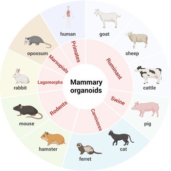
In recent years, mammary organoid systems have been extended beyond traditional model organisms to include livestock, companion animals, and other species. This expansion is particularly relevant to animal science, as it provides new experimental platforms for investigating lactation biology and developing strategies to improve production traits across species (Fig. 2). Current studies have established mammary organoids from a wide range of animal species, supported by optimized culture conditions tailored to different tissue sources, matrix requirements, and growth factor combinations (Fig. 2 and Table 1).
Animal species in which mammary organoid models have been established. Summary of mammary organoid models reported in various animals. See Table 1 for the corresponding references. Created with BioRender.com.

In livestock species, mammary organoids derived from dairy goats can reproduce the bilayered epithelial structure and respond to hormonal stimulation by producing key milk components (Jia et al. Reference Jia, Zhang and Luo2024). Similar progress has been reported in sheep, where organoid systems support stable gene manipulation and exhibit active expression of genes involved in milk fat, lactose, and protein synthesis (Feng et al. Reference Feng, Ma and Bai2025). These findings highlight the potential of mammary organoids as functional platforms for cell-based milk production.
Beyond production biology, mammary organoids are increasingly used in comparative oncology. Spontaneous mammary tumors in companion animals, such as cats, share important features with human breast cancer, particularly in metastatic behavior. Organoid models derived from these tumors provide a valuable platform for investigating conserved signaling pathways and assessing therapeutic responses, thereby complementing conventional human-based models (Yamamoto et al. Reference Yamamoto, Elbadawy and Tsunedomi2025).
In addition, cross-species organoid systems are beginning to provide new insights into mammary gland evolution (Fig. 2). Comparative studies using organoids from multiple species have revealed differences in branching morphogenesis and highlighted the role of signaling pathways, such as Rho associated coiled-coil containing protein kinase, in shaping species-specific developmental patterns (Kim et al. Reference Kim, Sinha and Sears2024). Importantly, these cross-species models may help bridge the gap between fundamental biology and practical applications in animal production systems.
Cell resource for mammary organoids
The successful establishment of mammary organoids largely depends on the choice of starting cell source, which determines their structural organization, differentiation capacity, and downstream applications. Current approaches mainly rely on adult stem or progenitor cells (ASCs), pluripotent stem cells (PSCs), patient-derived tissues, and single-cell-derived systems. Each source offers distinct advantages but also imposes specific constraints, making cell selection a key step in experimental design (Table 1).
ASCs remain the most commonly used source due to their capacity for self-renewal and lineage-restricted differentiation (Schutgens and Clevers Reference Schutgens and Clevers2020; Zhao et al. Reference Zhao, Chen and Dowbaj2022). Advances in culture conditions, particularly the incorporation of Wnt signaling, have significantly improved their expansion in vitro (Jardé et al. Reference Jardé, Lloyd-lewis and Thomas2016; Rosenbluth et al. Reference Rosenbluth, Schackmann and Gray2020). Organoids derived from ASCs generally retain key epigenetic features and exhibit relatively mature phenotypes, making them well suited for modeling tissue-specific functions (Jin et al. Reference Jin, Xue and Liu2024). However, these systems are largely limited to epithelial components and do not fully capture the complexity of the native mammary microenvironment, including stromal, immune, and vascular elements. In addition, their differentiation potential is constrained by tissue origin, which limits their use in studying broader developmental processes (Rosenbluth et al. Reference Rosenbluth, Schackmann and Gray2020; Wang et al. Reference Wang, Chen and Zhang2025; Whelan et al. Reference Whelan, Muir and Nakagawa2018).
PSCs provide a more flexible alternative for modeling early developmental events. While embryonic stem cells have strong differentiation potential, their use is limited in practice, and iPSCs have become the primary platform (Schutgens and Clevers Reference Schutgens and Clevers2020; Yang et al. Reference Yang, Hu and Kung2023). Through stepwise differentiation protocols, iPSCs can be guided toward mammary epithelial or progenitor-like states and subsequently self-organize into organoid structures (Dai et al. Reference Dai, Wang and Yang2022; Qu et al. Reference Qu, Han and Gao2017). A key advantage of this system is its adaptability to human-specific and patient-derived applications, particularly when combined with genome-editing tools (Dai et al. Reference Dai, Wang and Yang2022; Dekkers et al. Reference Dekkers, Whittle and Vaillant2020; Qu et al. Reference Qu, Han and Gao2017). However, PSC-derived organoids often display immature phenotypes, and achieving full functional maturation remains a challenge. Despite this, their broad developmental potential makes them particularly useful for studying lineage specification and early morphogenesis (Dai et al. Reference Dai, Wang and Yang2022; Heinzelmann et al. Reference Heinzelmann, Piraino and Costa2024; Qu et al. Reference Qu, Han and Gao2017).
Patient-derived tissues offer the most direct link to clinical biology. Organoids established directly from surgical specimens or biopsy samples, commonly referred to as patient-derived organoids (PDOs), preserve the architectural features, genetic alterations, and cellular heterogeneity of the original tumors more effectively than conventional cell lines (Bose et al. Reference Bose, Clevers and Shen2021; Sachs et al. Reference Sachs, De Ligt and Kopper2018). This makes them highly valuable for translational research, particularly in drug screening and precision medicine (Table 1). PDOs can be expanded into living biobanks and used to predict individual responses to therapy (Schutgens and Clevers Reference Schutgens and Clevers2020; Yin et al. Reference Yin, Zhou and Zhang2025). However, they do not fully reproduce the systemic and immune context of the tumor microenvironment.
Recent advances in single-cell isolation and culture technologies have enabled the generation of mammary organoids from individual cells, thereby providing a powerful system for studying clonal dynamics, lineage relationships, and cellular heterogeneity. For example, organoids derived from single Lgr5+ mammary stem cells are capable of forming complex epithelial structures, whereas lineage-tracing studies have shown that basal cells can regenerate bilayered gland-like architectures, while luminal cells more commonly form simpler acinar structures (Jamieson et al. Reference Jamieson, Dekkers and Rios2017; Zhang et al. Reference Zhang, Adileh and Martin2017). These observations highlight the distinct regenerative capacities of different mammary epithelial populations. In practice, single-cell-derived organoid culture typically requires ROCK inhibitors, such as Y-27632, to improve cell survival following dissociation. Although clonal organoid formation can be technically demanding and may show lower efficiency than bulk-cell culture, the defined cellular origin and relative genetic uniformity of these models make them particularly well suited for investigating tumor initiation, clonal evolution, and subtype-specific drug responses (Dekkers et al. Reference Dekkers, Whittle and Vaillant2020; Jamieson et al. Reference Jamieson, Dekkers and Rios2017; Sachs et al. Reference Sachs, De Ligt and Kopper2018; Zhang et al. Reference Zhang, Adileh and Martin2017).
Overall, ASCs are best suited for tissue-specific modeling, PSCs for developmental studies, PDOs for clinical applications, and single-cell systems for clonal analysis. The choice of cellular origin defines not only the structural fidelity of mammary organoids but also their suitability for specific biological and translational applications.
ECM resource for mammary organoid culture
Control over the ECM has emerged as a central determinant of organoid fidelity. Unlike conventional 2D systems, organoid cultures depend on matrix environments that simultaneously provide structural support, biochemical signaling, and mechanical regulation (Hughes et al. Reference Hughes, Postovit and G.a2010; Walters and Gentleman Reference Walters and Gentleman2015). As a result, ECM design is no longer a passive scaffold choice but an active variable that shapes tissue architecture and function (Aisenbrey and Murphy Reference Aisenbrey and W.l2020; Huang et al. Reference Huang, Zhang and Zhang2025; Hughes et al. Reference Hughes, Postovit and G.a2010). Matrigel has served as the standard, largely due to its ability to support diverse organoid types through a rich mixture of laminin, collagen IV, and growth factors (Kim et al. Reference Kim, Kim and Gao2017). However, its widespread use also defines a major bottleneck for the field. Its undefined composition, batch variability, and tumor-derived origin limit reproducibility, constrain mechanistic interpretation, and complicate translational applications (Aisenbrey and W.L Reference Aisenbrey and W.l2020; Hughes et al. Reference Hughes, Postovit and G.a2010; Kozlowski et al. Reference Kozlowski, Crook and H.t2021; Taghizadeh et al. Reference Taghizadeh, Taghizadeh and Kim2025). In this sense, Matrigel represents both an enabling technology and a barrier to further progress (Table 1).
Efforts to move beyond Matrigel reflect a broader shift toward engineered microenvironments. Fully synthetic hydrogels offer precise control over composition and mechanics, enabling systematic interrogation of matrix-dependent processes (Aisenbrey and W.L Reference Aisenbrey and W.l2020; Gjorevski et al. Reference Gjorevski, Sachs and Manfrin2016). Their lack of intrinsic bioactivity necessitates additional functionalization, and their biological relevance remains incomplete (Aisenbrey and W.L Reference Aisenbrey and W.l2020; Kozlowski et al. Reference Kozlowski, Crook and H.t2021). Recombinant protein- and peptide-based matrices attempt to bridge this gap by introducing defined biochemical cues, but their scalability and cost remain limiting factors (Table 1).
Decellularized ECM occupies an intermediate position between natural and engineered systems. By preserving native structural organization and signaling components, it provides a more physiologically grounded environment (Guo et al. Reference Guo, Liu and Zhang2024; Kim et al. Reference Kim, Kim and Gao2017; Zhu et al. Reference Zhu, Yuhan and Yu2023). At the same time, source-to-source variability and limited availability may restrict its broader adoption. Therefore, decellularized ECM has gained attention as a more biologically relevant alternative (Hasenauer et al. Reference Hasenauer, Bevc and McCabe2025; Mollica et al. Reference Mollica, Booth-creech and Reid2019). Achieving fully defined, tunable, and biologically relevant ECM platforms will be critical for improving reproducibility and for turning organoids into quantitatively interpretable models.
Growth factors and culture media in mammary organoid culture
If ECM determines the structural context, growth factors define the signaling landscape of organoid systems. Mammary organoid cultures often rely on serum-free, chemically defined media in which combinations of supplements and growth factors replace the complex signals present in vivo. In most mammary organoid culture systems, advanced Dulbecco’s Modified Eagle Medium (DMEM)/F-12 was used as the basal medium (Table 1). Core supplements such as B27 reduce variability, while metabolic regulators including N-acetylcysteine and nicotinamide help maintain redox balance and cellular homeostasis (Dekkers et al. Reference Dekkers, Van Vliet and Sachs2021; Sachs et al. Reference Sachs, De Ligt and Kopper2018). These components stabilize the system, whereas the growth factor network actively drives cellular behavior.
Epidermal growth factor (EGF) and fibroblast growth factor (FGF) signaling form a central regulatory axis, coordinating proliferation, survival, and tissue expansion through pathways such as MAPK and PI3K/Akt and through interactions with Wnt signaling (Chu et al. Reference Chu, Li and Ding2010; Jung et al. Reference Jung, Lee and Park2019; Roukens et al. Reference Roukens, Frederiks and Seinstra2021). Additional cues refine lineage specification. Neuregulin-1 supports luminal differentiation, whereas estrogen signaling is essential for maintaining the growth and functional identity of hormone-responsive epithelial cells (Jardé et al. Reference Jardé, Lloyd-lewis and Thomas2016; Rosenbluth et al. Reference Rosenbluth, Schackmann and Gray2020). Consistent with these findings, Rosenbluth et al. and Jardé et al. demonstrated that human mammary organoids could maintain epithelial lineage diversity, highlighting the importance of neuregulin-1 in preserving cellular identity (Jardé et al. Reference Jardé, Lloyd-lewis and Thomas2016; Rosenbluth et al. Reference Rosenbluth, Schackmann and Gray2020). Importantly, these signaling inputs do not act independently. The combined effects of N-spondin 1, Noggin, and Neuregulin1 determine organoid growth, lineage balance, and functional maturation. These findings highlight that precise control of signaling environments is essential for maintaining both the stability and functional identity of mammary organoids.
For lactation induction, the culture system needs to shift from an expansion-promoting medium to a differentiation-inducing medium. This usually involves lactogenic hormones such as prolactin, insulin, and glucocorticoids, which promote secretory differentiation and milk-component synthesis. For instance, lactogenic stimulation using prolactin has been used to model milk production and involution-like processes in mouse and ruminant mammary organoids (Jia et al. Reference Jia, Zhang and Luo2024; Sumbal et al. Reference Sumbal, Chiche and Charifou2020). These findings indicate that culture media should be tailored to the intended application, with future studies prioritizing chemically defined and functionally interpretable formulations.
Applications of mammary organoids
Breast cancer modeling
Mammary organoids have transformed experimental approaches to breast cancer by providing models that retain patient-specific features while remaining experimentally tractable (Fig. 3). Compared with conventional 2D systems, patient-derived mammary organoids enable rapid establishment from limited clinical materials and preserve key aspects of tumor heterogeneity and molecular identity (Huang et al. Reference Huang, Mei and Wan2024; Sachs et al. Reference Sachs, De Ligt and Kopper2018). These features enable organoids to capture subtype-specific tumor behaviors that are often lost in traditional cell culture systems. For example, Dekkers et al. established long-term cultures of patient-derived breast cancer organoids that maintained histopathological characteristics and intertumoral heterogeneity across molecular subtypes (Dekkers et al. Reference Dekkers, Van Vliet and Sachs2021). Such findings have strengthened the use of mammary organoids as platforms for investigating breast cancer heterogeneity, disease progression, and treatment resistance.
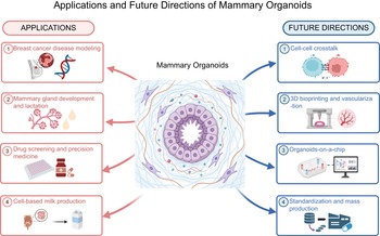
Applications and potentials of mammary organoids. Mammary organoids are applied in disease modeling, mammary development and lactation studies, drug screening, precision medicine, and cell-based milk production. Future advances will focus on multi-organoid systems, vascularization, organoids-on-a-chip, and standardized scalable production. Created with BioRender.com.

Figure 3 Long description
The infographic illustrates the applications and future directions of mammary organoids. On the left, under ′Applications′, four areas are listed: 1) Breast cancer disease modeling, 2) Mammary gland development and lactation, 3) Drug screening and precision medicine, 4) Cell-based milk production. Each application is connected to the central image labeled ′Mammary Organoids′. On the right, under ′Future Directions′, four areas are listed: 1) Cell-cell crosstalk, 2) 3D bioprinting and vascularization, 3) Organoids-on-a-chip, 4) Standardization and mass production. Each future direction is connected to the central image. The central image features a stylized representation of mammary organoids, surrounded by arrows indicating connections to the applications and future directions.
Beyond descriptive modeling, organoids enable causal interrogation of tumor biology. Genome editing enables the precise introduction of specific mutations into organoids derived from normal tissues, thereby facilitating the controlled, stepwise modeling of tumorigenesis. Recently, Dekkers et al. used CRISPR/Cas9-mediated editing to disrupt breast cancer-associated tumor suppressor genes, including TP53, PTEN, RB1, and NF1, in normal mammary organoids (Dekkers et al. Reference Dekkers, Van Vliet and Sachs2021, Reference Dekkers, Whittle and Vaillant2020). This approach demonstrated how specific genetic alterations drive malignant phenotypic changes, including altered tissue organization, increased proliferative capacity, and tumor-like behavior. Such studies provide direct genotype-phenotype evidence that is difficult to obtain in vivo and highlight the value of organoids for reconstructing disease progression rather than simply observing tumor states (Drost et al. Reference Drost, Van Jaarsveld and Ponsioen2015; Wang et al. Reference Wang, Yang and Wang2026).
Mammary gland development and lactation
Beyond cancer research, mammary organoids provide a controlled system for studying normal mammary gland development and lactation. By adjusting hormonal and growth-factor inputs, organoids can reproduce key developmental transitions, including ductal morphogenesis, alveolar differentiation, and post-lactational involution (Zhu et al. Reference Zhu, Jia and Zhang2022). In fact, mammary organoids have helped define how extracellular cues coordinate tissue architecture and secretory function. For example, FGF2 and EGF promote ductal branching, partly through MAPK-related signaling, whereas prolactin activates lactogenic differentiation, including milk-protein expression and lipid-droplet accumulation, through janus kinase (JAK)-signal transducer and activator of transcription (STAT)-related pathways (Aggeler et al. Reference Aggeler, Park and Bissell1988; Kreuzaler et al. Reference Kreuzaler, Staniszewska and Li2011; Simian et al. Reference Simian, Hirai and Navre2001).
Cross-species organoid systems extend these mechanistic studies into comparative and agricultural biology. By enabling parallel studies across species, they provide a route to identify conserved and species-specific regulators of mammary development and lactation (Finot et al. Reference Finot, Chanat and Dessauge2021; Kim et al. Reference Kim, Sinha and Sears2024). For example, dairy goat mammary organoids, combined with single-cell transcriptomics and in vivo genetic perturbation, helped identify luminal hormone-responsive cells and prolactin receptor-related signaling as key regulators of secretory cell maturation during mammary regenerative remodeling (Li et al. Reference Li, Zhang and Luo2025). Similarly, organoid models from goat, bovine, ovine, and murine mammary tissues provide comparative platforms for examining species-specific regulation of milk synthesis and mammary gland renewal (Feng et al. Reference Feng, Ma and Bai2025; Finot et al. Reference Finot, Chanat and Dessauge2021; Jia et al. Reference Jia, Zhang and Luo2024; Kim et al. Reference Kim, Sinha and Sears2024).
Drug screening and precision medicine
One of the most immediate translational impacts of mammary organoids lies in drug development. By preserving patient-specific genomic and phenotypic features, organoids provide more predictive models of therapeutic response than conventional cell lines. PDOs enable functional testing of drug sensitivity across individuals and tumor subtypes, supporting the development of personalized treatment strategies (Corsini and Knoblich Reference Corsini and J.a2022; Di Paola et al. Reference Di Paola, Calafato and Piccaluga2025; Zhao et al. Reference Zhao, Feng and Xia2025). For example, comparative drug testing using luminal, human epidermal growth factor receptor (HER2)-positive, and triple-negative breast cancer organoids revealed distinct subtype-specific response patterns and resistance mechanisms, supporting the identification of rational combination therapies and patient stratification strategies (Chitrangi et al. Reference Chitrangi, Vaity and Jamdar2023; Guillen et al. Reference Guillen, Fujita and Butterfield2022; Liu et al. Reference Liu, Fang and Yuan2026; Sachs et al. Reference Sachs, De Ligt and Kopper2018). These findings highlight the potential of organoid-based assays to complement genomic profiling by providing direct functional evidence of drug response.
Integration with engineering platforms further extends these capabilities. Microfluidic systems and bioprinted models introduce spatial and dynamic complexity, enabling more realistic modeling of drug delivery, microenvironmental interactions, and treatment response (Li et al. Reference Li, Yu and Zhou2024; Wang et al. Reference Wang, Ning and Zhao2024). These platforms can incorporate ECM barriers, stromal cells, immune components, and dynamic perfusion, thereby improving the modeling of drug penetration and tumor–stroma interactions (Blanco-fernandez et al. Reference Blanco-fernandez, Rey-vinolas and Bagci2022; Mollica et al. Reference Mollica, Booth-creech and Reid2019; Subia et al. Reference Subia, Dahiya and Mishra2021). Such systems have improved our understanding of how microenvironmental factors influence therapeutic efficacy and resistance. Importantly, emerging studies suggest that organoids may also support adaptive treatment strategies. For example, combined circulating tumor DNA monitoring with sequential breast cancer organoid drug screening demonstrated that secondary organoid testing could identify alternative therapeutic options following the emergence of treatment-associated genetic alterations (Liu et al. Reference Liu, Fang and Yuan2026).
Mammary organoids in cell-based milk production
Beyond biomedicine, mammary organoids are beginning to contribute to cellular agriculture, particularly for cell-based milk production. This emerging application reframes mammary organoids from experimental models into functional bioengineering platforms capable of reproducing key aspects of lactation in vitro (Kwon et al. Reference Kwon, Jung and Kothuri2024). Unlike conventional mammary epithelial cell cultures, mammary organoids retain tissue-level organization and coordinated secretory activity. Under appropriate hormonal stimulation, they can synthesize major milk components, including caseins, lactose, and lipids, providing evidence that core lactogenic programs can be maintained outside the animal (Sumbal et al. Reference Sumbal, Chiche and Charifou2020; Yuan et al. Reference Yuan, Xie and Bai2023).
Organoid systems also provide unprecedented opportunities to dissect and engineer milk production. Because genetic, nutritional, and environmental variables can be precisely controlled, organoids offer a tractable platform for investigating the regulation of milk composition, including protein content, lipid metabolism, and bioactive factor production (Finot et al. Reference Finot, Chanat and Dessauge2021; Sumbal et al. Reference Sumbal, Chiche and Charifou2020; Yao et al. Reference Yao, Ranadheera and Shen2024). For example, Jia et al. established a goat mammary organoid system that maintained a bilayer epithelial architecture resembling native mammary tissue. Upon lactogenic induction, these organoids accumulated lipid droplets and sustained milk-production activity for more than 3 weeks. Proteomic analyses further revealed that lactating organoids exhibited molecular signatures highly similar to those of mammary tissue in vivo, supporting their utility as models for studying milk synthesis and secretory regulation. Despite these advances, substantial challenges remain before cell-based milk production becomes commercially feasible. Matrix standardization, long-term maintenance of secretory function, scalable bioreactor systems, and downstream processing strategies will all be required to translate mammary organoids from experimental platforms to practical dairy biomanufacturing systems (Kim et al. Reference Kim, Kim and Gao2017; Kwon et al. Reference Kwon, Jung and Kothuri2024).
Future perspectives in mammary organoid applications
Decoding mammary-specific cell–cell interactions and tissue hierarchy
A major challenge in current mammary organoid systems is the incomplete representation of the cellular diversity present in native mammary tissue. Most established models are dominated by epithelial cells and only partially capture interactions with fibroblasts, adipocytes, immune cells, vascular cells, and neuroendocrine components (Narkhede et al. Reference Narkhede, Shevde and S.s2017; Schutgens and Clevers Reference Schutgens and Clevers2020). As a result, many fundamental questions in mammary biology remain difficult to address, including how stromal populations regulate branching morphogenesis, how immune cells contribute to involution and tissue remodeling, and how niche-derived signals influence mammary stem-cell fate (Chumduri et al. Reference Chumduri and M.y2021; Pan et al. Reference Pan, Chen and Deng2025; Utz et al. Reference Utz, Turpin and Lampe2020). Future efforts should move beyond epithelial-centric models toward multicellular mammary ecosystems. Such systems will not only improve physiological fidelity but also enable mechanistic dissection of cell–cell communication networks that govern mammary development, lactation, regeneration, and tumor progression.
Reconstructing mammary tissue architecture and vascular integration
A major limitation of current mammary organoid systems is their inability to fully recapitulate the structural and cellular complexity of the native mammary gland. Most organoids rely on diffusion-mediated nutrient transport and lack vascular, stromal, adipose, and immune compartments, which restricts long-term survival, functional maturation, and physiological relevance (Nwokoye and Abilez Reference Nwokoye and Abilez2024; Zhang et al. Reference Zhang, Wan and Kamm2021). These limitations are particularly important for mammary biology, as mammary development, lactation, involution, and tumor progression are all regulated by dynamic interactions among epithelial, vascular, stromal, and immune cell populations.
Future efforts should therefore focus on reconstructing the multicellular architecture of mammary tissue rather than simply increasing organoid size. Vascularization represents a critical step toward this goal, as perfusable networks could improve nutrient delivery, support long-term culture, and enable more physiologically relevant modeling of endocrine signaling and drug distribution. Recent advances in 3D bioprinting provide promising tools for engineering such complexity (Abilez et al. Reference Abilez, Yang and Guan2025). By enabling spatially controlled deposition of cells and biomaterials, bioprinting allows the incorporation of endothelial cells, fibroblasts, adipocytes, and immune cells into defined tissue architectures (Miao et al. Reference Miao, Pek and Tan2025; Wang et al. Reference Wang, Ning and Zhao2024).
Several studies have demonstrated the feasibility of applying these approaches to mammary systems. Reid et al. generated bioprinted tumoroids and chimeric mammary organoids to investigate tumor–epithelial interactions (Reid et al. Reference Reid, Palmer and Mollica2019). In a complementary approach, Mollica et al. demonstrated that bioinks based on decellularized ECM (dECM) derived from mammary tissue could provide tissue-specific biochemical cues that supported epithelial organization and tumor growth (Mollica et al. Reference Mollica, Booth-creech and Reid2019). More recently, volumetric bioprinting combined with bovine mammary dECM-based scaffolds supported lactation-related functions, including β-casein secretion and milk fat globule production (Blanco-fernandez et al. Reference Blanco-fernandez, Rey-vinolas and Bagci2022; Hasenauer et al. Reference Hasenauer, Bevc and McCabe2025). These studies suggest that mammary-specific biomaterials may be as important as vascularization itself for achieving functional maturation.
Modeling dynamic mammary physiology using organoids-on-a-chip
A persistent limitation of conventional mammary organoid cultures is their inability to reproduce the dynamic physical and endocrine environments that regulate mammary gland development and function in vivo. Organoids-on-a-chip (OrgOCs) offer a promising strategy to address this challenge by integrating mammary organoids with microfluidic platforms that enable precise control of fluid flow, biochemical gradients, and mechanical cues (Wang et al. Reference Wang, Ning and Zhao2024; Papamichail et al. Reference Papamichail, Koch and Veerman2025). Rather than simply improving nutrient delivery, these systems provide opportunities to investigate how dynamic environmental signals regulate mammary epithelial behavior and tissue function.
For mammary biology, OrgOCs may be valuable for modeling endocrine regulation and tissue remodeling. Controlled hormone delivery could recapitulate cyclical estrogen and progesterone dynamics, pregnancy-associated endocrine shifts, and lactogenic stimulation, thereby facilitating the study of developmental transitions that are difficult to model in conventional culture systems. Similarly, microfluidic flow and mechanical stimulation may help recreate ductal pressure, extracellular fluid dynamics, and microenvironmental conditions associated with lactation, involution, and tumor progression (Li et al. Reference Li, Yu and Zhou2024b; Papamichail et al. Reference Papamichail, Koch and Veerman2025). The integration of real-time biosensors further enables continuous monitoring of cellular behavior and drug responses (Subia et al. Reference Subia, Dahiya and Mishra2021; Wang et al. Reference Wang, Ning and Zhao2024). Future development should focus on integrating endocrine regulation, multicellular complexity, and sustained functionality into a unified platform, thereby enabling more faithful recapitulation of mammary physiology and disease processes.
Remaining challenges and future directions
Despite substantial progress, several key challenges continue to limit the development of physiologically faithful mammary organoid models. Among these, one challenge is achieving stable long-term functional maturation. Although many mammary organoids can reproduce aspects of ductal morphogenesis and lactogenic differentiation, sustained hormone responsiveness, maintenance of lineage diversity, and long-term secretory activity remain difficult to achieve. In particular, the progressive loss of basal cell populations and changes in cellular composition during extended culture continue to limit physiological fidelity (Rosenbluth et al. Reference Rosenbluth, Schackmann and Gray2020).
Reproducibility also remains a major concern. Variability in cell sources, ECM composition, and culture protocols can generate substantial differences between laboratories, complicating data comparison and limiting translational applications. The development of chemically defined matrices, standardized culture systems, and quantitative functional benchmarks will therefore be essential for improving experimental robustness. The next generation of mammary organoid models will likely move beyond isolated epithelial structures toward integrated tissue systems that incorporate multicellular interactions, vascular support, dynamic endocrine regulation, and functional lactation competence. Achieving this goal will require the convergence of stem cell biology, biomaterials science, bioengineering, and systems biology (Zhou et al. Reference Zhou, Wu and Wang2023).
Conclusion
In summary, mammary organoid technology has rapidly advanced over the past decade, evolving from simple 3D culture systems to more sophisticated models that partially recapitulate the structural and functional features of the mammary gland. These systems have significantly improved our ability to study mammary development, lactation biology, and breast cancer, offering clear advantages over traditional 2D cultures and animal models in terms of physiological relevance and experimental flexibility.
The diversity of cell sources, together with continuous improvements in ECM design and culture conditions, has expanded the applicability of mammary organoids across multiple research fields. Moreover, the development of organoids from livestock species further extends their importance in animal science, providing new opportunities to enhance milk production and mammary health.
Despite these advances, several challenges remain. Current models still lack full tissue complexity, including vascular, immune, and endocrine interactions, and issues such as limited reproducibility and incomplete functional maturation need to be addressed. Emerging technologies, including multi-organ systems, 3D bioprinting, and OrgOCs, offer promising solutions but require further optimization before widespread adoption. Future efforts should focus on improving model standardization, integrating multiple cell types and organ systems, and enhancing functional fidelity. As these systems continue to evolve, mammary organoids are likely to become a central platform linking fundamental biology with translational and industrial applications.
Author Contributions
M.W.: writing the original draft. M.W., M.S., L.C., D.Y., L.C., Y.Y., and S.P.: literature and data collection. H.S.: funding acquisition and supervision, and editing the draft. All authors contributed to the article and approved the submitted version.
Funding Statement
This study was jointly supported by the National Natural Science Foundation of China (32472917 and U23A20231).
Conflicts of Interest
The authors declare that there are no conflicts of interest.


 Open access

