Introduction
In infertility treatments using controlled hormonal ovarian hyperstimulation, most oocytes retrieved from ovaries are mature metaphase-II arrested oocytes, but about 10–20% remain immature, at metaphase-I or at prophase-I (germinal vesicle stage, GV). Immature oocytes may spontaneously reach metaphase-II in vitro, but some never become mature (Cha & Chian, Reference Cha and Chian1998).
The process from germinal vesicle breakdown of prophase-I oocytes to meiosis-I and meiosis-II occurs in the ovarian follicle and is named oocyte maturation. These nuclear and chromosome events are coordinated with cytoplasmic maturation. The acquisition of meiotic competence occurs at the time of antrum formation, but the oocyte remains arrested at prophase-I due to high levels of cyclic adenosine monophosphate. Meiotic resumption is triggered by the preovulatory surge of luteinizing hormone (LH), which is also responsible for ovulation. As LH receptors reside on theca cells, LH triggers the oocyte to resume meiosis by indirect mechanisms, mainly through the activation of synthesis and release of growth factors (Mehlmann, Reference Mehlmann2005).
Studies on oocyte maturation have mainly focussed on molecular processes (Nogueira et al., Reference Mehlmann2003; Mehlmann, Reference Mehlmann2005; Han et al., Reference Han, Vaccari, Nedachi, Andersen, Kovacina, Roth and Conti2006; Tosti, Reference Tosti2006; Di Luigi et al., Reference Di Luigi, Weitzman, Pace, Siano, Maier and Mehlmann2008; McGinnis et al., Reference McGinnis, Carroll and Kinsey2011), gene-expression profiles (Assou et al., Reference Han, Vaccari, Nedachi, Andersen, Kovacina, Roth and Conti2006; Virant-Klun et al., Reference Virant-Klun, Knez, Tonazevic and Skutella2013) and oocyte–somatic cell interactions (Li et al., Reference Li, McKenzie and Matzuk2008; Li & Albertini, Reference Li and Albertini2013). Several descriptive studies on the ultrastructural composition on human oocytes at different maturation stages have been reported previously (Hertig & Adams, Reference Hertig and Adams1967; Zamboni et al., Reference Virant-Klun, Knez, Tonazevic and Skutella1972; El Shafie et al., Reference El Shafie, Sousa, Windt and Kruger2000; Sathananthan & Trouson, Reference Sathananthan and Trouson2000; Khalili et al., Reference Liang, Soyal and Dean2013; Shahedi et al., Reference Shahedi, Khalili, Soleimani and Morshedizad2013a), some of them have been after vitrification of immature (Shahedi et al., Reference Shahedi, Hosseini, Khalili, Norouzian, Salehi, Piriaei and Nottola2013b; Palmerini et al., Reference Palmerini, Antinori, Maione, Cerusico, Versaci, Nottola, Macchiarelli, Khalili and Antinori2014) or mature oocytes (Khalili et al., Reference Khalili, Maione, Palmerini, Bianchi, Macchiarelli and Nottola2012).
To our knowledge there are no data on the three-dimensional quantitative ultrastructural analysis of human oocytes, either immature or mature. Such data could be useful to understand the cellular mechanisms that underlie oocyte maturation, which in turn might be important for the establishment of optimal in vitro handling protocols for oocytes after follicle retrieval, and for the development of novel methods of in vitro oocyte maturation (IVM), with less aggressive hormonal stimulation protocols (Tan & Child, Reference Tan and Child2002; Picton et al., Reference Picton, Harris, Muruvi and Chambers2008).
In this study, we aimed to systematically quantify the distribution of organelles in oocytes at prophase-I using a stereological analysis.
Materials and methods
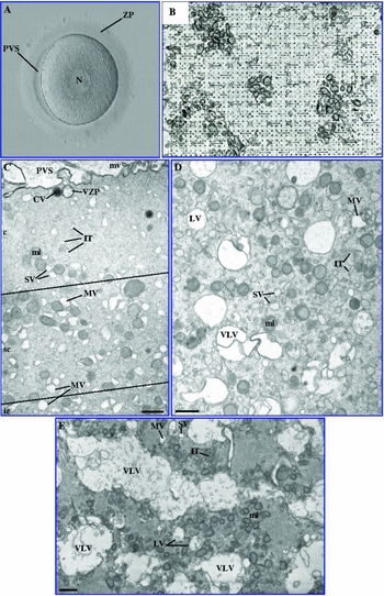
According to the National Law on Medically Assisted Procreation (http://dre.pt/pdf1sdip/2006/07/14300/52455250.pdf Law 32/2006) and the National Council on Medically Assisted Procreation guidelines (http://www.cnpma.org.pt/Docs/Profissionais_Requisitos_CentrosPMA.pdf CNPMA, 2008), seven immature GV oocytes (Fig. 1A ), from a cohort presenting normal oocytes, were used after patient informed and written consent of five cases enrolled in intracytoplasmic sperm injection (ICSI) treatment cycles.
(A) Image taken by inverted microscopy. Prophase-I (GV) live oocyte with zona pellucida (ZP), perivitelline space (PVS) and centrally located nucleus (N). (B) Stereological grid. (C–E) Images taken by transmission electron microscopy. (C) GV cortex (c), subcortex (sc) and inner cytoplasm (ic). (D, E) Inner cytoplasm. There were no distinct subregions on the distribution of SER vesicles and tubules, but only intermixed areas of predominance of SER vesicles and tubules or SER-VLV. (E) Region of the inner cytoplasm to show the richness in SER vesicles and tubules. These organelles occupied 6.1% of the oocyte relative volume and 6.23% of the relative volume of the inner cytoplasm. (F) Region of the inner cytoplasm to show the richness in SER-VLV. These occupied 6.9% of the oocyte relative volume and 10.1% of the relative volume of the inner cytoplasm. Microvilli (mv), cortical vesicle (CV), vesicle with zona pellucida-like materials (VZP), smooth endoplasmic reticulum (SER) isolated tubules (IT), mitochondria (mi), SER small vesicles (SV), SER medium vesicles (MV), SER large vesicles (LV), SER very large vesicles (VLV). Bars: 1 μm.

Stimulation protocol
Women underwent controlled ovarian hyperstimulation (Huirne et al., Reference Huirne, Homburg and Lambalk2007; Pinto et al., Reference Pinto, Oliveira, Cardoso, Teixeira da Silva, Silva, Sousa and Barros2009) using a gonadotrophin releasing hormone short antagonist protocol with cetrorelix (Merck Serono, Geneva, Switzerland) or ganirelix (Organon, Oss, The Netherlands). For stimulation, recombinant follicle stimulating hormone was used (Puregon-Organon; Gonal-F-Merck Serono). For triggering final oocyte maturation, human chorionic gonadotrophin was administered (5000–10000 IU; Pregnyl-Organon). Estradiol serum levels were assayed 1 day before or at the day of human chorionic gonadotrophin administration.
Oocyte handling
Retrieved oocytes were washed in G-MOPS-Plus (VitroLife, Kungsbacka, Sweden) or Sperm Preparation Medium (SPM; Medicult Origio, Jyllinge, Denmark), and incubated in in vitro fertilization (IVF) medium (Medicult) or G-IVF-PLUS (VitroLife) for 1–4 h (37ºC, 6% CO2, 5% O2 in air). Cumulus–oocyte complexes were denuded under light mineral oil (Liquid paraffin: Medicult; or Ovoil-100: VitroLife) with 80 UI hyaluronidase (ICSI Cumulase: Medicult; or Hyase-10×: VitroLife). Immature GV oocytes were selected under an inverted microscope (Nikon DIAPHOT 200; Nikon, Tokyo, Japan) and live cell images were taken (×10 and ×20 magnification objectives) with the use of the Cronus-3 imaging system (Research-Instruments Ltd, Cornwall, UK).
Transmission electron microscopy
Oocytes were fixed with Karnovsky (2.5% glutaraldehyde, 4% paraformaldehyde, 0.15 M sodium cacodylate buffer) (Sigma-Aldrich, St. Louis, USA; Merck, Darmstadt, Germany) for 30 min at room temperature and then for 2 h at 4ºC, washed in 0.15 M sodium cacodylate buffer, pH 7.3 (Merck), post-fixed with 2% osmium tetraoxide (Merck) in buffer containing 0.8% potassium ferricyanide (Merck) for 2 h at 4°C, washed in buffer (10 min), serially dehydrated in ethanol (Panreac, Barcelona, Spain), equilibrated with propylene oxide (Merck) and embedded in Epon (Sigma). Semithin and ultrathin sections were prepared with a diamond knife (Diatome, Hatfield, Switzerland) in a LKB ultramicrotome (Leika Microsystems, Weltzlar, Germany). Using a table of random numbers for initial cutting, GV oocytes were then serially sectioned and ultrathin sections collected and analysed each 15 μm. Ultrathin sections were collected on 100-mesh formvar carbon-coated copper grids (Taab, Berks, UK), stained with 3 % aqueous uranyl acetate (20 min) (BDH, Poole, UK) and Reynolds lead citrate (10 min) (Merck). Ultrathin sections were observed under a JEOL 100XII transmission electron microscope (TEM) (JEOL, Tokyo, Japan) operated at 60 kV (Sousa & Tesarik Reference Shahedi, Khalili, Soleimani and Morshedizad1994; El Shafie et al., Reference El Shafie, Sousa, Windt and Kruger2000).
Stereological and statistical analysis
For each microscope grid, a systematic sampling was performed with photographs taken at alternate TEM field spaces when at least 50% of the field was occupied by the oocyte cytoplasm. Images were taken at ×5300 magnification and printed at three time's higher magnification (×15,900).
A classical manual stereological technique based on point-counting with an adequate stereological grid was used (Fig. 1B ). Printed photographs were placed under the grid and the number of grid points placed over each organelle was noted. The relative volume (Vv) of each organelle was obtained by applying the formula (Weibel et al., Reference Weibel, Kistler and Scherle1966) Vv (organelle, oocyte) = [number of points (organelle)/number of points (oocyte)] × 100 (%).
Organelles included were: mitochondria, dictyosomes, cortical vesicles, vesicles containing zona pellucida-like materials, multivesicular bodies, lysosomes (lipofuscin bodies or refractile bodies), annulate lamellae, smooth endoplasmic reticulum (SER) very large vesicles (SER-VLV), SER large vesicles (SER-LV: vesicles slight larger than mitochondria), SER medium vesicles (SER-MV: more or less of the size of mitochondria), SER small vesicles (SER-SV), SER isolated tubules (SER-IT), total SER with exception of the SER-VLV (SER-LV, SER-MV, SER-SV, SER-IT), and the nucleus.
Additionally, each oocyte was divided into three regions from the oolemma up to the cell centre: cortex (5 μm), subcortex (5–10 μm) and inner cytoplasm (>10 μm). These measures were performed in the printed version. The same stereological procedure was then adopted, applying the formula Vv (organelle, cortex/subcortex/inner cytoplasm) = [number of points (organelle)/number of points (cortex/subcortex/inner cytoplasm)] × 100 (%).
Statistical analysis was performed with Microsoft Excel 2007 and SPSS version 22.0. Results are presented as means, standard error of the mean (SEM = standard deviation/n1/2) and coefficient of variation (CVar = standard deviation/mean).
Normal distribution was tested with Kolmogorov–Smirnov test. As expected, the samples did not follow the normal distribution, and non-parametric tests were used. Kruskal–Wallis test and Mann–Whitney U-test with Bonferroni correction were used to compare the means of Vv (organelle, oocyte), Vv (organelle, cortex), Vv (organelle, subcortex) and Vv (organelle, inner cytoplasm) as appropriate. Statistical significance level was set at P < 0.05.
Results
Stereological analysis was made in a total of 327 photographs: 142 with representation of the oocyte cortex (Fig. 1C ), 162 with representation of the oocyte subcortex (Fig. 1C ) and 297 with representation of the oocyte inner cytoplasm (Fig. 1D ).
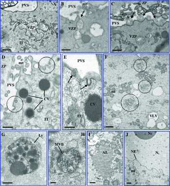
The common organelles described in human oocytes were observed. Additionally we identified receptor-mediated endocytosis and two organelles first described in human GV oocytes: (i) SER-VLV, with an elongated and irregular outline appearance that appeared concentrated more in the inner cytoplasm (Fig. 1E ); and (ii) medium-sized vesicles, frequently with an irregular outline, which presented floccular fine fibrillar contents that probably corresponded to zona pellucida-like materials, as they were observed to be exocytosed into the perivitelline space where their structure resembled that of the zona pellucida (ZP) (Fig. 2A–C ). These vesicles were not observed in the inner cytoplasm and their structure was totally different from that of the dense cortical vesicles (Fig. 2D ); and coated vesicles that are a sign of receptor-mediated endocytosis (Fig. 2D, E ).
Images taken by transmission electron microscopy. (A–C) Vesicles containing zona pellucida-like materials (VZP). (A) VZP with irregular outline. (B) VZP fused with the oocyte membrane (arrow). (C) Contents of VZP exocytosed (arrows) in the perivitelline space (PVS). Microvilli (mv), zona pellucida (ZP), smooth endoplasmic reticulum (SER) isolated tubules (IT). (D, E) Coated vesicles encircled by dotted circles (D), and at a higher magnification (arrows) (E). Cortical vesicles (CV). (F) Dictyosomes (dotted circles). SER very large vesicle (VLV), mitochondria (mi). (G) lysosome (Ly). (H) Multivesicular body (MVB). (I) Annulate lamellae (AL). (J) nucleus (N), nucleolus (Nc), nuclear envelope (NE). Bars:1 μm (F, J), 0.5 μm (A–D, G, I), 0.2 μm (E, H).

The other organelles observed were: (i) mitochondria (Figs 1 and 2), which predominated in the inner cytoplasm; (ii) SER-LV, SER-MV, SER-SV and SER-IT (Fig. 1C, D ) that were observed uniformly distributed in the three oocyte regions; (iii) cortical vesicles (Fig. 2D ), which predominated in the oocyte cortex; (iv) dictyosomes (Fig. 2F ); (v) lysosomes (Fig. 2G ); and (vi) multivesicular bodies (Fig. 2H ), which were identified in the three oocyte regions. Annulate lamellae (Fig. 2I ) were observed in three oocytes, two localized in the inner cytoplasm and one in the subcortex. The nucleus (Fig. 2J ) was observed in the inner cytoplasm (centrally located), except in one oocyte where it was more eccentrically placed (occupying the subcortex and cortex). Rough endoplasmic reticulum (RER), SER tubular aggregates (aSERT) associated or not with mitochondria, SER vesicles associated with mitochondria and polyribosomes were not observed in the GV oocytes analysed.
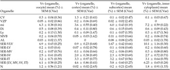
Quantitatively, SER-VLV (6.9%), mitochondria (6.3%) and the SER vesicles and tubules excluding the SER-VLV (6.1%) were the organelles that occupied the highest relative volumes in the GV oocyte cytoplasm. Each one of the remaining cytoplasmic organelles occupied a relative volume lower than 1%, with the exception of SER-SV (2.5%), SER-IT (3.2%) and the nucleus (1.2%) (Table 1).
Relative volume of organelles (Vv) per GV oocyte, oocyte cortex, subcortex and inner cytoplasm

Results presented as mean (%), SEM: standard error of the mean (standard deviation/n1/2), and CVar: coefficient of variation (standard deviation/mean).
CV: cortical vesicles; VZP: vesicles containing zona pellucida-like materials; mi: mitochondria; Di: dictyosomes; Ly: lysosomes; MVB: multivesicular bodies; AL: annulate lamellae; SER-VLV: smooth endoplasmic reticulum (SER) very large vesicles; SER-LV: SER large vesicles; SER-MV: SER medium-sized vesicles; SER-SV: SER small vesicles; SER-IT: SER isolated tubules.
When globally comparing the three oocyte regions using the Kruskal–Wallis test, significant differences were observed in the relative volume occupied by mitochondria (P = 0.005), cortical vesicles (P = 0.001), medium-sized vesicles containing ZP-like materials (P = 0.004), SER-VLV (P = 0.001) and the nucleus P = 0.036). However, no significant differences were found for the other organelles: dictyosomes (P = 0.366), lysosomes (P = 0.638), multivesicular bodies (P = 0.052), annulate lamellae (P = 0.365), SER-LV (P = 0.112), SER-MV (P = 0.318), SER-SV (P = 0.754), SER-IT (P = 0.853), or all SER vesicles and tubules with exception of the SER-VLV (P = 0.853).
Strict pairwise comparisons for the three regions using the Mann–Whitney U-test, Bonferroni-corrected, showed that for mitochondria, the relative volume was lowest in the cortex, which was significantly different from the subcortex (3.6% versus 6%, P = 0.039) and the inner cytoplasm (3.6% versus 7.2%, P = 0.018). No differences were found between the subcortex and inner cytoplasm (P = 0.426). The relative volume occupied by cortical vesicles was significantly highest in the cortex, compared with the subcortex (1.3% versus 0.1%, P = 0.006) and to the inner cytoplasm (1.3% versus 0.1%, P = 0.006), with no differences between the subcortex and inner cytoplasm (P = 1.0). Similarly, regarding the medium-sized vesicles containing ZP-like materials, the relative volume was significantly higher in the oocyte cortex, with a few of these vesicles being observed in the subcortex (0.2% versus 0.02%, P = 0.036) and none in the inner cytoplasm (0.2% versus 0%, P = 0.015), with no differences observed between the subcortex and inner cytoplasm (P = 1.0). The relative volume occupied by SER-VLV was significantly higher in the inner cytoplasm compared with the cortex (10.1% versus 0.9%, P = 0.006) and the subcortex (10.1% versus 1.67%, P = 0.006), with no differences between the cortex and subcortex (P = 0.54). Concerning the nucleus, although statistically significant differences were found when globally comparing the three regions, strict pairwise comparisons failed to detect differences, either between the cortex and subcortex (P = 1.0), cortex and inner cytoplasm (P = 0.084) or subcortex and inner cytoplasm (P = 0.168).
Discussion
Human oocyte maturation is a complex process both at the cellular and at the molecular levels. Despite recent advances in the understanding of molecular processes involved in oocyte maturation (Nogueira et al., Reference Mehlmann2003; Mehlmann, Reference Mehlmann2005; Assou et al., Reference Han, Vaccari, Nedachi, Andersen, Kovacina, Roth and Conti2006; Han et al., Reference Han, Vaccari, Nedachi, Andersen, Kovacina, Roth and Conti2006; Tosti, Reference Tosti2006; Di Luigi et al., Reference Di Luigi, Weitzman, Pace, Siano, Maier and Mehlmann2008; Li et al., Reference Li, McKenzie and Matzuk2008; McGinnis et al., Reference McGinnis, Carroll and Kinsey2011; Li & Albertini, Reference Li and Albertini2013; Virant-Klun et al., Reference Virant-Klun, Knez, Tonazevic and Skutella2013), there are no previous quantitative studies on the composition and distribution of organelles in the human oocyte, and only a few studies have been dedicated to the detailed morphological ultrastructure of human GV oocytes (Zamboni et al., Reference Virant-Klun, Knez, Tonazevic and Skutella1972; Sathananthan et al., Reference Sathananthan, Selvaraj, Girijashankar, Ganesh, Selvaraj and Trouson2006; Mao et al., Reference Mao, Lou, Lou, Wang and Jin2014; Palmerini et al., Reference Palmerini, Antinori, Maione, Cerusico, Versaci, Nottola, Macchiarelli, Khalili and Antinori2014).
In our study, receptor-mediated endocytosis and two organelles are first described in GV oocytes. First, coated vesicles were observed at the oocyte surface, which indicates that human oocytes at prophase-I exchange materials with the exterior by receptor-mediated endocytosis. Second, vesicles containing ZP-like materials were observed in the oocyte cortex. These were morphologically distinct from the dense cortical vesicles, which were not observed to suffer precocious exocytosis. The contents of these vesicles appeared to suffer exocytosis and once in the perivitelline space their structure resembled the fine structure of ZP fibrils. During oogenesis, several transcription factors act in sequence to govern the expression of the genes encoding ZP proteins (Liang et al., Reference Liang, Soyal and Dean1997; Cui & Kim, Reference Cui and Kim2007), with ZP glycoproteins being synthesized since primordial follicles and to be originated from materials secreted by both the oocyte and granulosa cells (Tesarik & Kopecny, Reference Sousa, Barros, Silva and Tesarik1986; Sousa et al., Reference Sousa and Tesarik2015). Thus, our present findings corroborate the prophase-I oocyte contribution to ZP formation. Third, SER-VLV were found mainly in the inner cytoplasm. These vesicles had an irregular outlined appearance as if derived from the fusion of smaller vesicles. They represent the organelle with the highest relative volume occupied in the oocyte, but we could not establish a morphological relationship to the other SER vesicles. As they were not observed in degenerative GV oocytes, we believe that these vesicles do not correspond to oocyte degenerative signs. Thus, as they were not observed in mature oocytes (El Shafie et al., Reference El Shafie, Sousa, Windt and Kruger2000), they might have an unknown function at prophase-I or might represent recycled redundant SER vesicles.
Our quantitative data showed that the distribution of some organelles in the ooplasm of GV oocytes is polarized, as significant differences in organelle relative volumes were observed between oocyte regions. Cortical vesicles and medium-sized vesicles containing ZP-like materials were mostly observed at the oocyte cortex, whereas mitochondria were preferentially located in the subcortex and inner cytoplasm, and SER-VLV in the oocyte inner cytoplasm.
From previous descriptive studies on the ultrastructure of GV oocytes, cortical vesicles appeared either as concentrated in the oocyte cortex (Zamboni et al., Reference Virant-Klun, Knez, Tonazevic and Skutella1972; Sathananthan et al., Reference Sathananthan, Selvaraj, Girijashankar, Ganesh, Selvaraj and Trouson2006) or were distributed randomly (Mao et al., Reference Mao, Lou, Lou, Wang and Jin2014; Palmerini et al., Reference Palmerini, Antinori, Maione, Cerusico, Versaci, Nottola, Macchiarelli, Khalili and Antinori2014). Our quantitative data firmly supported that cortical vesicles are preferentially located in the oocyte cortex. The distribution of mitochondria was described to be uniformly dispersed (Zamboni et al., Reference Virant-Klun, Knez, Tonazevic and Skutella1972; Palmerini et al., Reference Palmerini, Antinori, Maione, Cerusico, Versaci, Nottola, Macchiarelli, Khalili and Antinori2014), concentrated in the subcortex and inner cytoplasm (Sathananthan et al., Reference Sathananthan, Selvaraj, Girijashankar, Ganesh, Selvaraj and Trouson2006) or in the inner cytoplasm (Mao et al., Reference Mao, Lou, Lou, Wang and Jin2014). Our stereological data confirm that mitochondria are preferentially located in the subcortex and inner cytoplasm.
In those previous studies no reference was made regarding SER-LV and SER-MV. In all cases the authors only observed SER-SV and SER-IT. These were described as uniformly distributed (Zamboni et al., Reference Virant-Klun, Knez, Tonazevic and Skutella1972), concentrated in the cortex and subcortex (Mao et al., Reference Mao, Lou, Lou, Wang and Jin2014) or concentrated in the subcortex and inner cytoplasm (Sathananthan et al., Reference Sathananthan, Selvaraj, Girijashankar, Ganesh, Selvaraj and Trouson2006; Palmerini et al., Reference Palmerini, Antinori, Maione, Cerusico, Versaci, Nottola, Macchiarelli, Khalili and Antinori2014). Our quantitative data confirm that SER vesicles and tubules are uniformly distributed. Although not statistically significant, these SER vesicles and tubules appeared concentrated more in the subcortex and mainly in the inner cytoplasm. We did not observe SER-SV associated with mitochondria, RER or polyribosomes. However, SER-SV associated with mitochondria have been described previously as randomly distributed (Zamboni et al., Reference Virant-Klun, Knez, Tonazevic and Skutella1972) or concentrated in the cortex and subcortex (Palmerini et al., Reference Palmerini, Antinori, Maione, Cerusico, Versaci, Nottola, Macchiarelli, Khalili and Antinori2014); RER has been referred to as randomly distributed (Zamboni et al., Reference Virant-Klun, Knez, Tonazevic and Skutella1972) or concentrated in the oocyte cortex and subcortex (Mao et al., Reference Mao, Lou, Lou, Wang and Jin2014); and ribosomes were found to be distributed uniformly, whereas polyribosomes appeared to be concentrated in the inner cytoplasm (Zamboni et al., Reference Virant-Klun, Knez, Tonazevic and Skutella1972). Annulate lamellae were described to appear only in the inner cytoplasm (Zamboni et al., Reference Virant-Klun, Knez, Tonazevic and Skutella1972) and our data showed that these are absent from the oocyte cortex but uniformly distributed through the subcortex (one case) and inner cytoplasm (two cases). In line with these previous studies, we also did not find aSERT in the cytoplasm of GV oocytes, which are a sign of oocyte maturation (Sousa et al., Reference Sousa, Barros, Silva and Tesarik1997; Sá et al., Reference Sá, Cunha, Silva, Luis, Oliveira, Tixeira da Silva, Barros and Sousa2011).
Dictyosomes were observed to be concentrated in the inner cytoplasm by all previous studies (Zamboni et al., Reference Virant-Klun, Knez, Tonazevic and Skutella1972; Sathananthan et al., Reference Sathananthan, Selvaraj, Girijashankar, Ganesh, Selvaraj and Trouson2006; Mao et al., Reference Mao, Lou, Lou, Wang and Jin2014; Palmerini et al., Reference Palmerini, Antinori, Maione, Cerusico, Versaci, Nottola, Macchiarelli, Khalili and Antinori2014), but our stereological study failed to find any significant difference between the three oocyte regions. Although not statistically significant, they appeared to be concentrated more in the subcortex and inner cytoplasm. Lysosomes were observed in the subcortex (Sathananthan et al., Reference Sathananthan, Selvaraj, Girijashankar, Ganesh, Selvaraj and Trouson2006) and inner cytoplasm (Palmerini et al., Reference Palmerini, Antinori, Maione, Cerusico, Versaci, Nottola, Macchiarelli, Khalili and Antinori2014). By our quantitative analysis, this organelle was uniformly distributed. Although not statistically significant, it appeared concentrated more in the inner cytoplasm. These previous authors did not mention the presence of multivesicular bodies, but in our study they were distributed uniformly. Although not statistically significant, they appeared concentrated more in the subcortex and inner cytoplasm.
Finally, the nucleus was referred to as being centrally located (Zamboni et al., Reference Virant-Klun, Knez, Tonazevic and Skutella1972) but other authors found it to be placed eccentrically (Sathananthan et al., Reference Sathananthan, Selvaraj, Girijashankar, Ganesh, Selvaraj and Trouson2006; Mao et al., Reference Mao, Lou, Lou, Wang and Jin2014; Palmerini et al., Reference Palmerini, Antinori, Maione, Cerusico, Versaci, Nottola, Macchiarelli, Khalili and Antinori2014). Our quantitative data showed that although not statistically significant, in the large majority of cases the nucleus resided in the inner cytoplasm and was located centrally.
In conclusion, quantitative stereological analysis revealed that some organelles display preferential locations inside the ooplasm of human GV oocytes: cortical vesicles and vesicles containing ZP-like material in the cortex, mitochondria and SER-VLV in the inner cytoplasm. Although not statistically significant, dictyosomes were more frequent in the subcortex and inner cytoplasm, lysosomes and multivesicular bodies in the inner cytoplasm, annulate lamellae in the subcortex, SER vesicles in the inner cytoplasm and SER-IT were distributed uniformly.
Quantitative stereological studies are needed on immature metaphase-I and mature metaphase-II oocytes, with analysis of correlations between ultrastructural and molecular data, to better understand human oocyte maturation. We believe that the integration of these quantitative morphological data and molecular information will allow a better understanding of human oocyte development and a more efficient design of further studies and protocols on oocyte IVM.
Disclosure statement
The authors have nothing to declare.
Acknowledgements
We would like to acknowledge Jorge Beires, MD, PhD, (Department of Gynecology and Obstetrics, Director of the Unit of Gynecology and Reproductive Medicine, Hospital de S. João, E.P.E, Porto, Portugal) and José Manuel Teixeira da Silva, MD, gynecologists, for oocyte retrieval. José Correia, MD, anaesthesist (Department of Anesthesiology, Hospital de S. João, E.P.E, Porto, Portugal). Cristiano Oliveira, MD, Gynecology and Obstetrics, Subspecialization in Reproductive Medicine and José Teixeira da Silva; MD, Gynecology and Obstetrics, Subspecialization in Reproductive Medicine (CGR) for patient evaluation and controlled ovarian hyperstimulation. Mariana Cunha, BSc, biologist, clinical embryologist – ESHRE and Paulo Viana BSc, biologist, clinical embryologist – ESHRE (CGR) for IVF laboratory work. Cláudia Osório, BSc, biologist, Ana Gonçalves, BSc, biochemist, and Nuno Barros BSc, microbiologist (CGR), for spermatology laboratory work.
Funding
This work was supported by institutional funds; UMIB is funded by National Funds through FCT-Foundation for Science and Technology, under the Pest-OE/SAU/UI0215/2014 Ana Sílvia Pires-Luís was attributed a grant from Scientific Research Projects ICBAS/Roche Pharmaceuticals for period of the work that she performed for this work.