Although primary suture is able to repair certain intracardiac defects, patches are needed in most cases. Synthetic (Dacron or Polytetrafluoroethylene) and biologic (autologous or xenograft pericardium) patch materials are used in the repair of intracardiac defects. Earlier studies have reported that some aneurysmal changes, calcification and substantial shrinkage may occur after the use of synthetic and autologous pericardium patches.Reference Mohri, Barnes, Rittenhouse, Reichenbach, Dillard and Merendino 1 , Reference McGoon 2 However, biologic materials (autologous and xenograft pericardial patch) have been mostly preferred in the last quarter of the century because of the low risk of infection or thrombosis and good biocompliance. Glutaraldehyde-preserved bovine pericardium patches were first used in cardiothoracic surgery in 1977 by Ionescu et alReference Ionescu, Tandon, Mary and Abid 3 in the prosthetic valve. Its easy manipulation, strength, and non-shrinking nature in intracardiac implantations after its use in the prosthetic valve have demonstrated that it could be an ideal patch material in the repair of intracardiac defects, and therefore it is being used in cardiovascular surgery. Thus, a retrospective study was designed with the aim of evaluating the short- and mid-term clinical and echocardiographic findings of glutaraldehyde-preserved bovine pericardium patches used in the repair of intracardiac defects between 2004 and 2010 in our clinic. Our database search found no articles published with such large numbers of patients.
Patients and methods
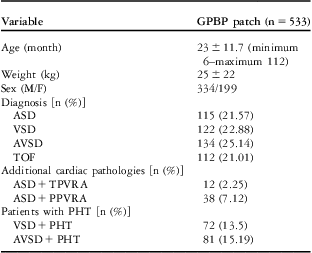
The design of the study was a retrospective, single-centre clinical trial. A total of 533 patients who have undergone operations for intracardiac defects between 2004 and 2010 were enrolled. Glutaraldehyde-preserved bovine pericardium patches (Peri-guard® and Supple Peri-guard®, Synovis Surgical Innovations, St. Paul, Minnesota, United States of America) were used in these patients to repair intracardiac defects (Fig 1a and b). Detailed patient characteristics and diagnoses are shown in Table 1.
( a and b ) The glutaraldehyde-preserved bovine pericardial patch are showing.

Baseline characteristics and diagnosis.

ASD = atrial septal defect; AVSD = atrioventricular septal defect; GPBP = glutaraldehyde-preserved bovine patch; n = number of patients; PHT = pulmonary hypertension; PPVRA = partial pulmonary venous return anomalism; TOF = tetralogy of Fallot; TPVRA = total pulmonary venous anomalism; VSD = ventricular septal defect
Data are presented as mean ± SD or median, with 25th–75th percentile range in brackets, unless otherwise stated
The trial was approved by the institutional ethics committee of the centre and was conducted in accordance with the Declaration of Helsinki. Informed consent was obtained from all patients or their parents or legal guardians.
Research methods
Transthoracic echocardiography was used routinely in the pre-operative diagnosis of patients and in the post-operative period. Cardiac catheterisation was performed when pulmonary hypertension was diagnosed with transthoracic echocardiography in atrial septal defect, ventricular septal defect, atrioventricular septal defect, and for coronary artery anomaly diagnosis in patients with tetralogy of Fallot. Glutaraldehyde-preserved bovine pericardium patches were used to repair intracardiac defects of these patients. The patients were followed up by transthoracic echocardiography on the 15th day post-operatively, then at 1-month, 3-month, and 6-month regular intervals until the end of childhood. The patients’ pre-operative, intra-operative, and early post-operative data were retrospectively recorded. After discharge from the cardiac unit, all follow-up data were retrospectively recorded in the cardiovascular surgery and paediatric cardiology departments.
Surgical procedure
The same anesthesia technique was performed in all patients. Median sternotomy was performed. Aortic cannulation, “bicaval” venous cannulation, vent cannulation, and aortic isothermic blood cardioplegia were performed in all patients. Operations were performed in mild (34°C) or moderate (28°C) hypothermia based on the type of intracardiac defects. Glutaraldehyde-preserved bovine pericardium patch materials were washed with %0.9 isotonic saline (Fig 1a and b). In patients with atrial septal defect and atrioventricular septal defect, a right atriotomy was performed. In patients with ventricular septal defect and tetralogy of Fallot, a right atriotomy or right ventriculotomy was performed. In patients with atrioventricular septal defect, single-patch techniques were used. Intracardiac defects were closed using the patch material by running suture technique with 5/0 polypropylene. In patients with tetralogy of Fallot, right ventricular outflow tract reconstruction was performed after removal of the cross-clamp.
Follow-up
The patients were followed up with transthoracic echocardiography on the 15th day post-operatively, then at 1-month, 3-month, and 6-month regular intervals until the end of childhood. The follow-up ranged from 26 to 124 months (mean: 126 ± 6 months).
Statistical analysis
Categorical data are given as absolute and relative frequencies (percentages). All data are given as means ± standard deviation.
Results
Baseline characteristics
The distribution of the patients according to the diagnosis is shown in Table 1. The patients ranged in age from 6 months to 112 months with a mean of 23 ± 11.7 months, and there were 334 male patients and 199 female patients (Table 1). The evaluation of the patients’ pre-operative clinical data with the Ross Scoring System showed a total score between 3 and 6 or 7 and 9.
Peri-operative data
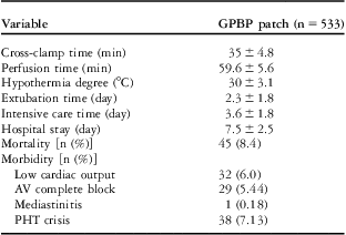
The mean cross-clamp time was 35 ± 4.8 minutes. The total perfusion time was 59.6 ± 5.6 minutes. The mean temperature during hypothermia was 30 ± 3.1°C. The mean extubation time was 2.3 ± 1.8 days. The mean intensive care time 3.6 ± 1.8 days, and the mean hospitalisation time was 7.5 ± 2.5 days. Peri-operative data of the patients are shown in Table 2.
Data from the patients who had intracardiac defect operation.

GPBP = glutaraldehyde-preserved bovine patch; n = number of patients; PHT = pulmonary hypertensive
Data are presented as mean ± SD or median, with 25th–75th percentile range in brackets, unless otherwise stated
Clinical outcomes
Patients with a total score of 0–2 or 3–6 according to the Ross Scoring System were followed up. Post-operative data of the patients are shown in Table 2. Low cardiac output, pulmonary hypertensive crisis, mediastinitis, and complete block were evaluated in early and late morbidity. Low cardiac output was diagnosed in 32 patients, and pulmonary hypertensive crisis developed in 38 patients – that is, 27 patients with atrioventricular septal defect and 11 patients with ventricular septal defect – in the post-operative period at the intensive care unit (Table 2). These patients were followed up and treated at the intensive care unit. Mediastinitis occurred in one patient (0.18%). This patient was treated medically and sternal revision was performed.
Atrioventricular blocks occurred because of the type of defect. During the early post-operative period, complete atrioventricular block occurred in inlet ventricular septal defect, atrioventricular septal defect, and tetralogy of Fallot. The incidence rate was 5.44%. The incidence rate according to the diagnosis of patients was 10.6% for ventricular septal defects, 10.4% for atrioventricular septal defects, and 2.17% for tetralogy of Fallot (Table 2).
In the evaluation of early and late mortalities, all mortalities were found to be in the early period. There were 45 (8.4%) mortalities. The mortality numbers according to the diagnosis were 11 (9.02%) for ventricular septal defect, 19 (14.1%) for atrioventricular septal defect, and 11 (9.8%) for tetralogy of Fallot. During the early post-operative period, fatal arrhythmia was the cause of mortality (Table 2). There were no patient mortalities in the late post-operative period.
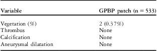
The patients were followed up by transthoracic echocardiography on the 15th day post-operatively, then at 1-month, 3-month, and 6-month regular intervals. Vegetation was diagnosed in two patients upon likelihood of thrombus, vegetation, calcification, and aneurysmal dilatation evaluated by transthoracic echocardiography (Table 3). Endocarditis treatment was commenced on the 7th day post-operatively on two of the patches used at the ventricular level upon diagnosis of vegetation by transthoracic echocardiography. A hospital-acquired infection was the cause of endocarditis. It was not related with the type of the patch. Patients with vegetation were medically treated.
Evaluation of the GPBP patch used in intracardiac defect in post-operative period with TTE.

GPBP = glutaraldehyde-preserved bovine patch; n = number of patients; TTE = transthoracic echocardiography
Data are presented as yes/none and with 25th–75th percentile range in brackets, unless otherwise stated
Other patch complications – thrombus, aneurysmal dilatation, and calcification – reported in previous studies, which could occur because of glutaraldehyde-preserved bovine pericardium on the patch, were not diagnosed in any of our patients (Table 3).
In our study, the number of early residual ventricular septal defects according to the diagnosis was six for ventricular septal defect and eight for atrioventricular septal defect. Patients with Qp/Os rates over 1.5 in residual defects were repaired by reoperation during the early period. The number of reoperated patients was nine – three for ventricular septal defect and six for atrioventricular septal defect. Late period recurrent ventricular septal defect was not found.
Discussion
A variety of patches may be used in cardiovascular surgery. In our study, we used glutaraldehyde-preserved bovine pericardium patches in 533 patients between 2004 and 2010 in the repair of intracardiac defects. An ideal patch in the repair of intracardiac defects should be non-immunogenic, durable, of low permeability, easily manipulatable during usage, easily available, and inexpensive, causing no thrombus or calcification. Although autologous pericardium patches are easily available, inexpensive, and non-permeable, they have become less used over time. The main reasons include the complexity of usage in the pericardium, which is opened in the previous palliative cardiac surgery, inability to close the pericardium at the end of surgery when autologous pericardium is removed, and the difficulties in manipulating the autologous patch during usage owing to its softness, shrinking, aneurysmal formation, and calcification.Reference Mohri, Barnes, Rittenhouse, Reichenbach, Dillard and Merendino 1 , Reference Kirklin and Barratt-Boyes 4 Synthetic and glutaraldehyde-preserved bovine pericardium patches were introduced next. The advantages and disadvantages resulting from the research are taken into consideration in making a choice of synthetic (Dacron, polytetraflouroethylene) or glutaraldehyde-preserved bovine pericardium patches. The most important thing to consider when choosing a patch is making sure that the patches do not cause aneurysmal dilatation, fibrosis, calcification, or thromboembolic complications.
Glutaraldehyde-preserved bovine pericardium patches provide almost excellent results in various procedures during cardiac surgeries. Their flexibility and durability, offering less calcification, show us that glutaraldehyde-preserved bovine pericardium patches can be used as an ideal patch in the repair of intracardiac defects.Reference Us, Sungun and Sanioglu 5 – Reference Li, Guo, Zieger, Model, Eghbalieh and Brenes 12 However, we have seen that there is very limited research in the literature covering glutaraldehyde-preserved bovine pericardium patches in the repair of intracardiac defects.Reference Us, Sungun and Sanioglu 5 , Reference Crawford, Sade and Spinale 9 , Reference Hardin, Muskett, Canter, Martin and Spray 11 , Reference Li, Guo, Zieger, Model, Eghbalieh and Brenes 12 , Reference Neethling, Hodge, Clode and Glancy 15 Although the patch is 0.5 mm in size, it provides a reliable retention saturation,Reference Obermiller, Hodde, McAlexander, Kokini and Badylak 13 and it has a characteristic use during operation. Preserving bovine pericardium with glutaraldehyde enables durability, and, at the same time, decreases its antigenicity and tendency to infection. Long-lasting stability in clinic applications has been observed as a result of its increased durability. What is interesting here is that glutaraldehyde-preserved bovine pericardium patches's compliance with the tissue looks like natural artery and its compliance with other synthetic patches is much higher. There are some studies reporting that because of the technology used to produce these preserving bovine pericardium with glutaraldehyde valves, calcification formation is low and they support endothelialisation.Reference Cunanan, Cubbling, Dinh, Shen, Tran-Hata and Rutledge 14 These patches in fact do not truly endothelialise; they are covered with pannus formation. Although thrombus can be seen rarely thanks to this endothelialisation, a fractioned and unfractioned heparin is suggested.Reference Li, Guo, Zieger, Model, Eghbalieh and Brenes 12 Gabbay et al,Reference Gabbay, Bortoltti, Factor, Shore and Frater 16 in their experimental study in which they implanted glutaraldehyde-preserved bovine pericardium patches in the atrial wall of a dog, reported that the calcification occurring 12 months later had occurred only in the side of the patch in contact with blood. Fibrosis development looks like the calcification mechanism, yet it is very rare in the patches.Reference Li, Guo, Zieger, Model, Eghbalieh and Brenes 12 Aneurysmal formation is also low in these patches with the effect of resistance to infection and low degeneration.Reference Li, Guo, Zieger, Model, Eghbalieh and Brenes 12 The study conducted by Us et alReference Us, Sungun and Sanioglu 5 with a limited number of patients reported that glutaraldehyde-preserved bovine pericardium patches were superior to synthetic patches in their ease of use.
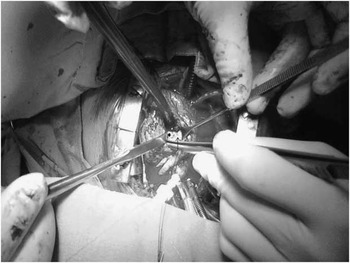
Our patients who underwent total correction surgery after diagnosis of tetralogy of Fallot have not been reoperated for 3 years because of the high right ventricular outflow tract gradient. The glutaraldehyde-preserved bovine pericardium patches were evaluated in surgery. No fibrosis, calcification, shrinking, or aneurysmal changes have been observed (Fig 2).
The glutaraldehyde-preserved bovine pericardial patch is showing at that closed VSD (*) and RVOT (↑) in reoperation. RVOT = right ventricular outflow tract; VSD = ventricular septal defect.

To conclude, owing to its useful features – elasticity, controllable softness, and easiness to shape and implant – and the confidence it gives to the surgeon during the operation, we have been using glutaraldehyde-preserved bovine pericardium patches in the repair of intracardiac defects since 2004 in our clinics. Glutaraldehyde-preserved bovine pericardium patches are an alternative to other patches and can be used safely.
Acknowledgements
None.
Financial Support
This research received no specific grant from any funding agency, commercial or not-for-profit sectors
Conflicts of Interest
None.
Ethical Standards
None.