Introduction
The Radiodonta Collins, Reference Collins1996, a clade comprising the anomalocaridids and their allies, is a lineage of soft-bodied stem-group euarthropods (Daley et al., Reference Daley, Budd and Caron2013a; Ortega-Hernández, Reference Ortega-Hernández2016) regarded as the oldest giant top predators in Paleozoic marine ecosystems (e.g., Whittington and Briggs, Reference Whittington and Briggs1985; Chen et al., Reference Chen, Ramsköld and Zhou1994). A series of ongoing discoveries from exceptionally preserved fossil deposits have revealed that radiodontans had a cosmopolitan distribution (e.g., Daley et al., Reference Daley, Budd and Caron2013a) and a long fossil record from the Cambrian Stage 3 (Daley and Legg, Reference Daley and Legg2015) to the Early Ordovician Tremadocian (Van Roy and Briggs, Reference Van Roy and Briggs2011; Van Roy et al., Reference Van Roy, Daley and Briggs2015), or even possibly to the Early Devonian Emsian (Kühl et al., Reference Kühl, Briggs and Rust2009). As apex predators, the feeding behaviors of radiodontans have become a major research topic (e.g., Briggs, Reference Briggs1979; Briggs and Mount, Reference Briggs and Mount1982; Whittington and Briggs, Reference Whittington and Briggs1985; Nedin, Reference Nedin1999; Daley et al., Reference Daley, Paterson, Edgecombe, García-Bellido and Jago2013b; Vinther et al., Reference Vinther, Stein, Longrich and Harper2014; Van Roy et al., Reference Van Roy, Daley and Briggs2015). However, the bulk of research on radiodontan feeding has focused on their frontal appendages, a pair of anteriormost raptorial appendages known to have attained considerable morphological disparity (Daley and Budd, Reference Daley and Budd2010; Aria and Caron, Reference Aria and Caron2015) and ecological adaptations to either macrophagous predation (e.g., Chen et al., Reference Chen, Ramsköld and Zhou1994; Daley and Edgecombe, Reference Daley and Edgecombe2014) or filter feeding (Vinther et al., Reference Vinther, Stein, Longrich and Harper2014; Van Roy et al., Reference Van Roy, Daley and Briggs2015).
In addition to their frontal appendages, radiodontans possessed a second feeding organ, an oral cone consisting of a ring of radially arranged plates developed around the mouth, from which the group gets its name. The oral cone, however, has attracted much less research attention; while at one point it was thought to be used for biting and chewing prey (Whittington and Briggs, Reference Whittington and Briggs1985), recent biomechanical modeling of the oral cone of Peytoia Walcott, Reference Walcott1911 (Hagadorn, Reference Hagadorn2009, Reference Hagadorn2010) and reconstruction of this structure in Anomalocaris Whiteaves, Reference Whiteaves1892 (Daley and Bergström, Reference Daley and Bergström2012) suggest that more likely it was involved in suction during the ingestion of food, rather than directly crushing prey. To date, only three radiodontan oral cone morphotypes have been recognized, primarily from fossil specimens from the Cambrian Stage 5 Burgess Shale, British Columbia, Canada (Daley and Bergström, Reference Daley and Bergström2012). These morphotypes include the ‘triradially’ symmetrical oral cone of Anomalocaris canadensis Whiteaves, Reference Whiteaves1892 (Daley and Bergström, Reference Daley and Bergström2012; Daley and Edgecombe, Reference Daley and Edgecombe2014) and the ‘tetraradially’ symmetrical oral cones of Peytoia nathorsti Walcott, Reference Walcott1911 (Whittington and Briggs, Reference Whittington and Briggs1985; Daley and Bergström, Reference Daley and Bergström2012), Hurdia victoria Walcott, Reference Walcott1912, and H. triangulata Walcott, Reference Walcott1912. The oral cones of both Peytoia and Hurdia comprise 32 plates with smooth surfaces and include four perpendicularly situated large plates separated by 28 small plates. The two morphotypes of oral cones can only be distinguished by the presence of a set of extra inner teeth within the central opening in Hurdia (Daley et al., Reference Daley, Budd, Caron, Edgecombe and Collins2009, Reference Daley, Budd and Caron2013a; see Discussion). In contrast, the oral cone in A. canadensis comprises three large plates intercalated by medium and small ones, all of which have scale-like nodes and furrowed folds toward their outer margins. In addition, an almost identical A. canadensis-type oral cone has also been identified in A. saron Hou et al., Reference Hou, Bergström and Ahlberg1995 from the Cambrian Stage 3 Chengjiang Lagerstätte, Yunnan, South China (Chen et al., Reference Chen, Ramsköld and Zhou1994, fig. 2; Daley and Bergström, Reference Daley and Bergström2012), as well as in an unidentified and isolated specimen from the Cambrian Stage 4 Emu Bay Shale, Kangaroo Island, South Australia (McHenry and Yates, Reference McHenry and Yates1993, fig. 8; Daley et al., Reference Daley, Paterson, Edgecombe, García-Bellido and Jago2013b, fig. 4). Other radiodontan oral cones described in the literature include examples of a partially preserved Hurdia sp. (Daley et al., Reference Daley, Budd and Caron2013a, fig. 24a) and Peytoia cf. P. nathorsti (Conway Morris and Robison, Reference Conway Morris and Robison1982) from the Cambrian Stage 5 Spence Shale and Drumian Wheeler Formation, both in Utah, USA, in addition to several other fragmentary or indeterminate examples (Masiak and Żylińska, Reference Masiak and Żylińska1994, fig. 7; Lieberman, Reference Lieberman2003, figs. 8.1, 9; Briggs et al., Reference Briggs, Lieberman, Hendricks, Halgedahl and Jarrard2008, figs. 1, 2.1). Structures present around mouth openings that are probably related to oral cones include the concentric ridges and triangular areas of Lyrarapax unguispinus (Cong et al., Reference Cong, Ma, Hou, Edgecombe and Strausfeld2014, fig. 1a–d, f) and L. trilobus (Cong et al., Reference Cong, Daley, Edgecombe, Hou and Chen2016, figs. 1.1–1.3, 1.5, 2.1–2.3), both from the Chengjiang Lagerstätte, and the circular plates of Schinderhannes bartelsi Kühl et al., Reference Kühl, Briggs and Rust2009 from the Early Devonian Emsian Hunsrück Slate. Moreover, a mouth apparatus exactly like Omnidens amplus Hou et al., Reference Hou, Bergström and Jie2006 from Chengjiang was recently identified in the ‘gilled lobopodian’ Pambdelurion whittingtoni Budd, Reference Budd1998 from the Cambrian Stage 3 Sirius Passet Lagerstätte, Greenland (Vinther et al., Reference Vinther, Porras, Young, Budd and Edgecombe2016), suggesting that its supposed radiodontan identity (Chen et al., Reference Chen, Ramsköld and Zhou1994, Reference Chen, Zhou, Zhu and Yeh1996; Chen, Reference Chen2004) may be problematic.
In this paper, we present a new radiodontan oral cone morphotype from the Guanshan Lagerstätte (Cambrian Series 2, Stage 4) in the Wulongqing Formation, Gaoloufang section, Kunming, eastern Yunnan, South China. The frontal appendages of two radiodontans, Anomalocaris kunmingensis Wang et al., Reference Wang, Huang and Hu2013 and Paranomalocaris multisegmentalis Wang et al., Reference Wang, Huang and Hu2013, have already been described from this Lagerstätte (Hu et al., Reference Hu, Zhu, Steiner, Luo, Zhao and Liu2010, Reference Hu, Zhu, Luo, Steiner, Zhao, Li, Liu and Zhang2013). The taxonomic identity of this new oral cone cannot be determined because it is preserved in isolation, not associated with frontal appendages or other body parts. Because the new oral cone combines features seen in both Anomalocaris and Peytoia oral cones, we regard it as a transitional form that has the potential to illuminate the morphological variations, feeding mechanisms, and evolution of these unique feeding structures.
Geologic setting
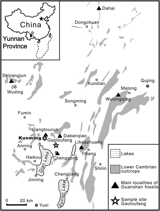
As with the better known Chengjiang Lagerstätte (e.g., Zhao et al., Reference Zhao, Hu, Caron, Zhu, Yin and Lu2012), the Guanshan Lagerstätte is a Burgess Shale-type deposit that is widely distributed in eastern Yunnan Province (Fig. 1) and has yielded abundant and diverse well-preserved soft-bodied fossils (Luo et al., Reference Luo, Li, Hu, Fu, Hou, Liu, Chen, Li, Pang and Liu2008; Hu et al., Reference Hu, Zhu, Steiner, Luo, Zhao and Liu2010, Reference Hu, Zhu, Luo, Steiner, Zhao, Li, Liu and Zhang2013; Liu et al., Reference Liu, Ou, Han, Zhang, He, Yao, Fu and Shu2012). Soft-bodied Guanshan fossils are found within yellowish-to-greenish mudstones intercalated with thinly bedded siltstone layers in the Wulongqing Formation (Hu et al., Reference Hu, Zhu, Steiner, Luo, Zhao and Liu2010, Reference Hu, Zhu, Luo, Steiner, Zhao, Li, Liu and Zhang2013). Two trilobite assemblage zones, the lower Palaeolenus Assemblage Zone and the upper Megapalaeolenus Assemblage Zone, have been identified within the Wulongqing Formation (e.g., Luo et al., Reference Luo, Li, Hu, Fu, Hou, Liu, Chen, Li, Pang and Liu2008); biostratigraphically, the Guanshan Lagerstätte belongs to the late period of the early Cambrian Canglangpuan Stage of South China (e.g., Lin, Reference Lin2008), dated to the global Cambrian Series 2, Stage 4 (Steiner et al., Reference Steiner, Zhu, Zhao and Erdtmann2005). Detailed discussion of the stratigraphy, sedimentology, and taphonomy of the Guanshan Lagerstätte can be found in Hu et al. (Reference Hu, Zhu, Steiner, Luo, Zhao and Liu2010, Reference Hu, Zhu, Luo, Steiner, Zhao, Li, Liu and Zhang2013).
Map to show the main fossil localities of the Guanshan Lagerstätte in eastern Yunnan Province, South China, including the Gaoloufang section, the sampling site in this paper. Fossil localities are from Hu et al. (Reference Hu, Zhu, Luo, Steiner, Zhao, Li, Liu and Zhang2013, fig. 3).

Materials and methods
Repository and institutional abbreviation
The single specimen described in this paper is housed in the Nanjing Institute of Geology and Palaeontology, Chinese Academy of Sciences, Nanjing, China (NIGPAS).
Locality and imaging
The isolated radiodontan oral cone (NIGPAS 162529) described here was collected from the Gaoloufang section at Guangwei in Kunming, Yunnan Province, China, one of the main Guanshan Lagerstätte localities (Fig. 1; Hu et al., Reference Hu, Zhu, Steiner, Luo, Zhao and Liu2010, Reference Hu, Zhu, Luo, Steiner, Zhao, Li, Liu and Zhang2013). Photographs of this fossil were made using a Nikon AF-S VR 105 mm f/2.8G macro lens mounted on a Nikon D300s digital single lens reflex camera, employing low-angle incident light to show the relief of structures; interpretative drawings were based on high-resolution photographs in Adobe Photoshop CS6. Our use of anatomical terminology follows Daley and Bergström (Reference Daley and Bergström2012).
Systematic paleontology
Phylum Euarthropoda Lankester, Reference Lankester1904
Order Radiodonta Collins, Reference Collins1996
Remarks
Monophyly of Radiodonta has been questioned recently because of the unstable phylogenetic position of Schinderhannes bartelsi Kühl et al., Reference Kühl, Briggs and Rust2009 (Ortega-Hernández, Reference Ortega-Hernández2016). Because Class Dinocarida Collins, Reference Collins1996, comprising Opabinia regalis Walcott, Reference Walcott1912 as the sister taxon of Radiodonta, received no support in recent cladistic analyses (e.g., Legg et al., Reference Legg, Sutton and Edgecombe2013; Van Roy et al., Reference Van Roy, Daley and Briggs2015; Yang et al., Reference Yang, Ortega-Hernández, Gerber, Butterfield, Hou, Lan and Zhang2015), it is not used in this study.
?Family Amplectobeluidae Vinther et al., Reference Vinther, Stein, Longrich and Harper2014
Remarks
Only a few nearly complete isolated specimens of the new radiodontan oral cone we describe have been discovered during the course of long and extensive excavations of the Guanshan Lagerstätte at Gaoloufang. Conversely, more than a hundred complete radiodontan frontal appendages have been collected from this section (Wang et al., Reference Wang, Huang and Hu2013; Hu et al., Reference Hu, Zhu, Luo, Steiner, Zhao, Li, Liu and Zhang2013). Within these frontal appendages, only a single specimen of Paranomalocaris multisegmentalis has been reported, and the remainder belong to Anomalocaris kunmingensis (Wang et al., Reference Wang, Huang and Hu2013; Hu et al., Reference Hu, Zhu, Luo, Steiner, Zhao, Li, Liu and Zhang2013). Thus, on the basis of this large-scale fossil sampling, our new radiodontan oral cone is most likely to be A. kunmingensis, placed in the family Amplectobeluidae Vinther et al., Reference Vinther, Stein, Longrich and Harper2014 in most recent phylogenetic analyses (Vinther et al., Reference Vinther, Stein, Longrich and Harper2014; Van Roy et al., Reference Van Roy, Daley and Briggs2015). However, the taxonomic identity of this new cone cannot be determined with certainty currently, and it is possible that it belongs to a third radiodontan species.
Radiodontan gen. indet. sp. indet.
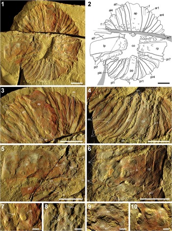
The single specimen of the new radiodontan oral cone described in this paper (NIGPAS 162529), counterpart only, from the Wulongqing Formation, Cambrian Series 2, Stage 4, Gaoloufang, in Kunming, eastern Yunnan Province, South China. (1) Photograph of entire specimen; (2) interpretative drawing of entire specimen; (3) anterior large plate and small plates; (4) posterior large plate and small plates; (5) left large plate and small plates next to it; (6) right large plate and small plates next to it. (7–10) Close-ups of scale-like nodes on large plates in (3–6), respectively. al1–al7=first to seventh small plates intercalated between the anterior large plate and the left large plate; ap=anterior large plate; ar1–ar7=first to seventh small plates intercalated between the anterior large plate and the right large plate; co=central opening; es=lateral extensions of plates; f=furrowed folds; is=inner spines of plates; ln=large scale-like nodes; lp=left large plate; ls=lateral spines of plates; pl1–pl7=first to seventh small plates intercalated between the posterior large plate and the left large plate; pp=posterior large plate; pr1–pr7=first to seventh small plates intercalated between the posterior large plate and the right large plate; rp=right large plate; sn=small scale-like nodes. (1–6) Scale bars=10 mm; (7–10) scale bars=2 mm.

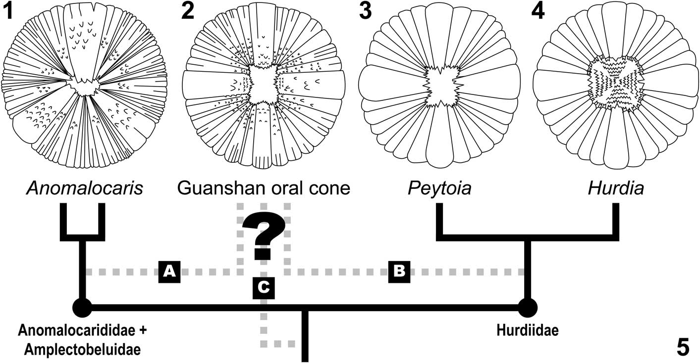
Reconstructions of the oral cones of different radiodontan taxa, not to scale. (1) Anomalocaris canadensis and A. saron, redrawn from Daley and Bergström (Reference Daley and Bergström2012, fig. 1c); (2) the new Guanshan radiodontan oral cone reported here, based on NIGPAS 162529; (3) Peytoia nathorsti, modified from Daley and Bergström (Reference Daley and Bergström2012, fig. 1a) and with an orbicular rather than taphonomically distorted rectangular shape (see text); (4) Hurdia victoria and H. triangulata, modified from Daley and Bergström (Reference Daley and Bergström2012, fig. 1b) and rotated 90°. Note that the lateral large plates are wider than the anteroposterior plates in (3, 4). (5) Cladogram based on the most recent radiodontan phylogeny from Van Roy et al. (Reference Van Roy, Daley and Briggs2015), showing the two main radiodontan clades Anomalocarididae+Amplectobeluidae and Hurdiidae in nodes, their representatives with oral cones known in solid lines, and three alternative positions of the Guanshan oral cone in dashed lines (A, B, and C). Two branches of Anomalocaris indicate its two species A. canadensis and A. saron. See text for discussion.

Material
A single, isolated, nearly complete radiodontan oral cone, NIGPAS 162529, counterpart only.
Description
Specimen NIGPAS 162529 is predominantly dorsoventrally preserved and just slightly laterally compressed, as indicated by the curved radial margins of plates and length differences between the left and right large plates (Fig. 2.1, 2.2). Minor distortion of the original symmetry, together with the imbricated arrangement of plates (Fig. 2.1, 2.2), suggests that this oral cone was a three-dimensional structure. The orientation of this new oral cone is interpretative due to its isolated preservation; because three of the four large preserved plates are similar in size and wider than the other one, we tentatively orient this specimen with the narrowest large plate in a posterior position (Figs. 2.1, 2.2, 3.2; see Discussion). This orientation is supported by reference to other known oral cones; in both Peytoia and Hurdia, the lateral large plates are wider than the anterior and posterior ones (Fig. 3.3, 3.4; see Discussion), while the anterior large plate is the widest in Anomalocaris (Fig. 3.1; see Discussion).
The new Guanshan oral cone is approximately circular and ‘tetraradially’ symmetrical, with a diameter of ~7.0 cm (Fig. 2.1, 2.2). The outer ring of the oral cone comprises 32 plates that surround an empty rectangular central opening (Fig. 2.1, 2.2); this central opening is ~1.5 cm long longitudinally and ~1.0 cm wide horizontally (Fig. 2.1, 2.2). Of the 32 plates, four large ones are located 90° from one another, in anterior, posterior, left, and right positions, respectively (Fig. 2.1, 2.2), while each of the two adjacent large plates is separated by seven small ones filling in each quarter of the outer ring (Fig. 2.1, 2.2). Of these, the left, right, and anterior large plates are of similar sizes, with a maximum length of ~3.3 cm and a maximum width of ~1.3 cm; the posterior one is significantly smaller, with a maximum length of ~2.0 cm and a maximum width of ~0.6 cm (Fig. 2.1, 2.2). The 28 small plates do not appear significantly different from one another in size, having a maximum length of ~2.7 cm and a maximum width of ~0.7 cm (Fig. 2.1, 2.2). Both large and small plates are overlapped adjacently by narrow lateral extensions (Fig. 2.3, 2.4).
Most of the small plates as well as the anterior and posterior large plates have furrowed folds that initiate from their outer margins (Fig. 2.1–2.4); these folds are absent from the left and right large plates (Fig. 2.1, 2.2, 2.5, 2.6). The longest of these furrowed folds reaches up to almost half the total plate length (Fig. 2.1–2.4). Clusters comprising up to 12 scale-like nodes develop on the surfaces of all large and small plates (Fig. 2.1–2.10). Although irregularly distributed, these nodes do share regular orientation with their tips projecting inward toward the central opening (Fig. 2.7–2.10). Most of these nodes are less than 2 mm in diameter, but a few reach between 3 mm and 5 mm (Fig. 2.2–2.7, 2.9, 2.10). Of these, smaller nodes are found on all large and small plates (Fig. 2.2–2.6, 2.8), while larger ones are only present on the anterior, left, and right large plates (Fig. 2.1–2.3, 2.5, 2.6), absent from the posterior one (Fig. 2.1, 2.2, 2.4, 2.8). Rows of similarly sized triangular spines, less than 1 mm in length, develop along the inner edges of all plates (Fig. 2.1, 2.2, 2.3–2.6); up to eight of these spines are present on the four large plates (Fig. 2.1, 2.2, 2.3–2.6), while just up to three are found on the small plates (Fig. 2.1–2.6). Similar spines are also present along the radial margins of large plates and their adjacent small plates (Fig. 2.1, 2.2, 2.5, 2.6).
Occurrence
From the Wulongqing Formation, late period of the early Cambrian Canglangpuan Stage (Cambrian Series 2, Stage 4), at Gaoloufang, Guangwei, in Kunming, eastern Yunnan, South China.
Discussion
Taphonomic notes
A critical assessment on potential taphonomic biases of radiodontan oral cones is essential given the limited characters we know from them. Doubt may be cast on the uniqueness of inner teeth in Hurdia. Intuitively, there is no good reason for the absence of a masticatory apparatus in other radiodontans, including Peytoia, Anomalocaris and the species represented by the new oral cone here, since pharyngeal teeth are present in a wide range of ecdysozoan groups (Smith & Caron, Reference Smith and Caron2015). The taphonomic scenario of faster degradation of these teeth is rejected by Daley et al. (Reference Daley, Budd, Caron, Edgecombe and Collins2009) because no evidence of extra inner teeth is present in any oral cones of Peytoia or Anomalocaris, and no isolated teeth are found from all Burgess Shale localities. While all radiodontan oral cones show a more or less convex shape and can potentially provide space for accommodating inner teeth, it should be acknowledged that the inner teeth of Hurdia are closely associated or articulated with the oral cone, which is a unique case in radiodontans we know. If extra inner teeth really exist in other radiodontans apart from Hurdia, these teeth are likely situated on the inner walls of buccal or pharyngeal cavities rather than being associated with the oral cone, and they might be either too small to be found or concealed within the rock matrix. The question whether extra inner teeth are present in other radiodontans can only be answered by more detailed reinvestigation or new fossil discoveries.
Another question worth considering is whether the absence of scale-like nodes in Peytoia and Hurdia oral cones is a natural feature or a taphonomic loss, as it has been suggested that surface microstructures on sclerotized plates could be effaced and that microscopic artifacts might be generated during taphonomic processes (e.g., radiodontan head sclerites; Zeng et al., Reference Zeng, Zhao, Yin and Zhu2017). In these cases, taphonomic effects act on microstructures of soft-bodied euarthropod sclerites from a specific locality or horizon of a fossil deposit and are not taxon selective. Fine scale-like nodes are well preserved in both articulated and disarticulated oral cones of A. canadensis recovered from the Burgess Shale (Daley and Bergström, Reference Daley and Bergström2012); thus, the possibility of a universal taphonomic effacement of these nodes can be eliminated. Considering the large number of oral cone specimens investigated from the Burgess Shale (Whittington and Briggs, Reference Whittington and Briggs1985; Daley et al., Reference Daley, Budd and Caron2013a), the smooth oral plates without scale-like nodes are most likely to be a natural feature of Peytoia and Hurdia.
Revision of Peytoia and Hurdia oral cones
Before making comparisons between the new Guanshan radiodontan oral cone and other known examples, we suggest some minor revisions to the most recent reconstructions of the oral cones of the Burgess Shale taxa Peytoia and Hurdia (Daley and Bergström, Reference Daley and Bergström2012, fig. 1a, b). Our first point concerns the outline of the Peytoia oral cone, while our second relates to the strict symmetry and orientation of the Peytoia and Hurdia oral cones.
The original outline of the Peytoia oral cone has been an open question because of potential taphonomic deformation (Daley and Bergström, Reference Daley and Bergström2012, p. 503; Daley et al., Reference Daley, Budd and Caron2013a, p. 779). Primarily drawing from one of the most complete and articulated Peytoia specimens (USNM 274143; Whittington and Briggs, Reference Whittington and Briggs1985, figs. 17, 20–24), a rectangular rather than a circular profile has been favored for oral cone reconstructions (Whittington and Briggs, Reference Whittington and Briggs1985; Collins, Reference Collins1996; Daley and Bergström, Reference Daley and Bergström2012, fig. 1a). However, the oral cones in another two articulated Peytoia specimens are circular in shape (USNM 57555, 274144, Whittington and Briggs, Reference Whittington and Briggs1985, figs. 44, 45, 47, 48, 50, 95–98), while the outlines of other isolated or articulated specimens are distorted. In examples we have measured, the length-width ratios of central openings (range=1.04–1.25, mean=1.15, standard deviation=0.08; logarithmic range=0.040–0.097, mean=0.060, standard deviation=0.030; based on USNM 57538, 57539, 57555, 253216, 274144, and 368584; Whittington and Briggs, Reference Whittington and Briggs1985, figs. 44, 45, 47, 48, 50, 60–67, 79, 80, 93, 95–98; ROM 51216; Collins, Reference Collins1996, fig. 5.2, 5.3) are statistically significantly less than those of the specimen used in reconstruction (length-width ratio=1.40, assuming a normal distribution, P[ratio ≥1.40]<.001; logarithmic length-width ratio=0.146, assuming a normal distribution, P[ratio ≥ 0.146]<.002; on the basis of USNM 274143; Whittington and Briggs, Reference Whittington and Briggs1985, figs. 17, 20–24), suggesting this is an abnormal, laterally compressed individual. We argue, on the basis of our measurements, that the oral cone of Peytoia more likely had a circular (Fig. 3.3) rather than rectangular outline. This result also implies that a circular oral cone outline characterizes all known radiodontans.
While the terms ‘triradial’ and ‘tetraradial’ are very useful in descriptions of radiodontan oral cones, they can be misleading when the strict symmetrical patterns of these structures are considered. Even though radially symmetrical oral structures are present across a range of ecdysozoan groups, including some stem-group euarthropods, and appear to be a primitive character (e.g., Smith and Caron, Reference Smith and Caron2015; Vinther et al., Reference Vinther, Porras, Young, Budd and Edgecombe2016), this was not necessarily the case for radiodontan oral cones. In the ‘triradial’ oral cone of Anomalocaris, the large anterior plate is larger than its two posterolateral counterparts (Daley and Bergström, Reference Daley and Bergström2012, p. 502, fig. 1C); thus, strictly speaking, this structure should be described as bilaterally symmetrical rather than ‘triradial.’ Similarly, in the ‘tetraradial’ oral cone of Peytoia, size differences are evident between the four large plates, with lateral plates being wider than the anterior and posterior plates. This variation in size can be seen in articulated (USNM 57555, 274143; Whittington and Briggs, Reference Whittington and Briggs1985, p. 582, figs. 17, 20–24, 95–98) and isolated specimens (USNM 57538, 57539, 253216; Whittington and Briggs, Reference Whittington and Briggs1985, figs. 60–67, 93; ROM 51216; Collins, Reference Collins1996, fig. 5.2, 5.3) and has been illustrated in reconstructions (Whittington and Briggs, Reference Whittington and Briggs1985, figs. 102b, 106c, 106f; Daley and Bergström, Reference Daley and Bergström2012, fig. 1a). Recognizing that the four large plates are of different sizes made it possible to confidently orient isolated Peytoia oral cones (USNM 57538, 253216; Whittington and Briggs, Reference Whittington and Briggs1985, figs. 60, 67), as implied in the original description. Thus, in a strict sense, as in Anomalocaris, the oral cone of Peytoia should be considered as biradially symmetrical rather than ‘tetraradial’ (Fig. 3.3). The same reasoning applies to Hurdia, which has the same Peytoia-type configurations of the outer ring of 32 plates (Daley et al., Reference Daley, Budd, Caron, Edgecombe and Collins2009; Daley and Bergström, Reference Daley and Bergström2012, Daley et al., Reference Daley, Budd and Caron2013a). In an almost complete, articulated specimen of Hurdia (Daley et al., Reference Daley, Budd and Caron2013a, fig. 21C, D) as well as in an oral cone associated with a pair of frontal appendages possibly preserved in the living orientation (Daley et al., Reference Daley, Budd and Caron2013a, fig. 22D), the lateral large plates are wider than the anteroposterior ones. Thus, we suggest that the orientation of the oral cone in Hurdia was most likely the same as in Peytoia, with wider large plates arranged laterally and narrower anteroposterior ones oriented longitudinally (Fig. 3.3, 3.4).
Comparative anatomy
The comparative anatomy of radiodontan oral cones is summarized (Table 1). In general, the new radiodontan oral cone from the Guanshan Lagerstätte combines features seen in Anomalocaris (Daley and Bergström, Reference Daley and Bergström2012; Daley and Edgecombe, Reference Daley and Edgecombe2014) and Peytoia (Whittington and Briggs, Reference Whittington and Briggs1985; Daley and Bergström, Reference Daley and Bergström2012), while lacking the additional rows of inner teeth that are unique to Hurdia (Daley et al., Reference Daley, Budd, Caron, Edgecombe and Collins2009, Reference Daley, Budd and Caron2013a; Daley and Bergström, Reference Daley and Bergström2012). The Guanshan oral cone resembles the oral cones of Peytoia and Hurdia because it has an outer ring of 32 plates comprising four perpendicularly situated large plates intercalated with seven small plates in each quarter. In Peytoia and Hurdia, however, the anterior and posterior large plates are narrower than the lateral ones with no apparent size difference, while in the Guanshan oral cone, the anterior large plate is as wide as the plates on the left and right and significantly wider than the posterior plate. The new Guanshan oral cone also exhibits features thought unique to Anomalocaris, including the presence of furrowed folds and scale-like nodes on plates, both of which are absent from Peytoia and Hurdia. Comparing to Anomalocaris, Peytoia, and Hurdia, which have no more than three spines on the inner margins of their large oral cone plates, the number of spines on the large plates of the Guanshan specimen is always at least five, reaching up to eight in some cases. The central opening of the Guanshan oral cone is also subrectangular, comparable to Peytoia, but whether observed differences between the subquadrate central opening in Hurdia and the irregular central opening in Anomalocaris are biological or taphonomic artifacts will require further study.
Anatomical comparisons of characters between radiodontan oral cones. Asterisks in the last column indicate that the extra inner teeth are not articulated with the oral cone, but they may still be present elsewhere in the buccal or pharyngeal cavity.

Feeding mechanisms
Morphological variations shown by radiodontan oral cones, which are exemplified by the new morphotype here, may reflect different feeding mechanisms (Daley and Bergström, Reference Daley and Bergström2012). However, in the absence of preserved frontal appendages, the feeding mechanism of the Guanshan oral cone cannot be unequivocally determined. A comprehensive understanding of radiodontan feeding mechanisms requires an integrative consideration of both frontal appendages and oral cones. Future studies could investigate whether the morphological variations shown by oral cones, including differences in plate configurations, presence or absence of furrowed folds, scale-like nodes, and extra inner teeth, are correlated with specific feeding behaviors such as macrophagous predation or filter feeding. Both new fossil discoveries (e.g., Daley and Bergström, Reference Daley and Bergström2012) and biomechanical modelling (e.g., Hagadorn, Reference Hagadorn2009, Reference Hagadorn2010) would play important roles in these investigations.
Evolutionary implications
The unique combination of features in the Guanshan oral cone represents an intermediate morphotype between the oral cones of Anomalocaris and Peytoia/Hurdia. Recent phylogenetic studies have revealed two main radiodontan lineages (Cong et al., Reference Cong, Ma, Hou, Edgecombe and Strausfeld2014; Vinther et al., Reference Vinther, Stein, Longrich and Harper2014; Van Roy et al., Reference Van Roy, Daley and Briggs2015), Anomalocarididae+Amplectobeluidae, including Anomalocaris, Amplectobelua Hou et al., Reference Hou, Bergström and Ahlberg1995 and Lyrarapax Cong et al., Reference Cong, Ma, Hou, Edgecombe and Strausfeld2014, and Hurdiidae, comprising Hurdia, Peytoia, and Aegirocassis Van Roy et al., Reference Van Roy, Daley and Briggs2015. Anatomical differences between Anomalocaris-type and Peytoia/Hurdia–type oral cones may thus represent a characteristic distinction between the two major radiodontan lineages. However, it remains unclear which oral cone type represents the primitive radiodontan condition.
Because the Guanshan oral cone combines features from both radiodontan lineages, its phylogenetic position would aid our understanding the evolution of oral cones. However, the current framework of radiodontan phylogeny is largely based on frontal appendage morphology, and only a few characters, which include plate configuration, furrowed folds, and scale-like nodes, can be meaningfully coded for the four types of oral cones we know (Table 1). In this case, lack of knowledge of the frontal appendages, in addition to the unique combination of character states in the Guanshan oral cone, makes it difficult to resolve its phylogenetic position from character state data. Recent attempts to resolve the phylogeny of radiodontans from limited character data have led to conflicting results. For example, the sister grouping of Peytoia and Anomalocaris resolved by a data set featured by ecdysozoan oral structures (Smith and Caron, Reference Smith and Caron2015, fig. 4) is contradicted with a Peytoia+Hurdia grouping in analyses based on comprehensive radiodontan characters including frontal appendages and oral cones (Cong et al., Reference Cong, Ma, Hou, Edgecombe and Strausfeld2014; Vinther et al., Reference Vinther, Stein, Longrich and Harper2014; Van Roy et al., Reference Van Roy, Daley and Briggs2015). Moreover, comparable character states in outgroup taxa such as scale-like nodes are inconsistent and are currently unhelpful in resolving character polarity. Without new anatomical or taxonomic data, a data set–based analysis remains unreliable in resolving the evolution of radiodontan oral cones.
Without a data set–based phylogeny, evolution of radiodontan oral cones can still be inferred by considering all possible scenarios in a phylogenetic framework. This becomes a question of determining the root in an unrooted three-taxon network made up of Anomalocaris, Peytoia+Hurdia, and the Guanshan oral cone (Fig. 3.5). Thus, three alternative evolutionary scenarios of radiodontan oral cones corresponding to three possible positions of the Guanshan oral cone can be reconstructed (A, B, C in Fig. 3.5; Table 2). Given the uncertain plesiomorphic character states, the possible character changes involved would include the increase or decrease in numbers of large and small plates and the gain or loss of furrowed folds and scale-like nodes (Table 2). Unfortunately, counting character state changes in each scenario only reaches an unresolved situation with the same steps (Table 2). Therefore, despite the possible morphological transformations being known, the exact evolutionary scenario of radiodontan oral cones cannot be determined with the limited data available at present. Incorporating additional fossil evidence such as the frontal appendages associated with the Guanshan oral cone and new oral cones of other radiodontans would resolve this issue. As discussed, it is likely that the new oral cone belongs to one established radiodontan species in the Guanshan Lagerstätte, Anomalocaris kunmingensis or Paranomalocaris multisegmentalis (Wang et al., Reference Wang, Huang and Hu2013) assigned to Amplectobeluidae and Anomalocarididae, respectively (Cong et al., Reference Cong, Ma, Hou, Edgecombe and Strausfeld2014; Vinther et al., Reference Vinther, Stein, Longrich and Harper2014; Van Roy et al., Reference Van Roy, Daley and Briggs2015), and is not a third hitherto unknown radiodontan. If this is the case, evolutionary scenario A (A in Fig. 3.5; Table 2) would be favored, where the Peytoia-type oral cone with a ‘tetraradial’ configuration is the plesiomorphic morphotype, and the Anomalocaris-type oral cone with a ‘triradial’ configuration and differentiated small plates is a derived feature of the clade Anomalocarididae+Amplectobeluidae.
Character state changes in different evolutionary scenarios corresponding to alternative phylogenetic positions of the Guanshan oral cone. Plesiomorphic states are followed by steps taken for each character in parentheses.

In conclusion, with a unique combination of Peytoia/Hurdia–type plate configuration and Anomalocaris-type plate structures, the new radiodontan oral cone from the Guanshan Lagerstätte augments our knowledge of the fossil record and morphological disparity of radiodontan oral cones and enlightens their potentially diverse feeding mechanisms and possible evolutionary pathways. Current radiodontan phylogeny is far from robust and would be improved by discoveries of other body parts associated with the frontal appendages.
Acknowledgments
J.-B. Caron and D. Erwin kindly facilitated access to the Burgess Shale collection at the ROM and USNM, respectively. Appreciation goes to Prof. J. Jin, Prof. Z. Zhang, and two anonymous reviewers for their most valuable comments and criticisms. This work was supported by the National Basic Research Programme of China (2013CB835006); the Strategic Priority Research Program (B) of the Chinese Academy of Sciences (XDB18000000); the National Natural Science Foundation of China (41472012); and the State Scholarship Fund from the China Scholarship Council.